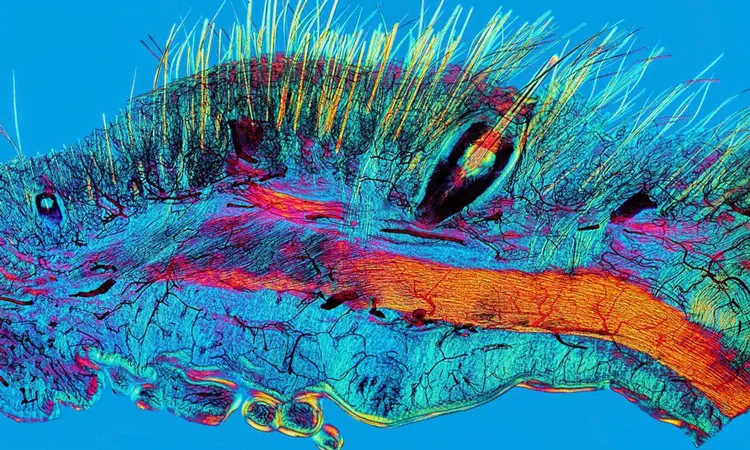
A polarized light micrograph of a section of cat skin showing hairs, whiskers and their blood supply, created from a vintage prepared slide dating before 1900.
Blood vessels were injected with dye (carmine; black) before fixing and sectioning the tissue in order to visualize the capillaries in the tissue. This was a newly developed technique at that time. The capillary bed seen here supplies the hairs and whisker. The fine hairs, thicker whisker, blood vessels and underlying muscle are all visible.
Image courtesy of David Linstead, Wellcome Images.
