Cory Dobson, Author at Sanford Burnham Prebys - Page 15 of 41
Institute News

Preuss School interns get an “A” grade at SBP

AuthorHelen I. Hwang
Date

August 4, 2017

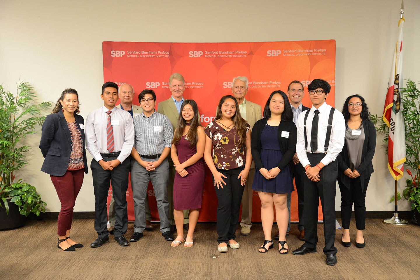
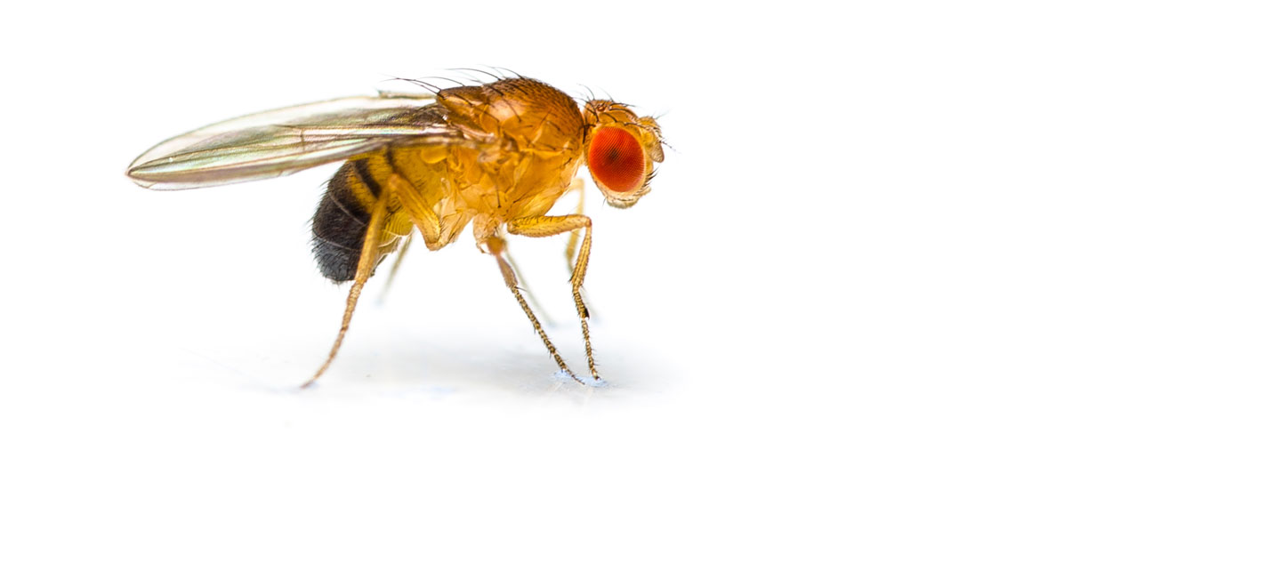
“I got to do things I never thought I could do,” said Yadira Gomez Rangel, 16, a rising junior at Preuss School in San Diego. “I got a chance to dissect a fly, which I didn’t think I could do,” she told the audience at Sanford Burnham Prebys Medical Discovery Institute (SBP), which included SBP Trustees Malin Burnham and Wain Fishburn as well as CEO Perry Nisen, MD, PhD

Rangel is one of seven students from the prestigious Preuss School, who completed a two-week internship. Students from the Preuss School, affiliated with UC San Diego, strive to become the first in their families to graduate from college. The SBP Preuss program is designed to introduce young scientists-in-training to medical research by working hand in hand with our scientists.

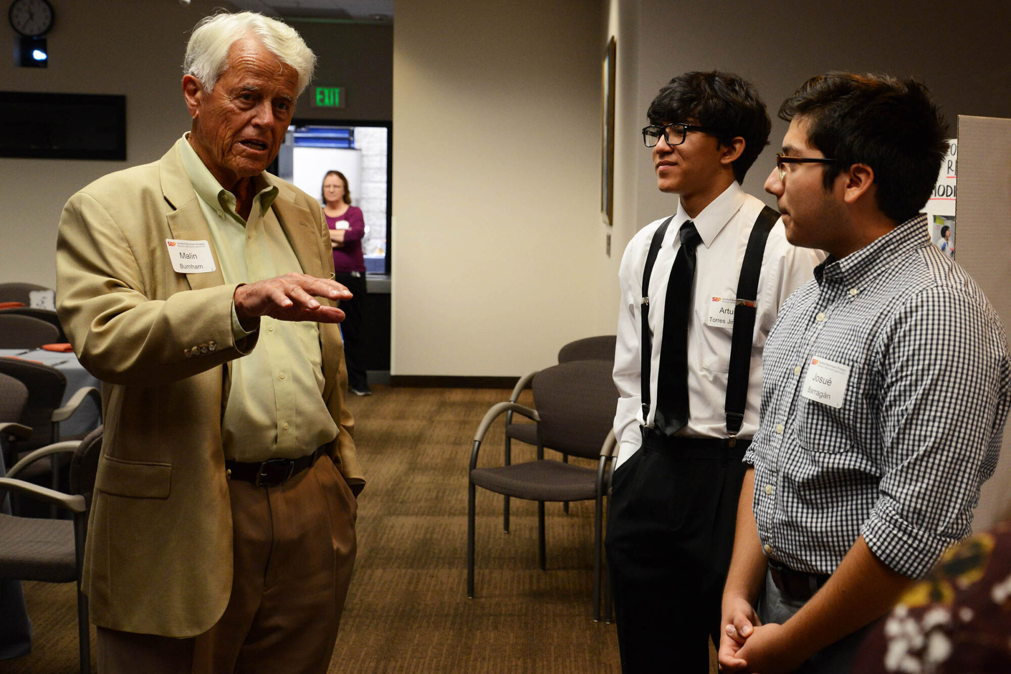
The group of 16-year-olds got a chance to rotate among four different labs at SBP. The other students included Michelle Villa Bardales, Josué Barragán, Edizandro Morales Herrera, Arturo Torres Jimenez, Jenny Nguyen and Natalie Nguyen. Students presented posters in English and Spanish, received a certificate and a stipend for their hard work.

Fishburn said the Preuss program at SBP was “inspirational” as he hoped the young teens would continue their path in science. At the celebratory luncheon with students, their families and SBP staff, Fishburn chatted with Tommy Le, a Preuss School graduate. Le was part of the SBP Preuss program for the two-week internship, followed by a six-week internship the following year, and is now doing a summer internship at SBP before entering UC San Diego in the fall where he’ll major in biochemistry.

Each summer, SBP also hosts a six-week internship for rising Preuss seniors, sponsored by the NIH CURE program. Two of the seven interns (who happen to be all female), Gizelle Avitia Mejica and Julieta Morales Ornelas, also completed the two-week Preuss program, which inspired them to apply again at SBP. “About 90 percent of what I learned in the lab I wouldn’t have been taught in the classroom,” says Mejica.  

During the internship, the teenagers studied several aspects of medical research. They examined the correlation between obesity and heart disease in fruit flies in the laboratory of Rolf Bodmer, Ph.D. Also, the kids studied zebrafish and tackled the challenge of curing diabetes in the laboratory of Duc Dong, Ph.D. They looked at how to use C. elegans worms to understand the aging process in the laboratory of Malene Hansen, Ph.D. Finally, in the laboratory of Jing Crystal Zhou, Ph.D., the young scientists learned about RNA modification, a process that occurs in all living organisms and can influence how diseases occur.

With hands-on training and in-depth laboratory involvement, the Preuss students gained invaluable skills and networking opportunities. The program is made possible by founding philanthropists Peggy and Peter Preuss and Debby and Wain Fishburn. Jimenez said, “It’s been a wonderful experience!”

Preuss School Internship Program with SBP Trustees


Institute News

SBP researchers awarded Padres Pedal the Cause collaborative grants

AuthorSusan Gammon
Date

July 31, 2017

Sanford Burnham Prebys Medical Research (SBP) is pleased to announce that it has been awarded five collaborative grants with the Moores Cancer Center at UC San Diego Health. The collaborative research projects are focused on cancers including B-cell lymphomas, colorectal cancer, pancreatic cancer and breast cancer. The awards are part of the $750,000 being distributed from proceeds raised by the 2016 cycling event.

“I am proud of our scientists and our partnership with Padres Pedal the Cause,” says Garth Powis, D. Phil., director of SBP’s NCI-designated Cancer Center. “Since its inception, Pedal the Cause has focused on creating a community event that engages cyclists and volunteers to raise money to advance innovative cancer research. We look forward to using these grants to make advances in our labs that will hopefully impact the health of cancer patients now and in the future.”

In November 2016, more than 1,500 riders, hundreds of volunteers, donors and sponsors took part in the cycling event. SBP was pleased to host water station for riders during the event, and many riders even stopped to take fun photos in front of our SBP bright orange backdrop.

SBP’s funded projects are listed below:

“Oncogenic Regulation of B-Lymphomagenesis by the Chromatin Modulator DOT1L”
Bing Ren, PhD (Moores Cancer Center at UC San Diego Health) Aniruddha Deshpande, PhD (Sanford Burnham Prebys Cancer Center)

“Decoding Colon Cancers Using Boolean Principles”
Pradipta Ghosh, MD (Moores Cancer Center at UC San Diego Health) Debashis Sahoo, PhD (Moores Cancer Center at UC San Diego Health), Manuel Perucho, PhD (Sanford Burnham Prebys Cancer Center)

“An Over-Expressed GPCR in Pancreatic Cancer Associated Fibroblasts as a Novel Therapeutic Target”
Paul Insel, MD (Moores Cancer Center at UC San Diego Health) Kristiina Vuori, MD, PhD (Sanford Burnham Prebys Cancer Center)

“Identification of Genes Critical for the Production of T-cells from Human Pluripotent Stem Cells for Development of “Off-the-Shelf” T-cells Immunotherapies”
Dan S. Kaufman, MD, PhD (Moores Cancer Center at UC San Diego Health) Sumit K. Chanda, PhD (Sanford Burnham Prebys Cancer Center)

“Targeting Cellular Mechanotransduction in Breast Cancer Metastasis”
Jing Yang, PhD (Moores Cancer Center at UC San Diego Health) Elena Pasquale, PhD (Sanford Burnham Prebys Cancer Center)

The fifth annual Padres Pedal the Cause event takes place November 11-12, 2017, at Petco Park and will feature courses of various distances for all skill levels, a stationary bike zone, virtual riding, a children’s ride as well as numerous volunteer opportunities for all those who want to make a difference in the fight against cancer.

Registration for the 2017 event is open. New this year, Padres Pedal is only the second cycling event to ride over the Coronado Bay Bridge.

For more information and registration please visit www.gopedal.org 

Join us on this year’s ride as a rider or volunteer. We’d love to have you on the team! Register today: Team SBP – Sanford Burnham Prebys 

Institute News

Cell Suicide: Caspases Call the Shots

AuthorSusan Gammon
Date

July 28, 2017

People are usually surprised to learn that many cells in our bodies essentially commit suicide. These cells don’t die because they are sick, but because they are following built-in signals that initiate their death. 

A few examples illustrate how this regulated cell death is really not surprising at all, but in fact is essential for proper development and health: 

  •  Developing fetuses generate many more cells than they can use. Excess cells that don’t contribute effectively to developing tissues must be eliminated to avoid accumulation of non-functional cells. 
  •  Many adult tissues contain cells that proliferate rapidly to replace old, worn-out cells. Without elimination of the old cells, tissues would balloon up  with non-functional cells. Adults may lose more than 50 billion cells every day due to this cell replacement process. 
  • Abnormal cells need to be eliminated to avoid development of diseases like cancer. The loss of ability to undergo regulated cell death is one  hallmark of malignant cancer cells.

What really is surprising about regulated cell death is the number of different ways cells can terminate themselves. This suggests that the different cell death pathways must communicate with each other to decide which mechanism will be used in a given situation. 

In a commentary in Cell Chemical Biology, Guy Salvesen, PhD, professor at  SBP reviews a recent report that partly solves the riddle of communication between two of these cell death pathways, called apoptosis and pyroptosis. 

Salvesen explains, “Apoptosis and pyroptosis are similar in that they both rely on protein-cleaving enzymes called caspases to carry out the cell death sentence. But there are big differences. Pyroptosis is a messy death that releases a lot of cell debris, activates the immune system and triggers inflammation. It does this via caspase-1, a version of the enzyme that chops up a protein called gasdermin-D (GSDMD) to create a small toxic piece of GSMD that mediates the process.  

“In contrast, apoptosis uses caspase-3 and caspase-7 to initiate cell death by damaging the cell nucleus and its DNA, causing a relative quiet cell death with minimal disruption to the rest of the body. These caspases also cleave GSDMD, but in way that doesn’t create the toxic fragment. Apoptosis is dominant—so once it’s triggered pyroptosis is put on the back burner.”

First author Marcin Poreba, PhD, a postdoc in the Salvesen lab, adds that, “The decision of a cell to use pyroptosis versus apoptosis depends to a large extent on the strength of the death signals the cell receives. In most cases, apoptosis will win out because of its ability to block the pyroptosis process. This is a good thing, since regulated cell death usually occurs as part of normal development and maintenance programs in which inflammation should be avoided. But in cases where pyroptotic stimuli are strong enough, for example in response to the need to eliminate cells infected by bacteria or viruses, the pyroptotic pathway can override apoptosis and terminate cells in a way that also recruits the immune system into the battle. It’s almost like a contest to see which caspases win the death race. 

Read the paper here.

Institute News

Are stem cells to blame for cancer re-growth?

AuthorBill Stallcup, PhD
Date

July 16, 2017

The scientific and popular media are both full of excitement about the use of stem cell therapies for replacing diseased or damaged tissues. In a new twist to this story, researchers are wondering if small populations of stem cells present in tumors (known as cancer stem cells) may be responsible for the ability of cancers to survive and re-establish themselves, even after the malignancies are apparently eliminated by a combination of surgery, chemotherapy and radiotherapy.

In a new report in the Journal of Clinical Oncology, Robert Wechsler-Reya, PhD, director of the Tumor Initiation and Maintenance Program at SBP, Luis Parada, PhD, from Memorial Sloan Kettering Cancer Center and Peter Dirks, MD, PhD, from Toronto’s Hospital for Sick Children, review the evidence that cancer stem cells may contribute to brain tumor re-appearance. As background, Wechsler-Reya explains that, “There are several good theories about how tumors can develop resistance to therapy and then re-appear after therapy ends. One theory proposes that the unique regenerative properties of cancer stem cells underlies the ability of tumors to rebound.”

So why wouldn’t aggressive cancer therapies destroy cancer stem cells along with the other tumor cells? Significantly, many cancer drugs are designed to be effective against rapidly-proliferating tumor cells. In contrast, both normal stem cells and cancer stem cells often exhibit low rates of proliferation. This allows them to sit quietly on the sidelines, dodging the lethal effects of the therapy and then ramping up their proliferation post-therapy to re-populate the damaged tissue. In the case of normal stem cells this leads to repair of damaged organs, while in the case of cancer stem cells it leads to re-growth of tumors.

According to Wechsler-Reya, some of the best evidence for the existence and power of cancer stem cells comes from studying brain tumors in mice. “Researchers have analyzed several types of mouse brain cancers by separating the tumor cells into pools that carry different markers (like sorting a bag of M&Ms into piles containing the different colors). These separate pools are then injected back into the brains of new mice to compare their tumor-growing abilities. Cells with markers thought to characterize brain tumor stem cells (e.g. the red M&Ms) can produce new tumors even when very few cells are injected. In contrast, the majority of tumor cells (all the other colors of M&Ms) have very poor ability to produce new tumors, highlighting the unique regenerative power of the cancer stem cell population.”

Researchers can now do similar experiments with human brain cancers by injecting tumor cells into special mice that lack an immune system and thus can’t reject the human cells. These studies show that human brain tumors also contain cancer stem cells that can regenerate tumors when transplanted in low numbers. These mouse studies may therefore serve as valuable tools for understanding human brain tumor stem cells and for devising ways to deal with them. For example, researchers hope that cancer stem cells may prove vulnerable to new types of targeted therapies that don’t depend on rapid tumor cell proliferation for their success. Such therapies would destroy cancer stem cells along with the other tumor cells, hopefully avoiding tumor re-growth.

Read a copy of the paper here.

Institute News

Genes and proteins go hand-in-hand

AuthorBill Stallcup, PhD
Date

July 14, 2017

Thanks to huge improvements in DNA sequencing technology, scientists have identified almost all the genes present in humans. Despite this achievement, there are still thousands of genes whose functions remain a mystery. Since genes are basically blueprints for making the proteins needed to run our cellular machinery, connecting genes with the specific functions of their encoded proteins is a critical next step in using genomic information to solve health-related problems.

Bridging the gap between gene sequence and protein function is the topic of a study published in the Journal of Biological Chemistry by Yu Yamaguchi, MD, PhD, professor at SBP. According to Yamaguchi, “There has been a long-standing mystery concerning the processing of hyaluronic acid (HA), a large sponge-like molecule required to maintain proper spacing between neighboring cells. We had already learned a lot about how cells make HA, but the other equally important side of the equation is how HA is broken down, which is needed to prevent HA build-up that can cause tissue fibrosis.”

The Yamaguchi lab knew that HA degradation must be accomplished by enzymes that cut HA into smaller pieces for further processing. “However, none of the known HA-cutting enzymes had the ability to cut the very large HA that exists outside the cell”, explains postdoctoral fellow Hayato Yamamoto, MD, PhD, first author on the study. “We decided to search gene sequence libraries to identify other proteins that were not previously suspected to cut HA, but which were structurally similar to known HA cutters.”

Their search turned up transmembrane protein 2 (TMEM2), whose structure predicted that it would exist on the cell surface and would also be able to cut HA. “My job was then to determine whether or not this protein could live up to our predictions,” recalls Yamaguchi. “We were able to show experimentally that the protein really did exist on the cell surface and was able to cut large HA molecules into smaller fragments for further processing inside the cell.”

Read the paper here.

Institute News

Will flies help fix heart rhythm problems?

AuthorSusan Gammon
Date

July 12, 2017

Your heart beats about 35 million times in a single a year. That’s a whopping number of beats—each generated by electrical signals that make the heart contract. Occasionally problems with heart’s electrical system can cause irregular rhythms, or arrhythmias. Some types of arrhythmias are merely annoying; others can last long enough to affect how the heart works, or even cause sudden cardiac arrest.

Although certain arrhythmias can be successfully treated with medication, surgery and/or devices such as pacemakers, cardiac disorders and heart disease still account for more deaths than any other disease.

Finding new treatments for arrhythmias requires a deep understanding of how the heart beats, and specifically the intricate electrical system that prompts the heart to contract. It also requires a model to study. Is Drosophila melanogaster—a type of fruit fly—the answer?

Using flies to study the heart

Both the human heart and fly hearts have four chambers and both start out as linear tubes in embryos—but ours loops during development to form a more compact structure where as a the fly heart does not. Despite this structural difference there are many functional similarities between the fly and human heart.

“One of the similarities that we focus on in my lab is the way ion channels work—and don’t work—to fully understand how faulty ion channels contribute to heart arrhythmias,” says Karen Ocorr, PhD, assistant professor at SBP.

Ion channels are proteins found in cell membranes that allow specific ions such as potassium, sodium and calcium, to pass through cells. When ion currents travel into heart muscle cells, the muscle becomes depolarized, creating an electrical current that causes the heart to contract. A second set of channels are important in repolarization of the heart, which allows it to relax and refill with blood.

In her new paper published in PLOS Genetics, Ocorr describes how two repolarizing potassium ion channels called hERG and KCNQ control the rate and efficiency of fly heart contractions—similar to their role in human heart muscle. The research also shows that mutations in hERG and KCNQ lead to arrhythmias that worsen with age—as they do in humans.

“In humans, when hERG is compromised, either by drugs or inherited mutations, hearts can take longer than normal to recharge between beats, causing a potentially fatal condition called long QT syndrome. In fact, some anti-arrhythmia drugs actually cause long QT syndrome, hence the need for better, more specific therapies,” explains Ocorr.

The capacity for drugs to cause long QT syndrome has led the Food and Drug Administration (FDA) to recommend including the evaluation of new cardiac and non-cardiac drugs for this possible side effect. The FDA is the United States agency that provides licenses to market new drugs.

Interestingly, neither of the ion channels we identified in the fly heart play a major role in the adult mouse heart, ruling it out as useful model to screen for drug-related long QT effects,” says Ocorr.

“We are encouraged that Drosophila may become an easy, accurate tool to pre-clinically screen for adverse cardiac events associated new anti-arrhythmia therapies—potentially making the next drug discovery for patients happen sooner.”

Read the paper here

Institute News

Immune scavenger cells: Good guys or bad guys?

AuthorSusan Gammon
Date

July 9, 2017

The immune system is our main defense against foreign invaders (bacteria and viruses) and also against mutant cells that develop into cancer. Some of the first immune responders to these threats are scavenger cells called macrophages that destroy targets by internalizing and degrading them.

But macrophages can be tricked. When they are continually activated in a chronic disease like cancer, they can be “instructed” by cancer cells to perform functions that benefit the growing tumor instead of destroying it.

Recently, William Stallcup, PhD, professor at Sanford Burnham Prebys Medical Discovery Institute (SBP) published a study in Trends in Cell and Molecular Biology describing one way that cancer cells transform macrophages from tumor fighters into tumor helpers.

“We found that a protein called NG2 on macrophages is important for the ability of these scavengers to exit from blood vessels and migrate into tumors,” explains Stallcup. “When we knocked out NG2 on macrophages in mice, instead of allowing mouse brain tumors to grow faster (in the absence of the scavenging macrophages), the tumors actually grew much more slowly. This was somewhat surprising since we expect macrophages to help fight cancer.”

“It turns out that tumor cells can modify macrophage function, making them produce factors that stimulate the formation of tumor-nourishing blood vessels, says Pilar Cejudo Martin, PhD, a postdoc in Stallcup’s lab and first author of the paper. “By knocking out NG2, we blocked macrophage entry into tumors so that there were fewer macrophages for the tumors to use to their own advantage. So due to poor blood vessel development, tumor growth was slowed.”

So does this mean that blocking NG2 could be a general means of improving treatment of diseases involving chronic macrophage recruitment?

“It depends,” says Stallcup. “When we used a mouse model of multiple sclerosis (MS) to show that knockout of NG2 impaired macrophage entry into the damaged spinal cord, we found that recovery was hindered. That’s because macrophages are needed to produce factors that stimulate production of the cells responsible for damage repair.

“So while blocking macrophage NG2 might be useful for cancer therapy, it looks like this approach would be counter-productive for treating MS. Even though macrophages seem like attractive targets for therapy, they have such a large bag of tricks that we will probably have to deal with blocking their effects on a case-by-case basis,” adds Stallcup.

Read the paper here.

Institute News

SBP spin off company AivoCode receives funding to advance drugs to treat brain injuries

AuthorSusan Gammon
Date

June 22, 2017

Every year, over 10 million people worldwide injure their brain, and it’s the most common cause of death and disability in young people. There are currently no drugs available to limit the additional damage to the brain from swelling and inflammation after the injury or help repair the brain.

Novel technology developed in the lab of Erkki Ruoslahti, MD, PhD, distinguished professor at SBP, has led to spin off company called AivoCode that just received funding from the National Science Foundation to advance a new platform for site-specific delivery of drugs to treat acute brain injury.

The approach uses a peptide sequence of four amino acids, cysteine, alanine, glutamine and lysine (CAQK) that recognizes brain tissue. The CAQK peptide binds to the components of the meshwork surrounding brain cells called chondroitin sulfate proteoglycans. Amounts of these large, sugar-coated proteins increase following brain injury, and CAQK can carry drugs and nanoparticles to damaged areas in the brain.  The original proof-of-concept studies were performed on mouse models of acute brain injury and human brain tissue samples.

The technology may make it possible to use new types of drugs that would otherwise not reach their target in the brain. If the company is successful in bringing the technology to the clinic, it may improve the outcome for brain injury victims and provide significant healthcare savings.


Institute News

How tumors shape their environment for their benefit

AuthorSusan Gammon
Date

June 21, 2017

In a new review article published in Current Opinions in Cell Biology, Professors Jorge Moscat, Maria Diaz-Meco and their graduate student Miguel Reina-Campos summarize evidence from their labs and others of how tumor cells play a large part in converting their surroundings to promote cancer progression. The research is important for developing therapies aimed at the area surrounding a tumor—the stroma—to reduce the likelihood of drug resistance, which is the main problem that today’s cancer therapies face.

“Although tumor cells carry mutations that are responsible for their altered behavior, the ability of tumors to grow and metastasize is controlled to a surprising extent by contributions from elements supplied by normal host (patient) cells,” says Diaz-Meco. “Blood vessels that provide nutrients to tumors are a good example of host elements that are critical for tumor growth and metastasis, but other host components such as fat cells, fibroblasts, and immune cells also contribute in important ways to tumor progression. These host components are known collectively as the tumor stroma or tumor microenvironment (TME).”

Different types of host stromal cells in the TME also contribute to a tumor’s ability to use different energy sources, and in turn are themselves influenced by the tumor’s metabolic activity. Reina-Campos points to cancer associated fibroblasts (CAFs) and pancreatic stellate cells (PSCs) as two cell types that interact with pancreatic cancer cells. He explains, “Under nutrient stress, PSCs can use a cell process called autophagy to produce the amino acid alanine, while CAFs produce the amino acid glutamine. Both of these amino acids serve as rich energy sources for the pancreatic tumor cells, which otherwise would have to rely on glucose—a less potent nutrient source.”

In addition to providing nutrients, CAFs and PSCs are also the source of fibrous material known as the extracellular matrix (ECM). The ECM is a potent stimulus for tumor progression and metastasis and is a particularly abundant component of pancreatic tumors. The authors stress that the properties of CAFs and PSCs in the TME are different from those of normal fibroblast and stellate cells, leading to the important concept that the stromal cells are strongly influenced by signals from the tumor cells, and reciprocally (once educated by the tumor cells) can sustain their growth to higher levels of malignancy.

Immune cells are another key TME component whose properties are profoundly influenced by tumor cells, and in turn have a significant ability to promote tumor growth. Moscat explains, “Given that the normal job of immune cells is to seek out and destroy abnormal invaders, the ability of tumors to convert immune cells into tumor promoting agents is really striking. Several different types of immune cells are re-programmed by tumors as a means of suppressing an anti-tumor immune response and generating a tumor promoting response”.

Overall, these studies reveal a number of ways by which tumor cells signal to stromal cells and, conversely, by which stromal cells signal back to tumors. Further defining these tumor-to-stroma and stroma-to-tumor signals can lead to deeper understanding of mechanisms of tumor-stroma crosstalk. Ideally, such mechanisms could then be inhibited by drugs as a means of improving tumor therapy, possibly in combination with other novel forms of cancer therapy that boost immune function.

Read the paper online here.

Institute News

2017 BIO features SBP scientists

AuthorKristen Cusato
Date

June 19, 2017

Two SBP researchers will be sharing their knowledge and insights at the 2017 BIO International Convention, which will be held June 19th to the 22nd at the San Diego Convention Center.

Rolf Bodmer, PhD, professor in the Development, Aging and Regeneration Program, will be part of a panel on June 21st titled, “Reimagine Old Age: New Frontiers in the Science of Aging.”  Bodmer will highlight new biomedical research, as other panelists report on drugs that are presently in development and discuss how pharmaceutical companies are approaching new treatments for diseases related to aging.


“With our population aging, it becomes more important to understand and find ways to remedy the rapid decline and diseases that come with getting older,” Bodmer says. “We don’t necessarily need to live that much longer, but we need to find out how to have all of our organ systems work their best, to live healthier and happier as we age.”


Bodmer will also talk about the use of fruit fly models in his lab at SBP to study different aspects of aging, including cardiac function.


“Organisms like the fruit fly have an infinite box of genetic tools that can be used to figure out how our genetics and signaling pathways work. What we learn from them can help in the development of drugs for people with age-related diseases,” says Bodmer.


Rolf Bodmer, PhD


Reimagine Old Age: New Frontiers in the Science of Aging


June 21st 10:45am Room 6C upper level SDCC

Scott Peterson, PhD, professor in the Tumor Microenvironment and Cancer Immunology Program, will be on the “Microbiome 2.0: Going Beyond Bugs as Drugs” panel, also on June 21st.  Dr. Peterson’s focus will be on using prebiotics as a way of modulating or altering the gut microbiome to impact health.

Additional panelists will discuss new microbiome targets, novel approaches to regulate the gut-brain axis and how big data fits in.

“Beyond bugs as drugs means it’s no longer just about probiotics, now prebiotics have become part of the equation,” Peterson says. “We want to know if we can use pre-biotics to change someone’s microbiome to make them more responsive to certain drugs.”

Peterson says the translational aspect of microbiome research is starting to take form, and he is looking forward to talking about his research with potential new partners at BIO 2017 and beyond.

“NIH funding is hard to come by right now, and getting biotech interested in the microbiome will eventually benefit all of us.”

Scott Peterson, PhD

Microbiome 2.0: Going Beyond Bugs as Drugs

June 21st 4:15pm Room 6C upper level SDCC