Cory Dobson, Author at Sanford Burnham Prebys - Page 17 of 41
Institute News

Highlights from the Graduate School of Biomedical Sciences annual retreat

AuthorSusan Gammon
Date

May 24, 2017

On May 18-19, SBP’s Graduate School of Biomedical Sciences held its eighth annual retreat in San Marcos, Calif.

This year’s theme, “Using Collaborations to Enhance Your Research,” gave our PhD students an opportunity to share their research projects and explain how collaborators are helping them achieve their goals. At SBP, we encourage scientists to work with researchers in other disciplines to answer questions of broad scope and complexity—questions that would remain unanswered without collaboration.

At the retreat, each student presents their work and is scored by their fellow students, faculty and mentors on a scale of 1 to 9 for quality, content and clarity of presentation. When the scorecards are tallied, the winner is presented with the “Howling Coyote” award.

Wesley McKeithan, SBP Graduate School of Biomedical Sciences
Wesley L. McKeithan, graduate student in the lab of Mark Mercola, PhD

This year’s winner was Wesley L. McKeithan, a PhD candidate in the laboratory of Mark Mercola, PhD Wesley’s presentation, titled “Use of patient-derived long QT syndrome type 3 hiPSC cardiomyocytes to develop a new anti-arrhythmic therapeutic,” covered his scientific journey to find a treatment for this rare condition that can cause symptoms, which range from fainting to sudden death.

Highlights from the retreat included a presentation by keynote speaker Jerold Chun, MD, PhD, professor and senior vice president of Neuroscience Drug Discovery at SBP, who gave an inspirational success story of his research on lysophospholipid signaling that led to Gilenya®, the first oral drug used to treat multiple sclerosis.

Guest keynote speaker Bill Parks, PhD, professor at Cedars-Sinai Medical Center, led an entertaining interactive session on the do’s and don’t of collaboration—sharing his best experiences and some of the worst (without naming names).

Many congratulations to all of the student participants as well as Guy Salvesen, PhD, dean of the Graduate Program, for providing leadership and educational opportunities for the students.

Special thanks to:

  • Student coordinators, Katja Birker and Daniel Ojeda Juarez
  • Mary Bradley, manager of the graduate program, and Alexia Pimentel, program coordinator, for organizing the retreat
  • And to Pfizer, Genentech and Celgene for helping to underwrite the event


Institute News

Inspiring the next generation of researchers and physicians

AuthorDeborah Robison
Date

May 24, 2017

Summer scholars investigate future careers in science and medicine. 

Nearly 28 percent of high school freshmen declare interest in a STEM-related field. That’s roughly one million students each year. But unfortunately, of these students, 57 percent will lose interest by the time they graduate from high school.

Summer internships are often sought for the boost they give to a resume but they also provide valuable direction for students unsure of how to chart a course toward a scientific career.

As Central Florida’s high school graduates prepare to head off to college, there is a small group who may have a leg up on their peers. Students who donned lab coats last summer at SBP Lake Nona, took part in an immersive lab experience to try their hands full-time at what could be a future career in a STEM-related field. Already academic stars, the practical experience fueled their passion for research and medicine.

“I learned a great deal about bench research techniques which I believe helped me stand out to admissions officers,” said Andrew Tiu, a Bishop Moore High School graduate who is headed to Georgetown University in the fall. “My internship also helped me learn about the mentality and motivation behind scientific research. It truly gave me a taste of the work I hope to take up in the future.”

Andrew Tiu

Then there’s Kathleen Garvey from Trinity Preparatory School who plans to pursue a special focus in research at Johns Hopkins; she acknowledges her lab mentor Peter Crawford, PhD, for helping ignite that passion. “My internship made me certain about wanting to study the life sciences and actively pursue more research opportunities.”

Kathleen Garvey

Other interns were similarly motivated. Veronica Eslava (University) and Saumya Kapoor (Cypress Creek) will attend University of Florida; Prianca Nagda and Rakhi Patel (Trinity Prep) are destined for Georgetown and University of Miami; and Steven Jones (East River) will attend University of Central Florida.

Is a career in science for you? What students should know.

We asked our faculty, “What advice would you give to high school and college students who are interested in research as a career?” Here are their top ten tips:

1. Develop a solid science foundation during high school—no need to focus on highly specialized courses but build a base of math, chemistry, physics and biology.

2. Take elective courses that offer a lab experience. It can be anything that allows you to set up small experiments or to be involved in a science project.

3. Join extracurricular groups that have an interest in science.

4. Volunteer for a few weeks or a summer at a “real” research place, such as a laboratory at a university or college.

5. Read general science articles written for non-specialists such as Scientific American. See what area triggers your interest.

6. The earlier you get your hands wet in the lab, the better. Start generating data and publishing papers ASAP. In this field you will be judged by the quantity and quality of your publications.

7. Besides lots of biology and chemistry, biomedical research can also involve plenty of physics, math, and computer science—stay open to linking various disciplines.

8. Education is expensive and life science careers may need additional studies after obtaining a bachelor’s degree.

9. Talk to a lot of people that have pursued a science career.

10. Enroll in your dream subject but look for cross-training opportunities.

Institute News

Exciting diabetes and obesity research highlights from Medical City

AuthorDeborah Robison
Date

May 22, 2017

Center for Metabolic Origins of Disease

With more than one-third of adults in the U.S. considered obese, scientists are searching for new ways to treat obesity and associated health problems such as type 2 diabetes. Four researchers from Sanford Burnham Prebys Medical Discovery Institute (SBP) at Lake Nona have been invited to present new perspectives and insights at the American Diabetes Association’s 77th Scientific Sessions, to be held June 9-13, 2017, in San Diego. The conference is the world’s largest gathering of research experts and clinicians focused on diabetes research, prevention and care. The presentations will inform new treatment strategies for the nearly 30 million people diagnosed with diabetes.

Potential early therapeutic target for diabetes prevention
Obesity often leads to accumulation of fat in muscle and faulty machinery involved in taking up glucose from a meal to use it for energy, leading to type 2 diabetes. A recent advance from the laboratory of Daniel P. Kelly, MD, scientific director of SBP at Lake Nona, may lead to a way to stop this pre-diabetic state from advancing. Dr. Kelly will present findings on a recently discovered cellular glucose sensor in muscle that serves as a key connection between insulin resistance and accumulation of fat in muscle, which occurs in obesity-related diabetes. When the protein is inhibited in skeletal muscle cells, regulatory genes that influence glucose uptake and insulin signaling are enhanced. The team is now validating the pathway as a therapeutic target to prevent type 2 diabetes.

Fatty liver and type 2 diabetes
Peter Crawford, MD, PhD, director of SBP’s Cardiovascular Metabolism Program, is studying the root causes of nonalcoholic fatty liver disease (NAFLD), a condition that affects nearly 80 percent of people with type 2 diabetes. About 5 percent of NAFLD cases advance to liver cirrhosis – a disease characterized by scarring and fibrosis that could require liver transplant. Dr. Crawford is an expert on how the liver processes energy derived from food. At the ADA meeting, he will discuss how the interruption of normal fat metabolism can lead to enhanced scarring. Through ongoing research, he hopes to be able to specifically identify which diabetes patients are at risk of developing advanced liver disease and to develop therapies that protect against disease progression.

Brain nutrient sensors help maintain energy balance
Diabetes researcher Julio Ayala, PhD wants to understand how specialized regions in the brain control food intake, energy expenditure and body weight. His ADA presentation will focus on how nutrient-sensors that control the balance between energy-consuming and energy-producing processes in almost every cell in our bodies also play a very specific role in the brain. His research shows that hormones, such as glucagon-like peptide-1 (GLP-1) regulate the activities of these brain nutrient sensors to influence hunger, satiety and ultimately body weight. Defective sensors are implicated in obesity and could be a target for new therapeutic treatments.

Glucose Sensor in Macrophages
Insulin resistance is a key feature of type 2 diabetes. When present, the impairment prevents insulin from getting glucose into muscle where it’s used for energy, and instead causes blood sugars to become elevated. The events that drive the development and progression of insulin resistance are not known. Laszlo Nagy, MD, PhD, director of SBP’s Genomic Control of Metabolism Program, will present new research that suggests that the inflammatory process—and specifically a type of white blood cells called macrophages—are involved. He will present a novel hypothesis on the role of macrophages, defined in Greek as “big eaters”, and identify molecules involved in muscle growth and glucose metabolism. His research aims to reveal cellular interactions that could become new therapeutic targets to treat type 2 diabetes.

Institute News

What SBP Scientists are Researching to Battle Skin Cancer

AuthorHelen I. Hwang
Date

May 16, 2017

Skin cancer is one of the most common of all cancers, and melanoma accounts for about 1 percent of skin cancers. However, melanoma causes a large majority of deaths from that particular type of cancer. Alarmingly, rates of skin cancer have been on the rise in the last 30 years. Here in Southern California, our everlasting summer comes with a price. Exposure to sun increases our risk to melanoma.

Melanoma occurs when the pigment-producing cells that give color to the skin become cancerous. Symptoms might include a new, unusual growth or a change in an existing mole. Melanomas can occur anywhere on the body.

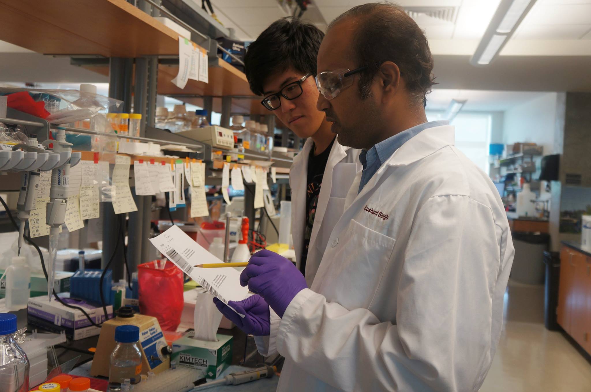
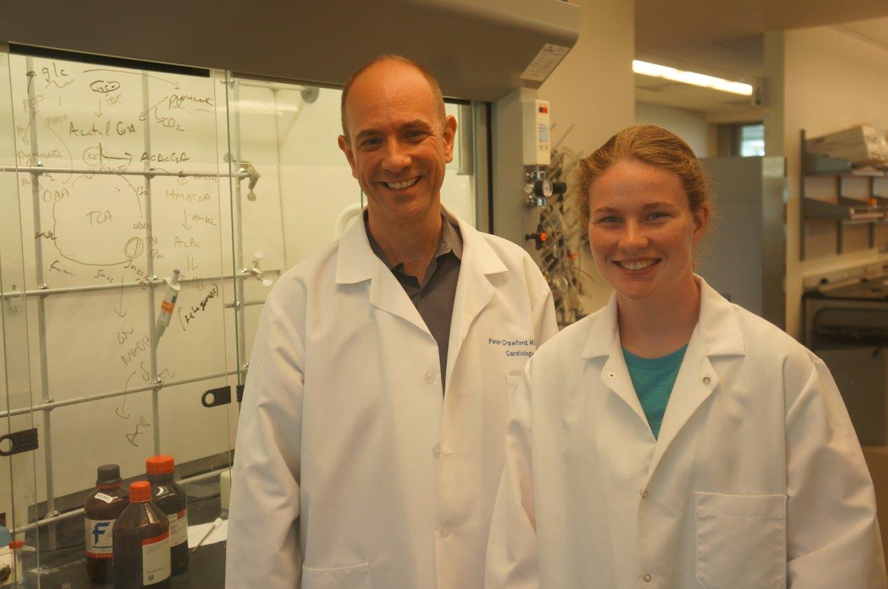
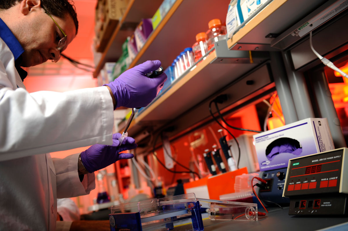
At Sanford Burnham Prebys Medical Discovery Institute (SBP), we have several researchers working on the causes of melanoma and discovering new ways to treat this deadly disease.

Here is a roundup of SBP’s latest research:

Key findings show how melanoma develops in order to identify potential therapeutic targets

Ze’ev Ronai, PhD
Professor and SBP Chief Scientific Advisor

Ronai’s laboratory has been studying how rewired signaling networks can underlie melanoma development, including resistance to therapy and metastatic propensity. One player in that rewiring is a protein called ATF-2, which can switch from its usual tumor-preventive function to become a tumor promoter when combined with a mutation in the human gene called BRAF.

Ronai’s work on a protein, ubiquitin ligases, led to the identification of RNF125 as an important regulator of melanoma resistance to a common chemotherapy drug. RNF125 impacts melanoma resistance by its regulation of JAK2, an important protein kinase which could play an important role in melanoma resistance to therapy.

Work on the ubiquitin ligase Siah2 identified its important role in melanoma growth and metastasis, and its contribution to melanomagenesis. Melanoma is believed to be a multi-step process (melanomagenesis) of genetic mutations that increase cell proliferation, differentiation, and death.

Work in the lab also concern novel metabolic pathways that are exploited by melanoma for their survival, with the goal of identifying combination drug therapies to combat the spread of melanoma. Earlier work on the enzyme PDK1 showed how it can be a potential therapeutic target for melanoma treatment.

Immunotherapy discovery has led to partnership with Eli Lilly

Linda Bradley, PhD
Professor, Immunity and Pathogenesis Program, Infectious and Inflammatory Diseases Center

Bradley’s group is focused on understanding how anti-tumor T cells can be optimized to kill melanoma tumors. They discovered an important molecule (PSGL-1) that puts the “break” on killer T cells, allowing melanoma tumors to survive and grow. Using animal models, they removed this “break” and T cells were able to destroy melanoma tumors. They have extended their studies and found that in melanoma tumors from patients, T cells also have this PSGL-1 “break”. Bradley’s lab has partnered with Eli Lilly to discover drugs that can modulate PSGL-1 activity in human disease that may offer new therapies for patients.

Knocking out a specific protein can slow melanoma growth 

William Stallcup, PhD
Professor, Tumor Microenvironment and Cancer Immunology Program

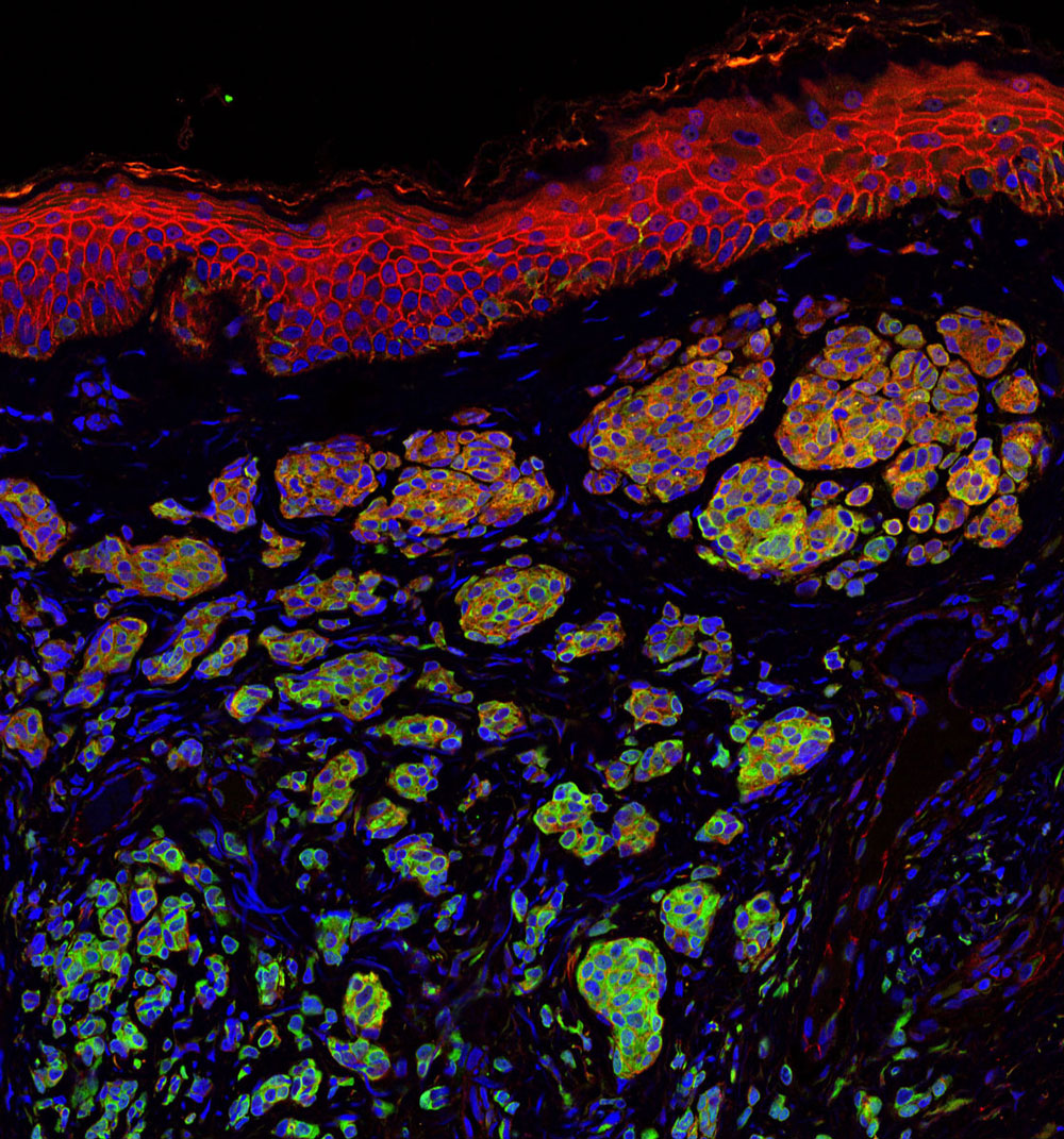
The danger of melanomas is their metastasis to organs, such as the brain, in which surgical removal is not effective. By injecting melanoma cells into the brains of mice, we have shown that the NG2 protein found in host tissues makes the brain a much “friendlier” environment for melanoma growth.

Specifically, NG2 is found on blood vessel cells called pericytes and on immune cells called macrophages. The presence of NG2 on both cell types improves the formation of blood vessels in brain melanomas, contributing to delivery of nutrients and thus to accelerated tumor growth. Genetically knocking out NG2 in either pericytes or macrophages greatly impairs blood vessel development and slows melanoma growth.

Mysterious molecule’s function in skin cancer identified

Ranjan Perera, PhD
Associate Professor, Integrative Metabolism Program

Ranjan’s research uncovered the workings of a mysterious molecule called SPRIGHTLY that has been previously implicated in colorectal cancer, breast cancer and melanoma. These findings bolster the case for exploring SPRIGHTLY as a potential therapeutic target or a biological marker that identifies cancer or predicts disease prognosis.

 Drug discovery to help babies has led to a clinical trial at a children’s hospital

Peter D. Adams, PhD
Professor, Tumor Initiation and Maintenance Program

Approximately 1 in 4 cases of melanoma begins with a mole, or nevus. Genetic mutations can cause cells to grow uncontrollably. By investigating how this occurs, we can understand why melanoma develops from some moles, but not others.

Babies born with a giant nevus that covers a large part of the body have especially high risk of melanoma, and the nevus cells can spread into their spine and brain. Adams’ research identified a drug that deters the cells from growing. The drug identified will be used in a clinical trial at Great Ormond Street Children’s Hospital in London, England that may help babies with this debilitating disease.

Discovery of a receptor mutation correlates with longer patient survival

Elena Pasquale, PhD
Professor, Tumor Initiation and Maintenance Program

Pasquale’s work has included whether mutations in the Eph receptor, tyrosine kinases, play a role in melanoma malignancy. Eph receptor mutations occur in approximately half of metastatic melanomas. We found that some melanoma mutations can drastically affect the signaling ability of Eph receptors, but could not detect any obvious effects of the mutations on melanoma cell malignancy.

Bioinformatic analysis of metastatic melanoma samples showed that Eph receptor mutations correlate with longer overall patient survival. In contrast, high expression of some Eph receptors correlates with decreased overall patient survival, suggesting that Eph receptor signaling can promote malignancy.

Institute News

Launching a career in science

AuthorDeborah Robison
Date

May 15, 2017

We recently caught up with Jacob Brown, PhD, a past Fishman Award winner, whose research career is really taking off.  Jacob studies the molecular mechanisms by which the gut and brain communicate to regulate food intake and energy expenditure. Working in the laboratory of Dr. Julio Ayala in the Center for Metabolic Origins of Disease, Jacob hopes to identify brain regions and signaling events that can be used to develop more effective obesity therapies.

You were recently awarded an NIH fellowship grant.  How did working at SBP contribute to the award?

While at SBP, I’ve collected intriguing preliminary data that I used to form the hypotheses and specific aims. Julio’s guidance helped to evolve my grant writing skills and my application was bolstered by incorporating the collaborative expertise of three other SBP faculty members.  

Strong grant writing skills are essential for research professionals. To what do you attribute your success?

Practice, practice, practice. I wrote seven grant applications within one and a half years of arriving at SBP. The process of writing and incorporating feedback from reviewers and lab members helped to hone my grantsmanship. This award has given me more confidence as I go forward to seek independent funding for my own lab.

What research questions are you investigating?

How do gut-derived hormones and fats cause satiety and weight loss? I am investigating a specific protein and lipid generated by the gut after eating sugar and fat to determine how they reduce food intake and body weight. While one engages the brain and the other engages receptors outside of the brain, I am investigating whether they can work together to form a greater anti-obesity response. The overall goal is to leverage the discoveries of this project to improve current obesity therapies.

Why did you choose science as a career?

As the youngest of seven kids, I was exposed to the hobbies and careers of my older siblings. These were diverse since my parents encouraged us to try out different things. They gave me a physiology picture book when I was in fourth grade. At the time I wanted to either be a rock star or doctor.  In college, I chose to major in physiology and was drawn to lab research.

What advice can you offer to recent post-docs?

Choosing your principle investigator is one of the most critical decisions. Find someone who is approachable and maintain open discussions with your PI about presenting your research at conferences, writing grants and developing a well-rounded set of professional skills.  Managing the grant writing process has been key in my development because it helped me think about the science from multiple angles and patch potential gaps before the reviewer sees them.  I owe a lot to Dr. Ayala’s mentoring style.

What fascinates you most about your research?

Where do I start? What interests me most is how our body’s tissues communicate with each other and how this communication regulates basic physiology in health and in disease states like obesity. It is incredible that one molecule can change a behavior as complex as eating. Eating behavior is influenced by more than just access to food; it’s also influenced by motivation, memories, mood and the reward the food represents. With my two daughters always on my mind, my main motivation is to help prevent the obesity epidemic from continuing into the next generations.

Institute News

Metabolomics reveals new insight into human health and fitness

AuthorKyle C. Ziegler
Date

May 9, 2017

Researchers and graduate students from across the country recently gathered for the fourth annual metabolomics symposium organized by Sanford Burnham Prebys Medical Discovery Institute (SBP) and the University of Florida’s Southeast Center for Integrated Metabolomics (SECIM). Metabolomics is a burgeoning technology platform that provides advanced research capabilities to analyze and quantify metabolite levels found in the human body.  Metabolites are the enormous set of small molecules that are the “currency of life”.  Measuring the amounts of metabolites in blood and elsewhere can enable early detection of disease and yield powerful insight on how the body responds to a particular drug treatments. In the future, this kind of molecular profiling may be used by physicians to help make personalized medicine a reality. Noted speakers shared how metabolomics advances their research interests.

  • There are literally hundreds of thousands of chemically distinct entities that are present in every cell in the brain and each can be modified based on the exposure or overuse of drugs. Jonathan Sweedler, PhD, from the University of Illinois, uses advanced technologies to characterize small molecules such as metabolites in single brain cells. This pioneering capability quantifies neuropeptides and maps signaling pathways involved in wide range of brain functions and behaviors.
     
  • Cancer cells are defined by their ability for uncontrolled growth. Gary Patti, PhD, from Washington University at St. Louis used metabolomics technologies to pinpoint a new source of energy that cancer cells use to multiply. Patti’s team found that cells use a by-product of glucose metabolism – lactate – long regarded as waste, as an energy source and a vehicle to transfer electrons to make building blocks essential for cell growth. The findings could lead to new drug targets to decrease the availability of ingredients necessary for tumor growth.
     
  • Charles Burant, MD, PhD, of the University of Michigan, studies the molecular underpinnings of cardiorespiratory fitness to understand individual response to exercise, diet and weight loss.  The overarching goal is to individualize interventions to improve a person’s metabolic health and longevity. Using metabolic and genetic profiling, Dr. Burant analyzes the genetic drivers that are responsible for differences in lung capacity, aging skeletal muscle and to gain insight into the underlying biology in diabetes, cardiovascular disorders and metabolic syndrome.
     
  • SBP researcher Steve Gardell, PhD, enlists metabolomics to study nicotinamide adenine dinucleotide (NAD) – a metabolite that plays a pivotal role in many different biological pathways.  Dr. Gardell uses genetic models and small molecules to drive up the levels of NAD in cells and explore the accompanying benefits on health, fitness and longevity. 
     
  • Loss of muscle mass occurs with aging or from disuse due to an acute illness.  The effect can lead to decreased mobility and physical functioning. Paul Coen, PhD, at the Translational Research Institute at Florida Hospital, uses metabolomics to find out why older adults don’t recover as well, even with exercise, after being inactive. Coen’s team is analyzing muscle biopsies from bedrest study participants to understand molecular changes in the mitochondria – the energy factories in muscle cells. Their goal is to develop therapies that re-stimulate energy production in muscles and aid recovery after disuse.


Institute News

You brought it to Bring It!

AuthorHelen I. Hwang
Date

May 3, 2017

Nearly 300 people joined SBP for a fabulous, fun-filled night at the “Bring It!” fundraiser at the Del Mar Fairgrounds on April 27, 2017.

With a “weird-science” 80s theme, guests came dressed in neon, Madonna outfits, characters from Ferris Bueller’s Day Off and Miami Vice, and the U.S. Olympic ice hockey team that won the gold against the Soviets in 1980.

In one of the most unique galas in the San Diego area, it was a night to remember. “It certainly was the most fun event ever,” said Juli Oh, one of the four committee co-chairs. Other Bring It! co-chairs included Matt Browne, Sarah and David Szekeres. Proceeds from the evening support groundbreaking biological research in cancer, neurodegenerative diseases, metabolic disorders, infectious and inflammatory diseases.

Guests also played Pac-Man, chowed down on poke in petri dishes, knocked back cryo mojitos, and ended on sweet notes such as mini cupcakes of the periodic table of elements and glow-in-the-dark cotton candy. They competed in heated trivia rounds and braved the stage for creative challenges, involving slinky belts, ping-pong balls, a kiddie pool and an explosive finale.

MC John Weisbarth hosted the event with a rad 80s cover band. SBP scientist Brooke Emerling talked about her research on breast and ovarian cancers and shared the story of how she is inspired to help a childhood friend, a 40-year-old single mom suffering from an aggressive form of breast cancer.

“SBP is a wonderful, wonderful nonprofit. If anyone actually wants to help patients, this is the place to start,” said David Szekeres.

For more photos, go to the SBP Facebook album.


Nuvasive team close up

Bring It game

Bring it science game


Nobel Sponsor
 

nuvasive logo


Curiosity Sponsor
 

Alexandria Real Estate logo


Science Sponsors
 

cooley logo

heron therapeutics logo


Innovation Sponsor
 

Arena logo

Marleigh and Alan Gleicher


Research Sponsors
 

Arnerich-Messena logo

barney & barney logo


bdo logo

cbre logo


CGS3 logo

Cushman-Wakefield logo


danaher logo

dla piper logo


Hercules capital logo

KPMG logo


lytx logo

Murfey Construction logo


Oxford Finance logo

pegasus logo


Reflow logo

retrophin logo


wells fargo logo

willis towers watson logo


Elizabeth Schwarzbach, PhD
Jonell and Gregory Tibbitts
Court and Julie Turner


Beer Underwriter
 

resident beer logo


Photography & Video Underwriters
 

Eyefoto logo
 

Krystal Productions logo


Arcade Underwriter
 

Square 1 Bank logo


Photo Booth Underwriter
 

Jeremy Anderson and Karen Deschaine


Event Favor Underwriter
 

Donnelly Financial logo

Institute News

How heart stress affects your entire body’s metabolism

AuthorJessica Moore
Date

April 27, 2017

Heart failure, in which the heart can’t pump enough blood to meet the body’s demands, is currently treated as a mechanical problem of the heart, using medicines that lower blood pressure, help the heart beat stronger, or reduce demand on the heart. But it’s becoming clear that it’s not just a mechanical problem—it’s also a metabolic one.

Even before the heart becomes weaker, it responds to the stresses that produce heart failure by changing the fuels it burns. That results in a reduction in the heart’s metabolic efficiency that worsens the heart’s condition. Since the heart burns so much fuel to keep beating, and consumes fats and carbohydrates along with other available fuels, any changes in its metabolic efficiency could impact metabolism throughout the body.

“Changes in systemic metabolism could also aggravate heart damage,” says E. Douglas Lewandowski, PhD, professor at Sanford Burnham Prebys Medical Discovery Institute. “Diabetes and metabolic syndrome increase the likelihood of heart failure. Understanding how weakening of the heart affects metabolism and vice versa could lead to new approaches to treatment.”

New therapies for heart failure are urgently needed—it affects almost 6 million people in the U.S. and has only a 50 percent five-year survival rate. It’s also debilitating and costly. Because it reduces the amount of blood that reaches the lungs and muscles, it makes everyday tasks exhausting. And since it causes major symptoms like shortness of breath, rapid or irregular heartbeat, severe weakness and dizziness, and lung congestion, it leads to over 4 million hospitalizations per year.

In a new study, Lewandowski’s lab investigated how the heart sends out metabolic signals in response to stress, long before it becomes physically weakened, and found something surprising in the way the body and other organs respond metabolically to cardiac stress. Publishing in Circulation: Heart Failure, they observed that, upon increasing demand on the heart, the initial whole body metabolic changes are favorable.

“Our results suggest that it may be possible to develop therapies that help prevent heart failure by sustaining beneficial changes in metabolism,” Lewandowski says.

Lewandowski’s team examined the effects of artificially stressing the heart on several indicators of metabolic health, and found that at early stages, they’re all good—increased glucose tolerance and insulin sensitivity and changes in fat that make it burn energy. Later, though, those measures showed worse metabolic health.

“Now, we want to figure out how this happens at the molecular level—what signals does the stressed heart send out that change metabolism in other organs?” Lewandowski comments. “Figuring out those details could reveal targets for heart-protecting therapies.”

Institute News

Battling infectious diseases with 3D structures

AuthorSusan Gammon, PhD
Date

April 25, 2017

Sanford Burnham Prebys Medical Discovery Institute (SBP) scientists are part of an international team led by Northwestern University Feinberg School of Medicine that has determined the 3D atomic structure of more than 1,000 proteins that are potential drug and vaccine targets to combat some of the world’s most dangerous emerging and re-emerging infectious diseases.

These experimentally determined structures have been deposited into the World-Wide Protein Data Bank, an archive supported by the National Institutes of Health (NIH), and are freely available to the scientific community. The 3D structures help expedite drug and vaccine research and advance the understanding of pathogens and organisms causing infectious disease.

“Almost 50 percent of the structures that we have deposited in the Protein Data Bank are proteins that were requested by scientific investigators from around the world,” said Feinberg’s Wayne Anderson, PhD, director of the project. “The NIH has also requested us to work on proteins for potential drug targets or vaccine candidates for many diseases, such as the Ebola virus, the Zika virus and antibiotic-resistant bacteria. We have determined several key structures from these priority organisms and published the results in high-impact journals such as Nature and Cell.

Teamwork with an international consortium

This milestone effort, funded by two five-year contracts from the National Institute of Allergy and Infectious Diseases (NIAID), totaling a budget of $57.7 million, represents a decade of work by the Center for Structural Genomics of Infectious Diseases (CSGID) at Feinberg, led by Anderson in partnership with these institutions:

  • University of Chicago
  • University of Virginia School of Medicine
  • University of Calgary
  • University of Toronto
  • Washington University School of Medicine in St. Louis
  • UT Southwestern Medical Center
  • J. Craig Venter Institute
  • Sanford Burnham Prebys Medical Discovery Institute
  • University College London

How the 3D structures are made

Before work begins on a targeted protein, a board appointed by the NIH examines each request. Once approved, the protein must be cloned, expressed and crystallized, and then X-ray diffraction data is collected at the Advanced Photon Source at Argonne National Laboratory. This data defines the location of each of the hundreds or even thousands of atoms to generate 3-D models of the structures that can be analyzed with graphics software. Each institution in the Center has an area of expertise it contributes to the project, working in parallel on many requests at once.

The bioinformatics group SBP, led by Adam Godzik, PhD, focuses on steps that have to be taken before the experimental work starts. Every protein suggested by the research community as a target for experimental structure determination is analyzed and an optimal procedure for its experimental determination is mapped out.

Experimental structure determination used to have a very high failure rate and the money and time spent on failed attempts is a major contributor to the total expense and time needed to solve protein structures. Both can be significantly improved using “Big Data” approaches, as researchers learn from thousands of successful and failed experiments in structural biology. The SBP bioinformatics group uses these approaches to improve success rates at CSGID, allowing our center to solve more structures at lower costs.

Until recently the process of determining the 3D structure of a protein took many months or even years to complete, but advances in technology, such as the Advanced Photon Source, and upgrades to computational hardware and software has dramatically accelerated the process. The Seattle Structural Genomics Center for Infectious Disease, a similar center funded by NIAID, is also on track to complete 1,000 3-D protein structures soon. Browse all of the structures deposited by the CSGID.

Anyone in the scientific community interested in requesting the determination of structures of proteins from pathogens in the NIAID Category A-C priority lists or organisms causing emerging and re-emerging infectious diseases, can submit requests to the Center’s web portal. As part of the services offered to the scientific community, the CSGID can also provide expression clones and purified proteins, free of charge.

This project has been supported by federal funds from the NIAID, NIH,  Department of Health and Human Services, under contract numbers HHSN272200700058C and HHSN272201200026C.

Institute News

Studying “triple threat” protein could lead to new brain cancer treatments

AuthorJessica Moore
Date

April 17, 2017

William Stallcup, PhD, professor at Sanford Burnham Prebys Medical Discovery Institute (SBP), recently published an overview on a protein called NG2 that plays an important role in glioma. Glioma is the most common form of brain cancer, with over 20,000 new cases in the U.S. each year. More than half of all gliomas are classified as glioblastoma, for which the average survival time is only 15 months. We spoke with Stallcup about the implications of NG2 research studies.

What is NG2 and why is it important in glioma?

NG2 is a proteoglycan—a protein on the cell surface with lots of sugars attached to it. It enhances signaling that causes cells to proliferate and move around more easily—exactly what you don’t want in cancer. NG2 is a triple threat because its actions in three cell types help brain tumors grow and spread—the cancer cells themselves, and cells that form new blood vessels that supply tumors with oxygen and nutrients, and immune cells called macrophages, which gliomas convert into their support system. We’ve shown that removing NG2 from any of these cell types slows down glioma growth in mice by 60% or more.

That suggests that blocking NG2 function would be a good way to treat glioma. Is it a good therapeutic target?

To answer that, I should first explain the challenges of treating brain cancer. Not all kinds of drugs can get into the brain, but small molecules can, and those drugs usually block enzymes or receptors. Because NG2 is a different kind of protein, we’d have to think about alternative strategies, like using inhibitory RNAs to reduce production of NG2.

NG2 may also be a good prognostic indicator, since NG2 expression by glioma cells correlates with their malignancy (i.e. the more NG2, the worse the outcome for the patient). Assessing how much NG2 is made by the tumor cells might help guide decisions about how aggressive the treatment strategy should be.

Just as importantly, understanding how NG2 interacts with other proteins to promote glioma growth could point to other ways to stop these tumors from growing. And new drugs are definitely needed—most gliomas are treated with surgery and chemo, which aren’t successful in advanced cases.

What led you to study NG2 and its function in brain cancer?

I actually discovered the protein when I was a postdoc, so my lab has been studying it for the last 30-plus years. A lot of our early work showed how NG2 supports proliferation and migration of immature brain cells during development. When NG2 was found to be present at high levels in glioma, we realized that our expertise put us in a great position to advance knowledge of this often devastating cancer.