Cory Dobson, Author at Sanford Burnham Prebys - Page 24 of 41
Institute News

Rheumatoid arthritis drug works by restoring balance to immune cells

AuthorJessica Moore
Date

September 29, 2016

In rheumatoid arthritis, the immune system’s patrollers—T cells—see the membrane that surrounds joints as a threat, and engage other immune cells to destroy it. Worse, those misguided T cells become hyperactive. Instead of dying when they should, they continue to fuel inflammation that breaks down the joint tissue.

Scientists led by Salvatore Albani, MD, PhD, an adjunct professor in the Immunity and Pathogenesis Program, have now found a way to return these manic, long-lived T cells to normal—and conveniently, in a drug called hydroxychloroquine, which is already approved to treat symptoms of rheumatoid arthritis. The new results are published in the European Journal of Immunology.

“Hydroxychloroquine is already being used to treat various autoimmune diseases including rheumatoid arthritis,” said Albani. “But, by revealing its mechanisms, we will now be able to develop a better drug for these disabling diseases.”

Rheumatoid arthritis is one of the most common autoimmune diseases, affecting 1.5 million adults in the U.S. Severe joint pain, swelling and stiffness make it challenging for patients to accomplish everyday tasks, and many are easily fatigued. Even with new treatments that have become available in recent years, only 20-40% of patients can keep symptoms at bay over the long term.


In the new study, Albani and his team wondered how overactive T cells survive in rheumatoid arthritis. They realized that the cells might require more energy and molecular building blocks than they could generate using standard metabolic pathways. An important cellular process called autophagy—the breakdown of large molecules and organelles for reuse, which is ramped up during starvation—was the first place they thought to look.

Just as they suspected, rates of autophagy were higher in the T cells of patients with rheumatoid arthritis, compared with healthy individuals. Applying hydroxychloroquine to those T cells restored their normal lifespans. The researchers also used a well-established mouse model of rheumatoid arthritis to show that hydroxychloroquine reduced joint swelling.

According to Albani, not all patients respond to hydroxychloroquine. “We think it may work best at early stages of the disease, when T cells are most important,” he said. “We plan to further explore that possibility, as well as ways to improve the outcomes of treating rheumatoid arthritis with autophagy inhibitors.”

The paper is available online here.

Institute News

Why share data from clinical trials? SBP’s CEO Perry Nisen weighs in

AuthorKristen Cusato
Date

September 26, 2016

Sharing clinical trial data with researchers who weren’t involved in the original study maximizes the value of patients’ participation, allowing more research questions to be answered than those of the original study. However, figuring out what data should be shared and how to do it has proven to be difficult.

The most recent issue of the New England Journal of Medicine devoted three perspective articles and an editorial on the topic of data sharing. Perry Nisen, MD, PhD, CEO of Sanford Burnham Prebys Medical Discovery Institute (SBP) and his colleagues discuss efforts to share clinical trial data and the hurdles that investigators still face.

“One of the risks is that there will not be a single simple system where these data can be accessed and analyzed, and the benefits of meta-analyzing data from multiple studies will be limited by cost and complexities,” said Nisen.

GlaxoSmithKline was a first mover in making anonymized patient-level data available from clinical trials. In 2013, the Clinical Study Data Request was established. The site is now managed by the Wellcome Trust, an independent, non-sponsor safe harbor, and includes more than 3,000 trials from 13 industry sponsors.


Nisen answers key questions about the future of clinical data sharing:

Q: Why should research sponsors go to the expense of sharing data?

Clinical data sharing is the right thing to do for science and society. First, it increases transparency of clinical trial data. It maximizes the contribution of trial participants to new knowledge and understanding. This allows researchers to confirm or refute findings, and enables them to generate other hypotheses. Scientific research globally is moving toward more transparency in clinical trial reporting and this is an important step toward building trust.


Q: What are the challenges to a one-stop shop for sharing all clinical trials data?

Protecting patient privacy and confidentiality is a major concern. Also, ensuring the data are used for valid scientific investigation, preventing erroneous claims of benefit or risk, and controlling the cost associated with anonymizing data in formats investigators can utilize effectively.

Other challenges inherent in data sharing include patient consent, data standards, standards for re-use, conflicts of interest, and intellectual property.


The editorial, also co-authored by Frank Rockhold, PhD, professor of biostatistics and bioinformatics at the Duke Clinical Research Institute, and Andrew Freeman, BSc, head of medical policy at GlaxoSmithKline, is available online here.

Institute News

New therapeutic target for Crohn’s disease

AuthorJessica Moore
Date

September 20, 2016

Research from the Sanford Burnham Prebys Medical Discovery Institute (SBP) identifies a promising new target for future drugs to treat inflammatory bowel disease (IBD). The study, published in Cell Reports, also indicates that another protein, protein kinase C (PKC) λ/ι, may serve as a biomarker of IBD severity.

“The intestine is protected by specialized cells, called Paneth cells, that secrete antimicrobial peptides,” said Jorge Moscat, PhD, deputy director and professor in the NCI-designated Cancer Center and senior author of the paper. “We found that maintaining normal numbers of Paneth cells requires PKC λ/ι, and that the amount of PKC λ/ι decreases as IBD gets worse. We also discovered a way to prevent Paneth cell loss—inhibiting a protein called EZH2, which could be a new therapeutic strategy for IBD.”

IBD, which includes Crohn’s disease and ulcerative colitis, affects 1.4 million people in the U.S. These chronic conditions are often debilitating, as they cause unpredictable abdominal pain and diarrhea. Because current medications only help control symptoms and not the underlying disease, 70% of Crohn’s patients and 30% of those with colitis must eventually undergo surgery. In addition, IBD increases risk of intestinal cancer by as much as 60%.

“We also examined the effect of PKC λ/ι on tumor formation,” said Maria Diaz-Meco, PhD, also a professor in the Cancer Center and co-author of the paper. “In contrast to some previous studies indicating that it might promote cancer development, we demonstrate that in the intestine, PKC λ/ι is protective.”

“We inactivated the PKC λ/ι gene in the intestine of mice, which caused them to have very few Paneth cells,” added Diaz-Meco. “Without Paneth cells, the intestine is more susceptible to bacterial infiltration, which leads to inflammation. Since inflammation favors cancer, it makes sense that PKC λ/ι is a tumor suppressor in this setting.”

To find a way to boost Paneth cell numbers and possibly treat IBD, the team looked for what drives the deficit in these protector cells. The key link was overactive EZH2, which turns off genes needed to generate Paneth cells.

“We used an in vitro model—‘mini guts’ in a dish—to show that blocking EZH2 helps return the number of Paneth cells to normal,” said Yuki Nakanishi, MD, a postdoctoral fellow in the Moscat/ Diaz-Meco lab and lead author of the work. “This demonstrates that inhibiting EZH2 could be a new way to slow the progression of IBD.”

Importantly, the team verified the relevance of their findings in intestinal biopsy samples from 30 patients with Crohn’s disease. Disease progression correlated with lower levels of PKC λ/ι.

“EZH2 inhibitors are currently being developed by the pharmaceutical industry to treat other cancers, so they could be tested for IBD relatively soon,” said Moscat. “But first, we need to do preclinical studies to test whether they block progression of the disease.”

The paper is available online here.

Institute News

Meet the 2016 Fishman Fund awardees

AuthorHelen I. Hwang
Date

September 16, 2016

When Joana Borlido, PhD, got the call that she won the inaugural Fishman Fund Fellowship late on a Friday afternoon, she called her parents, who had been waiting up past midnight in her native Portugal to find out if she had won the prestigious two-year postdoctoral fellowship along with a $5,000 career-development cash grant.

After all, her mother, a science teacher, had inspired Borlido to go into science by bringing home props of human body parts and subscribing to an immunology comic series. Yes, those biology cartoons do exist.

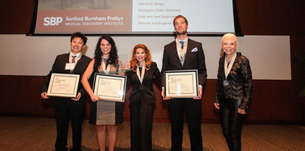
On September 15, the Fishman Fund ceremony honored Borlido and Fishman Fund Career Development recipients Bernhard Lechtenberg, PhD, and Jia (Zack) Shen, PhD

In celebration of SBP’s 40th anniversary, the significance of the Fishman Fund has been elevated with the addition of a two-year postdoctoral award called the Fishman Fund Fellowship. The Fishman Fund Fellowship is an incredible award that not only compensates exceptional postdocs at a higher income level than a typical fellowship, but also comes with a one-time $5,000 flexible career-development award, which they can use to further their professional endeavors. The established Fishman Fund Career Development awards of $10,000 will continue to be awarded as well.

Since the Fishman Fund’s inception in 2001, the Fishman Fund has grown from $1,500 cash awards. It was first established by Horowitz and Mary Bradley in honor of founders Dr. William and Lillian Fishman. In 2010, Jeanne Jones became the co-founder designee.

During the ceremony, Fishman Fund co-founder Reena Horowitz discussed the importance of supporting “the next generation of biomedical trailblazers.” She also said that the “Fishmans believed passionately in cultivating early-stage scientific talent. They knew that young investigators must focus intently on honing research skills and gaining professional experience.” Horowitz also took a moment to honor friends and supporters who have passed: Mary Bradley, Erna Viterbi, Pauline Foster, and Conrad Prebys.

Nina Fishman, daughter of SBP founders Dr. William and Lillian Fishman, read from a speech her father gave at the Institute. He said, “Each is encouraged to develop original ideas, which can be tested experimentally and subsequently qualify for grant support. Together we have the ‘critical mass’ of intellectuals and skills to accelerate progress in sciences.” She added that the Institute has demonstrated that a “relatively small but highly focused group can provide a nucleus where the whole is greater than the sum of its parts.”

Past winner Petrus de Jong, MD, PhD, shared how the Fishman Fund has boosted his career. With the grant, de Jong attended a drug delivery systems symposium with biotech entrepreneurs, participated in a research symposium in pancreatic cancer and received tailored career advice from senior scientists. “These events have been crucial for my personal career development, which would not have been possible without the Fishman Fund Award,” he said.

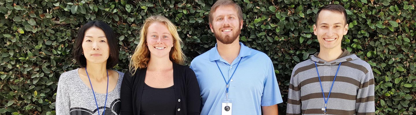
Fishman Fund Fellowship Awardee:

Joana Borlido, PhD, explores the mechanisms through which the channels that connect the cell nucleus (where the genome is stored) affect the immune system. She works in the laboratory of Maximiliano D’Angelo, PhDHer work will help elucidate the molecular basis for the onset and development of leukemia.

She completed her PhD in Oncology at the University of Cambridge in the U.K. and her BS in Biology at the University of Porto in Portugal.

If Borlido is not in the lab, she can usually be found playing foosball, which she admits she would play during all her free time if she could.

Fishman Fund Career Development Recipients:

Bernhard Lechtenberg, PhD, studies cellular signaling networks in normal human physiology and diseases in the laboratory of Stefan Riedl, PhDLechtenberg uses protein X-ray crystallography to study diseases such as cancer and ALS (Lou Gehrig’s disease). He received his BS and MS from the University of Luebeck in Germany and his PhD from the University of Cambridge in the U.K.

In his spare time, he and his wife Anne Hempel, also a SBP postdoc, participate in Ragnar Relay races, in which a team runs nonstop day and night to complete a course of almost 200 miles.

Jia (Zack) Shen, PhD, develops functional screens of for drugs that inhibit ubiquitin ligases (enzymes that tag proteins with a small protein called ubiquitin). Such drugs, alone and in combination with current chemotherapies, are considered important advances in the therapy of breast cancer patients. Shen works in the laboratory of Charles Spruck, PhD, and received his BS from Zhejiang Sci-Tech University and his PhD from the Chinese Academy of Sciences.

He and his wife love taking their 6-month-old daughter Olivia for walks in San Diego. Shen also plays soccer twice a week with a team at the Salk Institute.

With the generous support of the Fishman Fund, this stellar group of postdocs will benefit tremendously from well-earned rewards for their hard work and vision for “Science Benefiting Patients.”

Institute News

Meet the incoming class of 2016!

AuthorJessica Moore
Date

September 15, 2016

Sanford Burnham Prebys Medical Discovery Institute (SBP) welcomes the incoming graduate class of 2016:

Ayako Murao (B.A.Sc., Cal State San Marcos) will explore the mechanisms of cancer progression, especially melanoma, in the lab of Ze’ev Ronai, PhD

Katja Birker (BS, University of British Columbia) is joining the lab of Rolf Bodmer, PhD, where she will study heart development in fruit flies.

Joseph Fromm (BS, UCLA) will work under Nicholas Cosford, PhD, designing small molecule inhibitors of cell death to treat cancer.

Pavel Rhyzov (BS, Bonn-Rhein-Sieg University of Applied Sciences) will probe the structure and function of proteins involved in plague infectivity and cell death in the lab of Francesca Marassi, PhD

Since its launch in 2006, SBP’s Graduate School of Biomedical Sciences (GSBS) has awarded PhD degrees to 27 students who have gone on to careers in both academic research and biotechnology. In 2015 the Western Association of Schools and Colleges (WASC) granted accreditation to the GSBS, a distinction that assures the quality of our resources, policies, and practices to achieve educational goals.

“We’re excited to have these great new graduate students,” said Guy Salvesen, PhD, dean of GSBS and professor in the NCI-designated Cancer Center. “Graduate school can be a long haul, but our program allows students to start their research project immediately so that the average time to degree completion is 4.7 years compared to the national average of 6 years.”

“Our Institute provides access to the most sophisticated minds and technologies in biomedical sciences. There has never been a better time for aspiring scientists to explore careers in biomedical sciences, and SBP’s graduate program provides an outstanding environment to embark on this path,” adds Salvesen.


Could you be part of next year’s class? GSBS is accepting applications now.

Institute News

Un-blurring the lines in diabetes

AuthorJessica Moore
Date

September 14, 2016

You’re probably familiar with two types of diabetes—juvenile or type 1, which affects kids and young adults, and type 2, in older adults who are generally overweight and inactive. But the lines are less clear than they used to be. Now kids are developing type 2 diabetes, and some adult cases are actually more like a slowly progressing type 1. These fuzzy boundaries make it difficult to accurately diagnose all patients.

“Incorrect classification of diabetes is a major problem,” said Richard Pratley, MD, adjunct professor in the Integrative Metabolism Program, director of the Florida Hospital Diabetes Institute, and senior investigator at the Florida Hospital-SBP Translational Research Institute for Metabolism and Diabetes (TRI-MD). “Some patients can go months before their need for insulin is recognized, which allows damage to the pancreas to continue and increases the likelihood that they’ll develop complications.”

To make it easier to determine which kind of diabetes a patient has, Pratley’s lab looked at an emerging class of biomarkers called microRNAs—small RNAs that regulate the translation of other RNAs into proteins. These molecules are ideal indicators of disease because they are easily measured and remain intact after samples are collected.

“We observed that each subtype of diabetes has its own pattern of microRNAs that are increased or decreased,” Pratley explained. “With further validation in more patients, these results could lead to better diagnostics that, in combination with other standard lab tests, help distinguish the various forms.”

There are three conditions in which the body doesn’t produce enough insulin:

  • In type 1 diabetes, the immune system attacks the cells that make insulin—the beta cells of the pancreas.
  • In type 2, the body becomes resistant to insulin, so beta cells have to work extra hard to make more and more, which eventually wears them out.
  • Latent autoimmune diabetes of adults (LADA), like type 1, is caused by immune destruction of beta cells, but progresses much more slowly, and may also involve diet-related insulin resistance.

In the new study, published in Scientific Reports, Pratley’s team compared levels of microRNAs in the blood of patients with each diabetic condition. A total of eight microRNAs were significantly altered in the diabetic population compared to healthy controls.

“MicroRNA measurements alone weren’t enough to differentiate the three subtypes,” added Pratley. “But since each signature was unique, that information may improve our ability to diagnose each type of diabetic disease. Since microRNAs can be assayed noninvasively and cheaply, these tests may one day become commonplace in diabetes care.”

The paper is available online here.

Institute News

Six-day well-being program lowers inflammatory markers in blood

AuthorJessica Moore
Date

September 12, 2016

In a novel controlled clinical trial, a six-day Ayurvedic-based well-being program that featured a vegetarian diet, meditation, yoga and massages led to measurable decreases in blood levels of metabolites associated with inflammation, cardiovascular disease risk and cholesterol regulation.

The findings, published in Scientific Reports, represent a rare attempt to use metabolic biomarkers to assess the health benefits of integrative medicine and holistic practices. Scott Peterson, PhD, professor in the Bioinformatics and Structural Biology Program, contributed to the study, which was overseen by Deepak Chopra, MD, clinical professor in the Department of Family Medicine and Public Health at UC San Diego School of Medicine and a noted proponent of integrative medicine.

“It appears that a one-week Panchakarma program can significantly alter the metabolic profile of the person undergoing it,” said Chopra, whose foundation provided and managed funding for the study. “As part of our strategy to create a framework for whole systems biology research, our next step will be to correlate these changes with both gene expression and psychological health.”

“Many practices with roots in Ayurveda, such as meditation and yoga, are widely believed to be healthful, but their effects on human physiology are not well studied,” said Peterson.

The study involved 119 healthy male and female participants between 30 and 80 years of age who stayed at the Chopra Center for Wellbeing in Carlsbad, Calif. Slightly more than half were assigned to the Panchakarma intervention (the Chopra Center’s Perfect Health program, which typically costs $2,865 for a six-day treatment), while the remainder, who relaxed at a resort for the study period, served as controls. Blood plasma samples taken before and after the intervention or vacation were analyzed by liquid chromatography and mass spectrometry.

The researchers found that in the Panchakarma group, there was a measurable decrease in 12 specific cell membrane chemicals (phosphatidylcholines) correlating with serum cholesterol and inversely related to type 2 diabetes risk.

“These phospholipids exert broad effects on pathways related to inflammation and cholesterol metabolism,” said lead author Christine Tara Peterson, PhD, a postdoctoral fellow at UC San Diego School of Medicine. “Plasma and serum levels of the metabolites of phosphatidylcholine are highly predictive of cardiovascular disease risk.”

The authors suggested that given the very short duration of the trial, the serum profile changes were likely driven by the vegetarian diet component of Panchakarma. Further studies are needed to more fully understand the processes and mechanisms involved.


This post is based on a press release from UC San Diego. The paper is available online here.

Institute News

“No surrender” to CDG

AuthorHelen I. Hwang
Date

September 9, 2016

From a farmhouse in rural Iowa, Crystal Vittetoe is fighting for her two babies afflicted with congenital disorders of glycosylation, known as CDG. She and her family have raised over $37,000 from a single fundraiser, and the donations keep coming in. “If we don’t fight for research, we are surrendering to CDG,” says Vittetoe.

“What Crystal has done for our research at the Institute is incredible. She’s raised enough money to pay for half a postdoc’s salary to do research for one year, and now we need to find the other half,” says Hudson Freeze, PhD, director of the Human Genetics Program at Sanford Burnham Prebys Medical Discovery Institute (SBP). “We have so many projects we start and want to complete. We need more hands on the projects. And if a family needs help, we don’t turn anybody away,” he says.

CDG is a collection of genetic diseases that causes mental and physical developmental issues, which leads to severe damage to multiple organs like the liver, heart and intestines.

The Vittetoes have two young children with CDG—two-year-old daughter Everlee (in the photo above) and one-year-old son Breckyn. Vittetoe drove from Iowa to SBP in La Jolla, Calif., for the annual Rare Disease Day Symposium at SBP. There, she met other families, scientists, doctors as well as Freeze to learn about the latest research and treatments that can help their kids cope with her illness. Worldwide, there are less than 1,500 known cases of CDG where children are born with the genetic disorders.

Vittetoe realized from the family’s visit to SBP that much more research was needed to figure out why CDG happens and how to lessen the her children’s suffering. She was inspired to raise money for the Rocket Fund, in honor of John Taylor (Rocket) Williams IV who would’ve turned 10 years old this year. Sadly, he passed away at the age of two.

In the past year, Everlee has been hospitalized six times. During one episode, she was having an hourly seizure for 24 hours with the last one enduring for 3.5 hours. “It’s so stressful, no matter if she’s having a stroke-like episode or just needs fluids,” says Vittetoe.

With the help of family and friends, Vittetoe held a dinner and silent auction at Lebowski’s Rock ‘N Bowl in her hometown of Washington, Iowa with a population of just over 7,000. The three-hour inaugural event raised a phenomenal amount of money that even surprised Vittetoe. “We were blown away,” she said.

The bar donated 15% of the tab and a friend, who’s also a singer, volunteered the entertainment. Over 300 people contributed to a free-will dinner donation for delicious pork loin from the family’s hog farm and scrumptious sides whipped up by the children’s grandmother.

Substantial seed donations, along with gifts from local businesses, raised an enormous amount of funds at the silent auction. The Vittetoes have been farming in Iowa for generations, and Crystal’s husband Jonathan approached the local seed dealers who all said “yes” to helping out the kids. And of course, neighborhood farmers came to support the Vittetoes who always need seed for their crops.

People contributed checks from $10 to $5,000, and every dollar counted. Other families with CDG children drove over six hours from as far away as Minnesota and Illinois to show their support.

The giving doesn’t just stop with the fundraiser hosted by the Vittetoe family. Recently Crystal’s grandfather passed away in Colorado and the family asked for memorial donations to the Rocket Fund.

Vittetoe says, “It’s your babies and if you don’t do something, you’re just waving the white flag. We’re not waving the white flag. We just want to do something for them.”

Note:

The next SBP Rare Disease Day Symposium will be held on February 24, 2017. The day-long event will focus on Alagille syndrome, a genetic disorder that causes liver damage due to abnormalities in the bile ducts, which carry waste from the liver to the gallbladder and small intestine. For more information, click here.

Photo credit: Drish Photography.

Institute News

MDA grant speeds research toward better treatments for Duchenne muscular dystrophy

AuthorJessica Moore
Date

September 8, 2016

Pier Lorenzo Puri, MD, PhD, professor in the Development, Aging, and Regeneration Program, has focused his career on finding treatments to counter the progression of Duchenne muscular dystrophy (DMD). A new grant from the Muscular Dystrophy Association, totaling nearly $300,000 for three years of support, will help his team figure out how to improve on a drug that they helped advance to clinical trials.

DMD, which affects hundreds of thousands of boys and young men worldwide, causes progressive muscle weakness that is usually first apparent as a child learns to walk. All patients eventually rely on a wheelchair by their high school years, and few live past their early 20s.

In DMD, the absence of a structural protein called dystrophin leads to the progressive loss of muscle fibers, as they cannot maintain their integrity following contraction. The damage triggers both repair—generation of new muscle cells—and formation of fibrous deposits, which make the muscle stiff. At first, regeneration keeps pace with the damage so the muscle continues to work, but as time goes on, the balance tips to favor scarring.

Whether the muscle regenerates or becomes fibrotic is determined by specialized cells that reside between muscle fibers, called fibro-adipogenic progenitors. The drugs that were first shown to work by Puri’s lab, histone deacetylase inhibitors (HDACis), encourage the pro-regenerative activity of these cells, while preventing them from forming fibrotic scars and promoting fat infiltration.

One HDACi, called givinostat, is in a small clinical trial in Europe. Of the 19 patients enrolled, almost all are responding with more regeneration and less fibrosis and fat infiltration. The trial is expected to expand worldwide.

“Recently, we’ve found that not all interstitial cells switch to promoting fibrosis at once,” Puri explained. “That means only some of them need to be targeted by the drug. Since we’ve figured out how to distinguish the good ones from the bad ones, we’re going to use the grant funds to look further at what’s going on in each subpopulation and see how the drug affects them.

“This research could lead to new ways to specifically target the interstitial cells that need to be redirected to foster muscle renewal.”

Puri’s recent interview on local TV station KUSI about muscular dystrophy and his research is online here.

Institute News

Consortium awarded $15 million to unravel bipolar disorder and schizophrenia

AuthorSusan Gammon
Date

August 31, 2016

Sanford Burnham Prebys Medical Discovery Institute (SBP), the Johns Hopkins University School of Medicine, the Salk Institute for Biological Studies, and the University of Michigan will embark on a $15.4 million effort to develop new systems for quickly screening libraries of drugs for potential effectiveness against schizophrenia and bipolar disorder, the National Institute of Mental Health (NIMH) has announced. The consortium, which includes two industry partners, will be led by Hongjun Song, PhD, of Johns Hopkins and Rusty Gage, PhD, of Salk.

Bipolar disorder affects more than 5 million Americans, and treatments often help only the depressive swings or the opposing manic swings, not both. And though schizophrenia is a devastating disease that affects about 3 million Americans and many more worldwide, scientists still know very little about its underlying causes — which cells in the brain are affected and how — and existing treatments target symptoms only.

With the recent advance of induced pluripotent stem cell (iPSC) technology, researchers are able to use donated cells, such as skin cells, from a patient and convert them into any other cell type, such as neurons. Generating human neurons in a dish that are genetically similar to patients offers researchers a potent tool for studying these diseases and developing much-needed new therapies.

“IPSCs are a powerful platform for studying the underlying mechanisms of disease,” says Gage, a professor of genetics at Salk. “Collaborations that bring together academic and industry partners, such as this one enabled by NIMH, will greatly facilitate the improvement of iPSC approaches for high-throughput diagnostic and drug discovery.”

A major aim of this collaboration is to improve the quality of iPSC technology, which has been limited in the past by a lack of standards in the field and inconsistent practices among different laboratories. “There has been a bottleneck in stem cell research,” says Gage, a professor of genetics at Salk. “Every lab uses different protocols and cells from different patients, so it’s really hard to compare results. This collaboration gathers the resources needed to create robust, reproducible tests that can be used to develop new drugs for mental health disorders.”

The teams will use iPSCs generated from more than 50 patients with schizophrenia or bipolar disorder so that a wide range of genetic differences is taken into account. By coaxing iPSCs to become four different types of brain cells, the teams will be able to see which types are most affected by specific genetic differences and when those effects may occur during development.

First the researchers must figure out, at the cellular level, what features characterize a given illness in a given brain cell type. To do that, they will assess the cells’ shapes, connections, energy use, division and other properties. They will then develop a way of measuring those characteristics that works on a large scale, such as recording the activity of cells under hundreds of different conditions simultaneously.

“SBP’s Conrad Prebys Center for Chemical Genomics will play a key role in this initiative,” says Anne Bang, PhD, a director at the Center. “We will be developing assays and testing prototype drug compounds to see if they induce the desired response in iPSC disease models from the consortium. Our goal is to establish assays suitable for high throughput drug screening, ultimately leading to discovery of drugs for preclinical and clinical studies.”

Once a reliable, scalable and reproducible test system has been developed, the industry partners will have the opportunity to use it to identify or develop drugs that might combat mental illness. “This exciting new research has great potential to expedite drug discovery by using human cells from individuals who suffer from these devastating illnesses. Starting with a deeper understanding of each disorder should enable the biopharmaceutical industry to design drug discovery strategies that are focused on molecular pathology,” says Husseini K. Manji, MD, F.R.C.P.C., global therapeutic area head of neuroscience for Janssen Research & Development.

The researchers also expect to develop a large body of data that will shed light on the molecular and genetic differences between bipolar disorder and schizophrenia. And, since other mental health disorders share some of the genetic variations found in schizophrenia and bipolar disorder, the data will likely inform the study of many illnesses.

The National Cooperative Reprogrammed Cell Research Groups program, which is funding the research, was introduced by the National Institute of Mental Health in 2013 to overcome barriers to collaboration by creating precompetitive agreements that harness the unique strengths of academic and industry research.