Cory Dobson, Author at Sanford Burnham Prebys - Page 26 of 41
Institute News

Summer program sparks Preuss high schoolers’ passion for research

AuthorJessica Moore
Date

July 29, 2016

For two weeks this summer, eight rising juniors at The Preuss School at UC San Diego came to Sanford Burnham Prebys Medical Discovery Institute (SBP) each day to try their hands at laboratory techniques ranging from microinjection to dissection to real-time microscopy. This was the first time most had ever been in a research lab, and the experience opened their eyes to what science is like and the range of opportunities that studying biology can open.

“What we did here was much closer to real science than our labs in school,” said participant Bao Lam. “We got to try the methods that scientists use every day, and collect real data and draw conclusions from it.”

This exposure to scientific research is especially significant for students from The Preuss School, which serves low-income students who strive to become the first in their families to graduate from college.

The students rotated through four labs, where they worked with

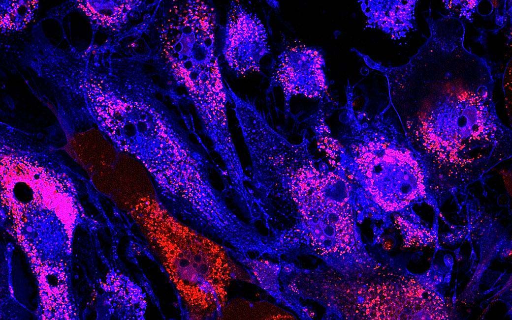
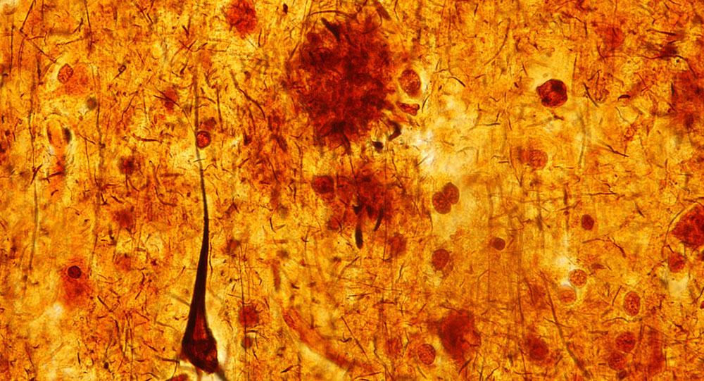
  • tiny soil roundworms, used to study aging
  • fruit flies, used to study heart disease and obesity,
  • zebrafish, used in research towards cellular reprogramming treatments for diabetes, and
  • mouse cells, used to study regulation of gene activity.

“I would definitely work in a lab again,” said Jennifer Vazquez-Gonzalez. “I gained an appreciation for how research can make a difference—the goal is to help patients.”

In the course of their rotations, the students interacted with many researchers, which helped give them a more realistic understanding of what scientists are like.

“I used to think all scientists were bald and boring,” said America Sanchez Radilla. “But the people we worked with were young and diverse and fun to be around.”

The group was also exposed to scientific career paths other than laboratory research. The program included a visit to the campus of Illumina, a leading producer of genetic sequencing equipment, and a panel discussion where they heard from professionals who support research in a variety of ways.

Those who especially enjoyed their time at SBP may come back next year for a more in-depth experience—the Institute also organizes a six-week program where students carry out their own project.

The program is made possible by philanthropists Peggy and Peter Preuss and Debby and Wain Fishburn.

Institute News

Research points to possible target to stop cancer stem cells

AuthorJessica Moore
Date

July 28, 2016

When you think of stem cells, you probably think of healing and regeneration—cells that can replace tissue lost to disease or injury. But tumors also arise from stem cells—a specific kind called cancer stem cells. Because these cells can divide indefinitely, while other cancer cells’ proliferation is more limited, therapies that get rid of them would eventually stop a tumor from growing, and from ever coming back.

Researchers in the laboratory of Dieter Wolf, MD, professor in the NCI-Designated Cancer Center, may have found a new way to do this, though in a roundabout way. While examining the function of two proteins found at high levels in tumors, they discovered that these factors are required for a type of metabolism that’s essential for cancer stem cells to survive.

“Our findings suggest that these components, eukaryotic initiation factors (eIFs) 3d and 3e, are novel targets for eliminating cancer stem cells,” said Wolf, senior author of the study, which was published in Cell Reports. “If we could find a way to turn these factors off, we could starve cancer stem cells.”

The details

eIF3d and eIF3e are part of the complex that initiates protein synthesis, also called translation. Specifically, they and other eIFs help bring the ribosome, which builds proteins by linking amino acids one by one, to messenger RNA, the molecules that carry the code the ribosome reads, specifying which amino acid should be added next.

Wolf’s team was interested in whether overproduction of eIF3d and eIF3e promotes cancer progression. To determine their function, they compared the amounts of all proteins made in cells lacking these factors to those in normal cells. A difference that stood out was in the levels of the protein complexes required to produce ATP, the cell’s energy currency, in mitochondria—eIF3d/e-deficient cells produced far lower amounts than normal.

“Cancer stem cells, unlike most tumor cells, rely on mitochondrial metabolism for energy,” Wolf explained. “Since tumor cells have much higher levels of eIF3d/e than normal cells, inhibiting those factors would preferentially block metabolism in cancer stem cells.

“Our results are somewhat surprising because eIF3 has long been thought to control the synthesis of all proteins. Instead, our data suggest that some parts of eIF3 selectively recruit certain mRNAs.”

Next steps

“We’re now examining how eIF3d/e affects cancer metabolism overall to see if these factors might be relevant to more than just cancer stem cells,” added Wolf. “Also, as a step towards advancing this research to the clinic, we’re designing screens to identify small molecule inhibitors of eIF3d/e.”

The paper is available online here.

Institute News

New potential way to slow advance of Alzheimer’s

AuthorJessica Moore
Date

July 27, 2016

Like weeds taking over a garden, the brains of Alzheimer’s patients become congested with clumps of protein. These clumps arise when a peptide called amyloid beta takes a shape that sticks to other amyloid beta molecules and converts them to the same sticky form, causing a chain reaction. The sticky form of amyloid beta is toxic, so as amyloid plaques accumulate, neuronal connections, and eventually whole neurons, are lost.

Research from the laboratory of Huaxi Xu, PhD, professor in the Degenerative Diseases Program, suggests a new possible way to minimize the generation of amyloid beta and slow the advance of this tragic disease. Alzheimer’s, which affects more than 5 million people and is the 6th leading cause of death in the US, destroys patients’ memory and, at later stages, their ability to communicate and understand their surroundings.

“Our results could eventually help us discover therapeutics that address the progression of Alzheimer’s disease,” said Xu. “That would be a big step forward—no such treatment has yet been approved.”

In the new study, published in the Journal of Neuroscience, Timothy Huang, PhD, a postdoc in Xu’s lab, examined the function of a receptor called SORLA because variants of the gene encoding it had been linked to early-onset Alzheimer’s. SORLA had also been shown to affect trafficking—transport from one cellular compartment to another—of amyloid beta’s precursor. Amyloid beta is generated only in acidic compartments, where the precursor is cut to yield the toxic form, so trafficking has a big impact on how much amyloid beta is made.

“We found that SORLA, with its partner SNX27, moves the amyloid precursor protein away from the acidic compartment, where it would be cut into amyloid beta, to the cell surface,” said Huang. “There, the amyloid precursor protein is cut in a way where it cannot be cut into amyloid beta.”

“Modulating trafficking of the amyloid precursor protein through SORLA could be a new way to treat Alzheimer’s,” added Xu. “Other strategies of decreasing levels of amyloid beta, such as inhibiting the enzyme that cuts the precursor, have failed in the clinic, so new approaches are needed.”

Xu and Huang next plan to investigate whether enhancing amyloid precursor trafficking via SORLA reduces loss of neurons and improves cognitive function in an animal model of Alzheimer’s.

The paper is available online here.

Institute News

Team led by Jamey Marth awarded $12.8M to develop new ways to prevent sepsis

AuthorJessica Moore
Date

July 26, 2016

A multidisciplinary team of scientists led by Jamey Marth, PhD, professor in the NCI-designated Cancer Center and director of UC Santa Barbara’s Center for Nanomedicine, is poised to undertake a major biomedical research initiative focused on the escalating problem of sepsis, the body’s abnormal response to severe infections.

The multi-investigator program will be supported by a five-year, $12.8 million research grant from the National Institutes of Health (NIH).

“Millions of people are diagnosed with sepsis each year worldwide, and on average 30 percent die from the complications of sepsis. No new effective treatments have been developed in decades,” said Marth.

Playing a lead role in the translational component is Jeffrey Fried, MD, an acute care physician at Santa Barbara Cottage Hospital and an expert in sepsis. Fried and Marth have collaborated over the past four years.

“With Dr. Fried’s expertise, we have already made unexpected discoveries pertaining to human sepsis,” Marth said.

“While we have made great strides at our hospital in reducing the mortality of sepsis by two-thirds over the past 11 years, we have reached a plateau of what we can accomplish without new treatments,” Fried explained. “Marth and his co-investigators have done seminal work in investigating the molecular basis of sepsis. This work should translate into the development of radically different and more effective approaches to treating sepsis in the future.”

Additional contributing biomedical scientists and clinicians include UC San Diego faculty member Jeffrey Esko, PhD, an expert in the mechanisms of blood-based diseases, and Dzung Le, MD, PhD, head of the clinical hematology and coagulation laboratory at UC San Diego’s Hillcrest and Thornton hospitals. Jeffrey Smith, PhD, also a professor in SBP’s NCI-designated Cancer Center, brings leading expertise in mass spectrometry methods applied to blood systems.

“I look forward to contributing to this potentially transformative research,” said Smith. “The proteomics analyses at SBP will link regulation of specific blood proteins to disease states, which should point to targets for future therapeutic development.”

The program will also benefit from the involvement of renowned scientists and clinicians on its advisory board. “Sepsis remains the leading killer of patients in intensive care units and there are no approved medications,” said advisory board member Victor Nizet, MD, PhD, chief of the Division of Host-Microbe Systems and Therapeutics at UC San Diego’s School of Medicine. “The highly innovative discoveries by Jamey Marth and his team have inspired a rethinking of how blood components respond to severe infection and suggest new ways to restore normal function and protect vital organs from injury.”

Marth noted the program’s potential to reduce the frequency of disability and death in patients diagnosed with sepsis. “We have an extraordinary opportunity to achieve major advances in the understanding and treatment of sepsis,” he said.

Venn diagram portraying relationships among causes and risk factors for sepsis

Venn diagram portraying relationships among causes and risk factors for sepsis. “SIRS” refers to systemic inflammatory response syndrome. Diagram provided by Jamey Marth.

This post is based on a press release from UC Santa Barbara.

Institute News

Promising target for blocking buildup of fatty plaques in arteries

AuthorJessica Moore
Date

July 22, 2016

Every 34 seconds, someone in the US has a heart attack or stroke. New research from the laboratory of Erkki Ruoslahti, MD, PhD, distinguished professor in the NCI-designated Cancer Center, could lead to treatments that lower that frequency.

Heart attacks and strokes are caused by a blocked artery, which cuts off blood supply to a part of the heart or brain. These blockages occur when atherosclerotic plaques—deposits of inflamed, fat-containing cells surrounded by fibrous material inside arteries—rupture and seed blood clots. In a study published in the Journal of Controlled Release, Ruoslahti’s team shows that a specific peptide blocks expansion of these plaques at advanced stages.

“Our findings demonstrate the relevance of a new target, p32, to slowing the deposition of plaque,” said Zhi-Gang She, PhD, staff scientist in Ruoslahti’s lab and co-lead author of the paper. “We’re hopeful that drugs that act on this protein would help lower the risk for heart attacks and stroke.”

The details

The new study used a peptide called LyP-1, a ring of nine amino acids that Ruoslahti’s group has worked with for many years. LyP-1 binds to p32, a protein that’s normally located inside cells, but is found on the surface of tumor cells and active macrophages.

“Macrophages drive plaque enlargement by taking up fats and promoting inflammation, and we knew from our other investigations that LyP-1 can trigger cell death in macrophages,” explained Ruoslahti. “We thought that LyP-1 might eliminate macrophages from plaques, which would slow the advance of atherosclerosis.”

Their results confirmed this expectation—the LyP-1 peptide greatly reduced the size of plaques in mice when it was administered at advanced stages.

“Eliminating macrophages from arterial plaque is like cutting off the roots of a plant,” said She. “Not only does that get rid of a portion of the plaque, but because macrophages feed it by taking up lipids, it also keeps the plaque from getting larger.”

Clinical relevance

“The peptide itself is not a candidate drug,” added Ruoslahti. “It can only be given by injection, which isn’t practical for a chronic disease like atherosclerosis. However, we have identified small molecules that interact with p32 in a similar way to LyP-1, so they could form the basis of a drug that’s taken as a pill.”

“The key to making sure this treatment strategy is safe is confirming that it doesn’t make the plaques more likely to rupture,” commented She. “We didn’t see anything indicating that LyP-1 makes plaques less stable, but future studies should explore that issue further.”

The paper is available online here.

Institute News

What can salamanders teach us about regeneration?

AuthorJessica Moore
Date

July 11, 2016

Salamanders can regenerate whole limbs, even as adults. While this amphibian superpower might seem irrelevant to human health, investigating the underlying biology may impart vital lessons about how the healing process can be redirected from scarring to replacing lost tissue. For this reason, Alessandra Dall’Agnese, a graduate student in the laboratory of Pier Lorenzo Puri, MD, PhD, professor in the Development, Aging, and Regeneration Program, recently wrote a review, published in BioEssays, comparing healing in salamanders to that in mammals.

“If we can figure out the means by which injured salamander limbs turn on developmental programs, we may be able to use that knowledge to create treatments that help the human body heal itself,” said Puri.

The review describes the key difference between how salamanders and humans heal following a major injury to a limb. Salamanders heal the wound, then form a regenerative center of proliferative, stem-like cells. In contrast, a regenerative center only forms in humans if the injury affects the tip of a finger or toe distal to the nail bed—otherwise, wound healing is followed by scarring.

“The important thing we’ve learned from salamanders is that there’s not an inherent limit to how much of a limb can be regrown,” explained Dall’Agnese. “The fact that mammals can only regrow the tips of digits instead suggests that there may be some property of the nail bed that fosters regeneration.

“From research in salamanders and mice, we know some of the factors that have to be turned on or off to enable regeneration. But we need a more detailed picture before we can start to develop therapies.”

“The possibility that we could discover salamanders’ secret to regeneration is a good example of why we should study a wide range of organisms,” Puri commented. “If we only studied animals closely related to us, we wouldn’t learn how to help our bodies do things they can’t normally do.”

The review is available online here.

Institute News

Siobhan Malany, PhD, selected to conduct novel medical research in space

AuthorDeborah Robison
Date

June 13, 2016

Siobhan Malany, PhD, director of Translational Biology at Sanford Burnham Prebys Medical Discovery Institute at Lake Nona (SBP) and founder of the Institute’s first spin-off company, Micro-gRx, Inc., has been awarded $435,000 to study atrophy in muscle cells in microgravity on the International Space Station (ISS). In microgravity, conditions accelerate changes in cell growth similar to what occurs in the aging and disease process of tissues. Using real-time analysis, Malany will be able to rapidly study cells for potential new therapeutic approaches to muscle degeneration associated with aging, injury or illness. Continue reading “Siobhan Malany, PhD, selected to conduct novel medical research in space”

Institute News

Cosimo Commisso explains cancer metabolism on NIH website

AuthorJessica Moore
Date

June 7, 2016

The most deadly of all cancers are driven by mutations in a family of genes known as RAS. In a new article on the website for the National Cancer Institute’s RAS Initiative, Cosimo Commisso, PhD, assistant professor in SBP’s NCI-designated Cancer Center, discusses how the metabolism of cancer cells might be different in different parts of solid tumors.

The RAS Initiative is a collaborative effort to explore innovative approaches for attacking the proteins encoded by mutant forms of RAS genes, which drive 30% of human cancers.

Institute News

Making ERK work as a therapeutic target for colorectal cancer

AuthorJessica Moore
Date

June 3, 2016

Colorectal cancer is the third most common cancer in the US, affecting 1.2 million people. Despite extensive research, the five-year survival rate remains below 15%, underscoring the need for new treatments.

One-third of colorectal cancers are driven by over-activation of extracellular signal-regulated kinases 1 and 2 (ERK1/2), which regulates proliferation, metabolism, and cell movement. However, drugs targeting the ERK1/2 pathway are not widely used to treat colorectal cancer because they don’t appreciably slow cancer growth. New research co-led by Petrus de Jong, MD, PhD, postdoctoral associate at SBP, points to a possible reason for this lack of effect, as well as a solution.

“We genetically deleted the ERK1/2 pathway in the lining of the mouse intestine, and we expected to see less cell proliferation,” said de Jong, a co-first author on the paper. “Instead, the opposite occurred. There was more cell growth and the cells were less organized.” de Jong works in the laboratory of Garth Powis, D.Phil., professor and director of SBP’s NCI-Designated Cancer Center, who also contributed to the investigation.

The new study, published in Nature Communications, shows that the increased cell growth caused by disabling ERK1/2 results from increased activity of a related kinase, ERK5. The team went on to show that inhibiting both pathways suppresses proliferation of human colorectal cancer cell lines and slows growth of tumor-like structures in vitro.

“Therapies aimed at targeting ERK1/2 likely fail because ERK5 compensates,” said Eyal Raz, MD, senior author and professor at the UC San Diego School of Medicine. “Previously, ERK5 didn’t seem important in colorectal cancer. This is an underappreciated escape pathway for tumor cells. Hence, the combination of ERK1/2 and ERK5 inhibitors may lead to more effective treatments for colorectal cancer patients.”

“If you block one pathway, cancer cells usually mutate and find another pathway that ultimately allows for a recurrence of cancer growth,” said Koji Taniguchi, MD, PhD, assistant project scientist at UC San Diego and the other co-first author. “Usually, mutations occur over weeks or months. But other times, as in this case, the tumor does not need to develop mutations to find an escape route from targeted therapy. When you find the compensatory pathway and block both, there is no more escape.”

The scientists suggested that other inhibitors of the ERK1/2 pathway should be tested with ERK5 inhibitors in both human colorectal cancer cells and mouse models to identify the most effective combination that could advance to clinical trials.

This post is a modified version of the press release from UC San Diego. Photo from Ed Uthman via Flickr.

The paper is available online here.