Development, Aging and Regeneration Archives - Sanford Burnham Prebys
Institute News

Scientists unite to get to the heart of AFib

AuthorSusan Gammon
Date

August 15, 2023

A collaborative study led by researchers at Sanford Burnham Prebys is paving the way to identifying gene networks that cause atrial fibrillation (AFib), the most common age-related cardiac arrhythmia.

The findings, published in Disease Models & Mechanisms, validate an approach that combines multiple experimental platforms to identify genes linked to an abnormal heart rhythm.

“One of the biggest challenges to solving the AFib genetic puzzle has been the lack of experimental models that are relevant to humans,” says Alex Colas, PhD, co-senior author and assistant professor in the Development, Aging and Regeneration Program at Sanford Burnham Prebys. “By working with colleagues who focus on AFib but in different systems, we have created a robust multiplatform model that can accurately pinpoint genes associated with this condition.”

AFib is characterized by an irregular, rapid heartbeat that causes a quivering of the upper chambers of the heart, called the atria. This condition is the result of a malfunction in the heart’s electrical system that can lead to heart failure and other heart-related complications, which include stroke-inducing blood clots.

AFib impacts more than 5.1 million people in the United States, with expectations of 15.9 million by 2050. It is more common in individuals over the age of 60 but can also occur in teenagers and young adults.

“There will never be a one-size-fits-all solution to AFib, since it can be caused by many different genes—and the genes that do cause it vary from person to person,” says Karen Ocorr, PhD, also a co-senior author and assistant professor in the Development, Aging and Regeneration Program at Sanford Burnham Prebys. “A better understanding of the gene network(s) that contribute to AFib will help us design tests to predict a person’s risk, and develop individualized approaches to treat this dangerous heart condition.”

To overcome the limitations of current AFib research models, Colas, Ocorr and researchers from UC Davis and Johns Hopkins University combined forces to assemble a multi-model platform that combines:

  • A high-throughput screen using atrial-like cells (derived from human-induced pluripotent stem cells) to measure how a gene mutation alters the strength and duration of a heartbeat.
  • A Drosophila (fruit fly) model—with heart genetics and development remarkably similar to human hearts—that permits analysis of gene mutations in a functioning organ.
  • A well-established computational model that uses computers to simulate the effects of gene mutations on the electrical activity in human atrial cells.

The accuracy of the multi-model platform was confirmed when each screened 20 genes, and all three platforms identified phospholamban, a protein found in the heart muscle with known links to AFib.

“This collaboration has greatly expanded our ability to understand AFib at the genetic level,” says Colas. “Importantly, the high-throughput screening component of the model will also allow us to rapidly and effectively screen for drugs that can restore a heart to its normal rhythm.”

He adds, “Hopefully this is just the beginning. There are many more cardiac diseases to which our system can be applied.”

Institute News

5 facts you need to know about atrial fibrillation (AFib)

AuthorMonica May
Date

February 14, 2019

It’s one of the most common heart rhythm disorders and a leading risk factor for stroke, but most people haven’t heard of it—that is, atrial fibrillation, also known as AFib or AF. Below are five facts everyone should know about AFib. 

  1. Nearly 10 percent of people over the age of 65 develop AFib, and it can be deadly. According to the Centers for Disease Control, it is estimated that 12.1 million people in the United States will have AFib in 2030. In 2019, AFib was mentioned on 183,321 death certificates and was the underlying cause of death in 26,535 of those deaths.
     
  2. There is no cure. Current treatments include surgery to remove the malfunctioning heart tissue; medications that reduce the risk of stroke by thinning the blood, such as warfarin or other anticoagulants; or medications that slow the heart rate or rhythm. But scientists currently don’t know the cause of AFib. There is no cure. 
     
  3. Increased stroke risk makes AFib lethal. The irregular heartbeats that characterize AFib can cause blood to pool in the heart, and clot. If a blood clot travels to the brain, stroke may occur. About 15 to 20 percent of strokes are due to AFib, according to the American Heart Association.
     
  4. The Apple Watch can detect—but not diagnose—the condition. The Apple Watch can take an electrocardiogram and send a notification if an irregular heart rhythm is identified. However, only a doctor can diagnosis AFib. Apple has teamed up with Johnson & Johnson to determine if the wearable technology’s ability to detect AFib earlier improves diagnosis and patient outcomes.
     
  5. Fruit flies could unlock new AFib treatments. Believe it or not, the heart of a fruit fly—which is a tube—models early heart development. In a human, this tube folds into the four chambers of the heart. Combined with their short life cycle and simple genome, fruit flies are an excellent model of heart disease that could unlock new treatments, including those for AFib. Listen to how SBP scientists are using fruit flies to study AFib.

Additional AFib resources: 

Institute News

Dietary restriction increases lifespan through effects on the gut

AuthorJessica Moore
Date

July 14, 2016

Dietary restriction, or limited food intake without malnutrition, has beneficial effects on longevity in many species, including humans. A new study from the Sanford Burnham Prebys Medical Discovery Institute (SBP), published today in PLoS Genetics, represents a major advance in understanding how dietary restriction leads to these advantages.

“In this study, we used the small roundworm C. elegans as a model to show that autophagy in the intestine is critical for lifespan extension,” said Malene Hansen, PhD, associate professor in SBP’s Development, Aging, and Regeneration Program and senior author of the study. “We found that the gut of dietary-restricted worms has a higher than normal rate of autophagy, which appears to improve fitness in multiple ways—preserving intestinal integrity and maintaining the animal’s ability to move around.”

Autophagy, or cellular recycling, is well known to play a role in lifespan extension. Autophagy involves breaking down the cell’s parts—its protein-making, power-generating, and transport systems—into small molecules. This both eliminates unnecessary or broken cell machinery and provides building blocks to make new cell components, which is especially important when starting materials are not provided by the diet.

In this study the research team wanted to understand how dietary restriction impacts autophagy in the intestine, whose proper function is already known to be important for long life.

“The strain of worms we used, called eat-2, is genetically predisposed to eat less, and they live longer than normal worms, so they provide an ideal model in which to investigate how dietary restriction extends lifespan,” said Sara Gelino, PhD, research associate in Hansen’s lab and lead author of the study. “We found that blocking autophagy in their intestines significantly shortened their lifespans, showing that autophagy in this organ is key for longevity.

“These results led us to examine how inhibiting autophagy impacts the function of the intestine. We found that while normal worms’ gut barriers become leaky as they get older, those of eat-2 worms remain intact. Preventing autophagy eliminated this benefit, which indicates that a non-leaky intestine is an important factor for long life.”

“How intestinal integrity relates to longevity is not clearly understood,” Hansen commented. “It’s possible that the decline in the gut’s barrier function associated with normal aging might let damaging substances or pathogens into the body.”

The research team also observed that turning off autophagy in the intestine made the slow-eating, long-lived worms move around less.

“The decrease in physical activity indicates that autophagy in one organ can have a major impact on other organs, in this case probably muscle or motor neurons,” said Hansen. “Finding the link between motility and autophagy in the intestine will require further research, but we speculate that inhibiting autophagy in the gut may impair the gut’s ability to metabolize nutrients or secrete hormones important for the function of other organs.”

While these results suggest that boosting autophagy in the gut is generally beneficial, Hansen cautions that further research is needed: “Before we can consider regulating autophagy to manage disease, we need to learn a lot more about how the process works both in a single cell as well as in the whole organism.”

Many of these future studies will also employ C. elegans. “Even though worms are much simpler than humans, many of the same basic mechanisms drive their biology. The knowledge we gain from this fast-paced research could eventually contribute to the development of new treatments that help people live longer, healthier lives,” added Hansen.

The paper is available online here.

Institute News

Former postdoc Suzanne Graham now leads business development at diagnostic company

AuthorJessica Moore
Date

July 8, 2016

Suzanne Graham, PhD, did postdoctoral research at SBP from 2004-2012, during which time she worked in two labs. With Fred Levine, MD, PhD, professor and director of the Sanford Children’s Health Research Center, she studied cell cycle control in pancreatic beta cells, and with Rolf Bodmer, PhD, professor and director of the Development, Aging, and Regeneration Program, she explored how metabolic syndrome affects heart function. She has served as senior manager of business development at Genection, which develops and sells genetic tests, in San Diego since June 2012.

What’s your job like— what do you do every day?

I’m responsible for managing relationships with clients (generally pharmaceutical companies)—making sure we’re getting the right data to them, answering their questions, and trying to get more business.

What drew you to business development?

I’m more sociable than the average R&D scientist—I wanted to be involved in lots of conversations, interfacing with all parts of the company.

What do you enjoy most about it and why? 

It’s exciting to help our clients find tools that help them meet their goals. For example, if a company is running a trial of a drug and they can tell it’s working for some patients but not others, I can help them either find a diagnostic test or work with our company to make one that helps them identify the likely responders.

Plus, since I’m working in industry, everything moves quickly. It’s really rewarding to see things through to completion.

What do you hope to accomplish in your career? 

Since I’m familiar with how genomics fits into pharmaceutical development, I’d love to move to a pharma company in a strategy role to help them expand into that area.

What did you gain from your postdoc experience at Sanford Burnham? 

My scientific training is instrumental in my work—it really helps me understand and define the questions our clients have, which is crucial for finding the right solution.

How did you find your first job after your postdoc? Was it challenging? 

I used a job placement company because it is VERY hard for postdocs to get into industry. They helped make my resume convey my skills in a way that speaks to biotech companies, understand what kind of jobs to look for, and create a strategy to connect with potential hirers.

What advice do you have for current postdocs looking for a job?

PhD scientists can fulfill a lot more roles than they might think. Almost any job related to biotech benefits from a deep understanding of science—even designing websites or doing social media for executives.

Institute News

Scientific breakthrough may limit damage caused by heart attacks

AuthorJessica Moore
Date

June 30, 2016

A research advance from the Sanford Burnham Prebys Medical Discovery Institute (SBP) and Stanford University could lead to new drugs that minimize the damage caused by heart attacks. The discovery, published in Nature Communications, reveals a key control point in the formation of new blood vessels in the heart, and offers a novel approach to treat heart disease patients.

“We found that a protein called RBPJ serves as the master controller of genes that regulate blood vessel growth in the adult heart,” said Mark Mercola, PhD, professor in SBP’s Development, Aging, and Regeneration Program and jointly appointed as professor of medicine at Stanford University, senior author of the study. “RBPJ acts as a brake on the formation of new blood vessels. Our findings suggest that drugs designed to block RBPJ may promote new blood supplies and improve heart attack outcomes.”

In the US, someone has a heart attack every 34 seconds. The ensuing loss of heart muscle, if it affects a large enough area, can severely reduce the heart’s pumping capacity, which causes labored breathing and makes day-to-day tasks difficult. This condition, called heart failure, arises within five years in at least one in four heart attack patients.

The reason heart muscle dies in a heart attack is that it becomes starved of oxygen—a heart attack is caused by blockage of an artery supplying the heart. If heart muscle had an alternative blood supply, more muscle would remain intact, and heart function would be preserved. Many researchers have therefore been searching for ways to promote the formation of additional blood vessels in the heart.

“Studies in animals have shown that having more blood vessels in the heart reduces the damage caused by ischemic injuries, but clinical trials of previous therapies haven’t succeeded,” said Ramon Díaz-Trelles, PhD, staff scientist at SBP and lead author of the study. “The likely reason they have failed is that these studies have evaluated single growth factors, but in fact building blood vessels requires the coordinated activity of numerous factors. Our data show that RBPJ controls the production of these factors in response to the demand for oxygen.

“We used mice that lack RBPJ to show that it plays a novel role in myocardial blood vessel formation (angiogenesis)—it acts as a master controller, repressing the genes needed to create new vessels,” added Diaz-Trelles. “What’s remarkable is that removing RBPJ in the heart muscle did not cause adverse effects—the heart remained structurally and functionally normal in mice without it, even into old age.”

“RBPJ is a promising therapeutic target. It’s druggable, and our findings suggest that blocking it could benefit patients with cardiovascular disease at risk of a heart attack. It may also be relevant to other diseases,” commented Pilar Ruiz-Lozano, PhD, associate professor of pediatrics at Stanford and adjunct professor at SBP, co-senior author. “Inhibitors of RBPJ might also be used to treat peripheral artery disease, and activators might be beneficial in cancer by inhibiting tumor angiogenesis.”

The paper is available online here.

Institute News

Siobhan Malany, PhD, selected to conduct novel medical research in space

AuthorDeborah Robison
Date

June 13, 2016

Siobhan Malany, PhD, director of Translational Biology at Sanford Burnham Prebys Medical Discovery Institute at Lake Nona (SBP) and founder of the Institute’s first spin-off company, Micro-gRx, Inc., has been awarded $435,000 to study atrophy in muscle cells in microgravity on the International Space Station (ISS). In microgravity, conditions accelerate changes in cell growth similar to what occurs in the aging and disease process of tissues. Using real-time analysis, Malany will be able to rapidly study cells for potential new therapeutic approaches to muscle degeneration associated with aging, injury or illness. Continue reading “Siobhan Malany, PhD, selected to conduct novel medical research in space”

Institute News

Former SBP postdoc Louis Lapierre now assistant professor at Brown University

Authorjmoore
Date

April 15, 2016

This is the first post in a series that will share what past SBP postdocs are doing now. 

Louis Lapierre, PhD, was a postdoctoral researcher in the laboratory of Malene Hansen, PhD, associate professor in the Development, Aging, and Regeneration Program from 2008 to 2014, where he studied the molecular mechanisms of aging using the microscopic roundworm C. elegans. Since January 2015, he has been an assistant professor in the Department of Molecular Biology, Cell Biology, and Biochemistry at Brown University. His lab researches the role of lipophagy in aging, the process by which cells recycle fats to enhance longevity.

What did you gain from your postdoc experience at Sanford-Burnham? What was the best thing about doing science here?

Conducting research at Sanford-Burnham was a great stepping-stone in my career development. After my first publication there, because I was in a lab led by an emerging scientist, I had the flexibility to carry out research that I personally cared about, which led to interesting and unexpected findings. Eventually, I obtained independent financial support that was critical for my transition to independence. Importantly, SBP’s location in La Jolla means that it is surrounded by highly skilled researchers and state-of-the-art infrastructure, which makes for a cutting-edge, competitive environment.

How did you find your first job after your postdoc? Was it challenging?

I interviewed for several positions during the first half of 2014. The faculty job search is grueling, but I feel privileged to have gone through it. The main challenge is the outstanding level of competition not only at research-intensive institutions, but also at lower tier schools.

Why do you say participating in the faculty selection process was a privilege?

I think it’s easy to forget that our accomplishments were possible because, at some point, someone believed in us. Receiving support from colleagues and mentors and then being selected for interview by a search committee is a privilege that only a few postdocs get to experience.

What advice do you have for postdocs who hope to find a faculty position?

During your postdoc, plan meticulously to position yourself in line with funding opportunities and aggressively pursue emerging topics. During the job search, understand your value on the market, develop an interesting research program with long-term potential and strategically market yourself at conferences. 

What do you enjoy most about your work and why?

I think the most fascinating part of basic research is the possibility of making new and exciting discoveries, and being at the forefront of knowledge. I also find mentoring highly rewarding because I get to make a difference in someone else’s life by building their confidence. This is especially gratifying when I get to work with students who have a passion for science, which is so important to thrive in this competitive environment.

What do you miss most about San Diego and why?

You only realize what you have when you lose it. Leaving San Diego was difficult because I knew nowhere else would be as good. I miss San Diego, not only for its incredible weather and beautiful beaches, but most importantly for the great friends I made there.

Institute News

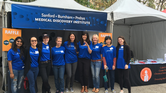
SBP helps students “worm” up to science at STEM Expo

Authorsgammon
Date

March 7, 2016

On Saturday, March 5, a keen group of SBP volunteers hauled wagons of lab coats, mutant worms and magnifying glasses to give the next generation of scientists—mainly kids in grades K-8—an opportunity to see first-hand how tiny worms named C. elegans are used to understand the aging process.

More

Institute News

Rare Disease Day symposium brings together experts on disorders of glycosylation

Authorjmoore
Date

March 2, 2016

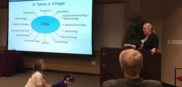
The Rare Disease Day symposium on February 26-27 featured many fascinating talks from experts on numerous aspects of congenital disorders of glycosylation (CDGs), from fundamental work on glycosylation pathways to animal models to diagnosis in the clinic. Following are summaries of each presentation:

Lawrence Tabak, D.D.S, PhD, deputy director of the NIH—After presenting his research on glycosylating enzymes in the 1980s, which helped lay the foundation for understanding the processes that are impaired in CDGs, Tabak discussed several initiatives by the NIH, including the Precision Medicine Initiative and efforts to increase reproducibility.

William Gahl, MD, PhD, director of the National Human Genome Research Institute (NHGRI)—Gahl highlighted several successes of the Undiagnosed Diseases Program. Most relevant to the field of CDGs was the discovery of the gene underlying a new type of CDG, in which an enzyme responsible for generating a necessary precursor for protein glycosylation (uridine diphosphate) is inactivated. This work also found that supplementation with uridine was an effective therapy.

Shengfang Jin, PhD, scientist at Agios Pharmaceuticals Inc.Jin presented her work on a mouse model of PMM2-CDG, which is caused by mutations in the gene for phosphomannomutase 2. Her research has identified a promising biomarker for PMM2-CDG, which is one of the more common types of CDG.

Richard Steet, PhD, associate professor at the University of Georgia—Steet’s lab is developing a new method of identifying which proteins are glycosylated by particular enzymes, which is important for understanding how each CDG-associated mutation leads to disease.

Reid Gilmore, PhD, professor at University of Massachusetts Medical School—Gilmore gave a detailed view of how two CDG-associated mutations, in isoforms of the same component (STT3A and STT3B) of a major glycosylating enzyme, oligosaccharyltransferase, impair protein glycosylation.

Robert Haltiwanger, PhD, professor at the University of Georgia—In another presentation on fundamental glycobiology, Haltiwanger described the function of two enzymes in the same pathway (fucosylation) inactivated in certain CDGs. Mutations in these enzymes underlie Peters plus syndrome and a single case of an unnamed severe CDG, respectively.

Marjan Huizing, PhD, staff scientist at the NHGRI—Using a mouse model of GNE myopathy, a progressive muscle disease caused by mutations in an enzyme required for protein sialylation, Huizing’s lab identified a therapy, supplementation with the sugar ManNAc, which is now in phase 2 trials, and identified a key biomarker. The mouse model also suggested that sialylation problems may be associated with certain kidney diseases, which is now under investigation.

Raymond Wang, MD, clinical geneticist at CHOC Children’s Clinic—Wang told the story of how he and scientific collaborators diagnosed an unusual case that initially appeared to be a CDG because of abnormal glycosylation. The disease-causing mutation was finally identified to be in mitochondrial translation, highlighting the similarities between CDGs and mitochondrial diseases.

David Beeson, PhD, professor at the University of Oxford—Beeson described a subset of congenital myasthenias caused by mutations in glycosylating enzymes, which have distinct symptoms from other myasthenias. These mutations likely cause this disorder by selectively impairing processing of the receptor by which muscle cells receive signals from nerves—the nicotinic acetylcholine receptor.

Lance Wells, PhD, professor at the University of Georgia— Wells summarized his work on the molecular basis of dystroglycanopathies, a subgroup of muscular dystrophies that arise from defects in O-mannosylation enzymes. Most recently, his lab resolved the puzzle of how mutations in an enzyme involved in a different form of glycosylation could cause this disease—they showed that the enzyme’s function had been incorrectly assigned.

Taroh Kinoshita, PhD, professor at Osaka University—Kinoshita is an expert on the addition of sugar-based anchors to lipids (GPI anchors), which link many proteins to the cell surface. He presented some of the extensive work from his team on how mutations in GPI-synthesizing enzymes cause disease, including identification of a therapy, vitamin B6, for seizures in GPI deficiencies.

Eva Morava, MD, PhD, professor at Tulane University Medical Center and the University of Leuven—Morava described preliminary results of a clinical trial of galactose supplementation to treat PGM1-CDG, in which patients are deficient in phosphoglucomutase-1 (this also impairs glucose metabolism). In these patients, galactose improves liver function and endocrine abnormalities and normalizes clotting factors.

Lynne Wolfe, MS, C.N.R.P. clinical research coordinator at the NHGRI—Wolfe discussed the CDG natural history study underway at the NIH—its goals and progress so far. The findings of this study will serve as a resource both for future diagnoses and for researchers in the field to correlate pathways with symptoms.

Tadashi Suzuki, D.Sci., team leader at the RIKEN Global Research Cluster—NGLY1 is different from other CDG-associated genes—it encodes a deglycosylating enzyme, which helps degrade glycosylated proteins that aren’t properly folded. Suzuki’s team has shown that inhibiting another deglycosylating enzyme, ENGase, prevents the formation of aggregates of misfolded proteins, suggesting that it could be a therapeutic target.

Hamed Jafar-Nejad, MD, associate professor at Baylor College of Medicine—Using fruit flies as a model, Jafar-Nejad’s lab is investigating how NGLY1 deficiency affects development. These flies replicate many of the features of human disease, including growth delay and impaired movement, so they could yield important insights into pathogenesis.

Institute News

Fine-tuning cellular energy increases longevity

AuthorJessica Moore
Date

February 25, 2016

New research from SBP has identified a protein that can extend the natural lifespan of C. elegans, a microscopic roundworm commonly used for research on aging and longevity. The findings, published in Cell Reports, expand what we know about the aging process and may lead to new ways to delay the onset of human age-related diseases such as cancer and neurodegenerative diseases. Continue reading “Fine-tuning cellular energy increases longevity”