On Thursday, October 30, 2014, Sanford-Burnham hosted more than 250 attendees at its 36th annual symposium to hear opinion-leading scientists discuss their latest findings on the microbiome. The microbiome is a relatively new frontier for research scientists with aims to understand how the trillions of microbes—bacteria, viruses, fungi, and others—that live in our nose, mouth, gut, and skin interact with human cells to influence health and disease. Continue reading “Sanford-Burnham’s 36th Annual Symposium: The Microbiome and Human Health”
Institute News
New molecular markers for prostate cancer identified
A team of scientists led by Sanford-Burnham’s Ranjan J. Perera, PhD, has identified a set of RNA molecules that are detectable in tissue samples and urine of prostate cancer patients, but not in normal healthy individuals. The study sets the stage for the development of more-sensitive and specific non-invasive tests for prostate cancer than those currently available, which could result in fewer unnecessary prostate biopsies with less treatment-related morbidity, according to a new study in The Journal of Molecular Diagnostics.
According to the American Cancer Society, prostate cancer is the second most common type of cancer in American men (behind skin cancer), and the second-leading cause of cancer death in men (after lung cancer). In 2014, more than 230,000 new cases of prostate cancer will be diagnosed. One in seven American men will get prostate cancer during his lifetime, and one in 36 will die from it. Since most men with prostate cancer have indolent (non-aggressive) disease for which conservative therapy or surveillance would be appropriate treatment, the clinical challenge is not only how to identify those with prostate cancer, but also how to distinguish those who would benefit from surgical or other aggressive treatment from those who would not.
Today, prostate cancer is primarily detected and monitored by testing for high concentrations of prostate-specific antigen (PSA) in blood samples. High PSA levels are often followed by a biopsy to confirm the presence of cancer, and whether it’s slow growing or aggressive. “While elevated PSA can be an alert to a lethal cancer, it can also detect less aggressive cancers that may never do any harm,” said Vipul Patel, MD, medical director of the Global Robotics Institute at Florida Hospital in Orlando, and co-author of the study. “Moreover, only 25 percent of men with raised PSA levels that have a biopsy actually have prostate cancer. Prostate cancer needs to be screened for; we just need to find a better marker.”
The researchers believe that they have identified a group of RNA molecules – known as long noncoding RNAs (lncRNAs) – that hold the potential for serving as better prognostic markers for prostate cancer. lncRNAs are noncoding RNA molecules that until recently were dismissed by scientists as non-functional noise in the genome. Now, lncRNAs are thought to regulate normal cellular development and are increasingly reported as contributing to a range of diseases, including cancer.
Detection of lncRNAs in urine
“We have identified a set of lncRNAs that appear to have an important role in prostate cancer diagnostics,” said Perera, associate professor and scientific director of Analytical Genomics and Bioinformatics at our Lake Nona campus. “The findings advance our understanding of the role of lncRNAs in cancer biology and, importantly, broaden the opportunity to use lncRNAs as biomarkers to detect prostate cancer.”
The study profiled the lncRNAs in three distinct groups: (1) human prostate cancer cell lines and normal prostate epithelial cells, (2) prostate adenocarcinoma tissue samples and matched normal tissue samples, (3) urine samples from patients with prostate cancer or benign prostate hypoplasia, and normal healthy individuals. In each case, the lncRNAs were elevated in prostate cancer patient samples, but not in patients with benign prostate hypoplasia or normal healthy individuals.
One advantage of lncRNAs is that the molecules can be detected in urine samples, which are more easily available than blood tests. One lncRNA, PCA3, was recently commercialized as a urine test to identify which men suspected of having prostate cancer should undergo repeat prostate biopsy. However, discrepancies have been found to exist between PCA3 levels and clinicopathologic features, said Perera. In the current study, PCA3 was detected in some, but not all of the study samples, suggesting that reliance on a single biomarker may be insufficient for prostate cancer detection, while combining additional markers may increase the specificity and sensitivity of the test.
“There is a tremendous unmet clinical need for better non-invasive screening tools for early detection of prostate cancer to reduce the overtreatment and morbidity of this disease,” added Patel. “Our findings represent a promising approach to meet this demand.”
Technical details of the study
The goal of the first experiment was to see whether lncRNAs are differentially expressed in prostate cancer by measuring total RNA from prostate cancer cell lines and normal epithelial prostatic cells using NCode human ncRNA array and SurePrint G3 human lncRNA microarrays. Hierarchical clustering revealed distinguishable lncRNA expression profiles. Thirty lncRNAs were up-regulated and the expression levels of three top-ranking candidates [XLOC_007697, LOC100287482, and AK024556 (also known as SPRY4-IT1)] were confirmed in prostate cancer cell lines by quantitative real-time polymerase chain reaction (qPCR) analysis. The SPRY4-IT1 was found to be up-regulated more than 100-fold in PC3 cells compared with prostatic epithelial cells.
In a second experiment, lncRNA expression was compared in pooled prostate cancer tissue samples and matched normal tissues from 10 frozen biopsy specimens. Hierarchical clustering of the differentially expressed lncRNAs was observed and 10 up-regulated lncRNAs were detected using microarrays. An additional set of 18 prostate cancer tissue samples was analyzed by qPCR and five lncRNAs were found to be significantly higher in prostate tumor tissues compared with matched normal tissues.
Researchers used qPCR to analyze total RNA isolated from urine in another experiment. Urine was collected from 13 prostate cancer patients and 14 healthy controls. All six lncRNAs were found to be significantly up-regulated in the urine samples from the prostate cancer patients compared with normal patient controls, while there were no differences between normal and benign prostatic hyperplasia patient samples.
In other studies focused particularly on SPRY4-IT1. Using both qPCR and highly sensitive droplet digital PCR, expression of SPRY-IT1 was found to be increased in 16 of 18 (89 percent) tissue samples from patients with prostatic adenocarcinoma, compared to normal tissue samples. The researchers developed chromogenic in situ hybridization (CISH) techniques to visualize SPRY4-IT1 expression in cancerous and matched normal tissue. Intense staining was seen in all adenocarcinoma samples, but not in normal prostatic tissue. Finally, the investigators showed that reduction of SPRY4-IT1 in prostate cancer cells through the use of small interfering RNA (siRNA) leads to decreased cell viability and cellular invasion as well as increased apoptosis, similar to what is seen in melanoma cells.
About the paper
“Long Noncoding RNAs as Putative Biomarkers for Prostate Cancer Detection,” by Bongyong Lee, Joseph Mazar, Muhammed Nauman Aftab, Feng Qi, John Shelley, Jian-Liang Li, Subramaniam Govindarajan, Felipe Valerio, Inoel Rivera, Tadzia Thurn, Tien Anh Tran, Darian Kameh, Vipul Patel, and Ranjan J. Perera, DOI: http://dx.doi.org/10.1016/j.jmoldx.2014.06.009. Published online ahead of The Journal of Molecular Diagnostics, Volume 16, Issue 6 (November 2014) published by Elsevier.
This is a post by our guest writer Janelle Weaver, PhD
Heart failure affects about five million people in the United States, and about half of these individuals die within five years of diagnosis. This condition occurs when the heart can’t pump enough blood to meet the body’s needs, and evidence suggests that abnormalities in energy metabolism play an important role. However, many past studies addressing the underlying molecular mechanisms have focused on severe, late-stage heart failure, potentially missing out on early events that could guide the development of treatment strategies for early disease stages. Continue reading “A signature for early-stage heart failure could improve diagnosis and prevent disease progression”
Institute News
A nuclear receptor that binds more than 5,000 sites in the genome—and promotes angiogenesis.
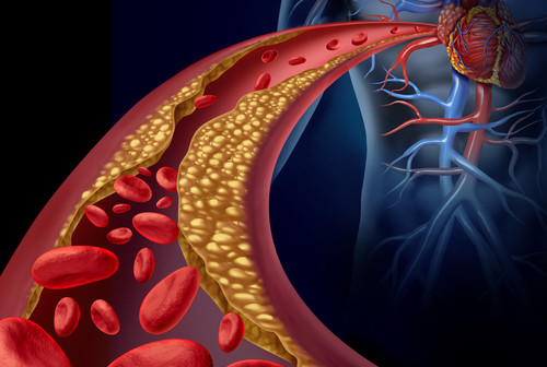
Type 2 diabetes has become a national epidemic, affecting nearly 26 million children and adults in the U.S. and approximately 170 million worldwide. According to the American Diabetes Association, $245 billion in costs are associated with diabetes, and 1 in 5 health-care dollars is spent caring for diabetics. A significant portion of the health costs associated with diabetes are those attributed to complications of the disease—including heart attacks, heart failure, stroke, dementia, chronic kidney disease, and amputations of the lower limbs. These complications emerge partly from hardening of the arteries caused by calcium deposits—a process known as arterial calcification—and are much more common in type 2 diabetics than in non-diabetics.
Dwight Towler, MD, PhD, professor and director of the Cardiovascular Pathobiology Program at Sanford-Burnham, has been actively researching the molecular causes of arterial calcification for more than a decade. Finding a way to prevent cardiovascular calcification could improve the vascular health of type 2 diabetes and prevent many of the associated medical complications.
In Towler’s previous work, he found that the assumption that arterial calcification was a natural, passive process that happens when cells die was incorrect. Instead, he showed that when the metabolism is disturbed—as in diabetes—calcium deposits are made by an active process that happens when key regulatory proteins erroneously trigger bone-formation genes in the arteries. Today, he is focused on those regulatory proteins, coded in the DNA by the Msx genes. Under normal conditions, Msx genes are essential for the formation of bones and teeth in the skull. But, in inflammatory conditions such as those associated with type 2 diabetes, the genes trigger the formation of calcium deposits in the arteries.
In his most recent study published on July 16 in the journal Diabetes, in collaboration with Dr. Robert Maxson of the University of Southern California, Towler’s research team examined the impact of Msx genes in mice genetically engineered to develop diabetes when fed high-fat diets. Previously, Towler showed how high-fat diets up-regulated the Msx genes in the aorta and coronary vessels of these mice, and caused calcium deposits via the Wnt paracrine signaling cascade. Now the question was: What would happen if Msx genes were absent in these mice?
“We were pleased to find that down-regulation of the Msx genes did indeed reduce the arterial calcification and vascular stiffness associated with diabetes,” said Towler. Our results are important because currently, there are no drugs to treat cardiovascular calcification. We have now identified four signaling pathways that represent targets for new drugs to intervene and inhibit the process.”
As a board-certified internist, Towler is committed to advancing these research findings to improve patient health and health care. “Our next step is to biochemically and genetically validate these pathways in human vascular disease—and identify drugs that improve vascular structure and function in mice. We are starting with lead compounds already tested in humans for other indications to see if we can repurpose those drugs to minimize the time it takes to get a treatment to the patients that suffer from this devastating complication of diabetes,” added Towler.
The study was performed in collaboration with the Norris Cancer Center, University of Southern California (CA), Washington University in St. Louis (MO), the Translational Research Institute for Metabolism and Diabetes (FL), and MD Anderson Cancer Center (TX).
Funding for the study was provided by NIH grants HL69229 and HL81138, the Barnes-Jewish Hospital Foundation, and Sanford-Burnham Medical Research Institute.