Nanomedicine Archives - Sanford Burnham Prebys
Institute News

Harnessing nanoparticles to fight drug-resistant infections

AuthorSusan Gammon
Date

June 7, 2018

The race against time to develop new antibiotics is more important than ever. A Wellcome Trust 2016 study predicted the present rate of emerging new virulent bacterial strains will outstrip the U.S. Food and Drug Administration approval rate for new antimicrobial agents by 2050—which means deaths from antimicrobial resistant strains will exceed deaths from cancer. Current studies show that in the United States, more than 23,000 deaths occur annually due to drug-resistant bacteria. Worldwide, the number is 700,000 deaths.

Erkii Ruoslahti, MD, PhD, distinguished professor at SBP, recently co-authored a study published in the journal Nature Communications that describes the first example of an effective gene therapeutic approach to fight lethal bacteria infections. The method uses a nanotherapeutic to deliver short interfering RNA (siRNA) that targets immune cells to bolster the immune system. The method was successfully used against a lethal bacterial infection of Staphylococcus aureus pneumonia in mice.

Until now, delivering siRNA-based therapeutics in the body has been difficult because of barriers built by both the body and by bacteria. These barriers have impeded the progress of gene therapeutic treatment of bacterial infections, including siRNA therapeutics.

To remedy this, the research team generated a porous nanoparticle host that protected siRNA payload from premature degradation in the blood stream. A peptide molecule engineered by Ruoslahti and Hong-Bo Pang, PhD at the University of Minnesota, was added to the surface of the nanoparticle that selectively attaches to immune cells called macrophages.

The researchers relied on the expertise of Ji-Ho Park, PhD, at the Korea Advanced Institute of Science and Technology (KAIST) to engineer a chemical coating, a fusogenic lipid, which allowed the nanoparticle to fuse with the cellular membrane and squirt the nanoparticle with its payload into the cell. The fusion mechanism also provided a means to induce dissolution of the silicon nanoparticles, releasing the siRNA therapeutic. This siRNA payload then reprogrammed the macrophage, activating it to engulf bacterial invaders.

The study was led by Michael Sailor, PhD, distinguished professor of chemistry and biochemistry at UC San Diego.

Institute News

New technology could deliver drugs to brain injuries

AuthorJessica Moore
Date

June 28, 2016

A new study led by scientists at the Sanford Burnham Prebys Medical Discovery Institute (SBP) describes a technology that could lead to new therapeutics for traumatic brain injuries. The discovery, published in Nature Communications, provides a means of homing drugs or nanoparticles to injured areas of the brain.

“We have found a peptide sequence of four amino acids, cysteine, alanine, glutamine, and lysine (CAQK), that recognizes injured brain tissue,” said Erkki Ruoslahti, MD, PhD, distinguished professor in SBP’s NCI-Designated Cancer Center and senior author of the study. “This peptide could be used to deliver treatments that limit the extent of damage.”

About 2.5 million people in the US sustain traumatic brain injuries each year, usually resulting from car crashes, falls, and violence. While the initial injury cannot be repaired, the damaging effects of breaking open brain cells and blood vessels that ensue over the following hours and days can be minimized.

“Current interventions for acute brain injury are aimed at stabilizing the patient by reducing intracranial pressure and maintaining blood flow, but there are no approved drugs to stop the cascade of events that cause secondary injury,” said Aman Mann, PhD, postdoctoral researcher in Ruoslahti’s lab and co-first author of the study with Pablo Scodeller, PhD, another postdoc in the lab.

More than one hundred compounds are currently in preclinical tests to lessen brain damage following injury. These candidate drugs block the events that cause secondary damage, including inflammation, high levels of free radicals, over-excitation of neurons, and signaling that leads to cell death.

“Our goal was to find an alternative to directly injecting therapeutics into the brain, which is invasive and can add complications,” explained Ruoslahti. “Using this peptide to deliver drugs means they could be administered intravenously, but still reach the site of injury in sufficient quantities to have an effect.”

The CAQK peptide binds to components of the meshwork surrounding brain cells called chondroitin sulfate proteoglycans. Amounts of these large, sugar-decorated proteins increase following brain injury.

“Not only did we show that CAQK carries drug-sized molecules and nanoparticles to damaged areas in mouse models of acute brain injury, we also tested peptide binding to injured human brain samples and found the same selectivity,” added Mann.

“This peptide could also be used to create tools to identify brain injuries, particularly mild ones, by attaching the peptide to materials that can be detected by medical imaging devices,” Ruoslahti commented. “And, because the peptide can deliver nanoparticles that can be loaded with large molecules, it could enable enzyme or gene-silencing therapies.”

This platform technology has been licensed by a startup company, AivoCode, which was recently awarded a Small Business Innovation Research (SBIR) grant from the National Science Foundation for further development and commercialization.

Ruoslahti’s team and their collaborators are currently testing the applications of these findings using animal models of other central nervous system (CNS) injuries such as spinal cord injury and multiple sclerosis.

The paper is available online here.


Institute News

Drug delivery to the placenta for healthier pregnancies

AuthorGuest Blogger
Date

May 6, 2016

Nearly 10% of  babies are born premature in the United States, according to the March of Dimes.  The underlying cause is often a poorly functioning placenta, the organ that nourishes and maintains the fetus. Continue reading “Drug delivery to the placenta for healthier pregnancies”

Institute News

A new approach to treating osteoarthritis

AuthorSusan Gammon
Date

January 20, 2015

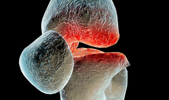
In a recent collaborative research study between two brothers—one a rheumatologist and the other a medical engineer—novel shaped nanoparticles were able to deliver anti-osteoarthritis drugs directly to the cells that drive the onset and progression of osteoarthritis (OA). The findings show promise to improve the treatment options for the nearly 21 million Americans, 25 years of age and older, that suffer from this chronic, often debilitating disease.

“We are excited to have developed nanoparticles which can efficiently and safely bring anti-OA drugs into the cells called chondrocytes that cause OA,” said Massimo Bottini, Ph.D., adjunct assistant professor in the Bioinformatics and Structural Biology Program at Sanford-Burnham. “Our method not only delivered the drug effectively, but stayed in the joint for a prolonged time without causing side effects. This is a significant improvement over previous attempts to deliver anti-OA drugs to affected joints.”

About osteoarthritis Under normal conditions, the extracellular matrix of the joint is maintained through a continual remodeling process in which low levels of different enzymes that produce and degrade cartilage are maintained. However, with increasing age and general wear and tear on joints, OA can occur when the enzymes that degrade cartilage are overproduced, creating an imbalance that leans toward the loss of collagen and joint impairment.

“For advanced OA, joint-replacement surgery is the only option for patients to regain comfortable and normal joint functions. For less severe cases, there is currently no medical therapy that can slow down or halt progression of the disease. This makes OA one of the largest unmet clinical needs in the field of rheumatology,” said Nunzio Bottini, MD, Ph.D., an associate professor in the Division of Cellular Biology at the La Jolla Institute for Allergy and Immunology (LIAI), who is also a practicing rheumatologist—and Massimo’s brother.

“The goal of treating OA is to restore the balance of the enzymes that control the matrix environment. Since there is no blood supply to the joint, drugs to treat the disease must be injected directly into the joint,” said Nunzio.

“Until now, scientists have tried using spherical nanoparticles to deliver anti-OA drugs. But the physical shape and size of the spheres predisposes them to diffuse into the synovial fluid and be flushed out of the joint before they can be effective,” said Massimo. “We have designed a one-dimensional linear nanoparticle made of graphite that is 100,000 times thinner than a human hair. This unique nanoparticle is engineered to travel through the negatively charged extracellular matrix and carry molecules to the nucleus of chondrocytes to turn off the genes that cause the disease.”

The study Using a mouse model for OA, the brothers injected the novel nanoparticle loaded with a gene inhibitor into the knees of affected mice. The nanoparticle delivered large amounts of the gene inhibitor to the cytoplasm and the nucleus of chondrocytes. Importantly, particles remained in the joint for two weeks compared to only few days for spherical nanoparticles.

“This is a significant improvement over previous attempts to deliver drugs to OA joints,” said Massimo. “Our next step is to further optimize the nanoparticle, see how long it remains in the body, and move to clinical studies in humans,” said Massimo.

Arthritis is a complex disease and integrated work between technologists—such as my brother Massimo—and biologists like me significantly increases the chance to make major treatment advances. Our next objective is to secure NIH funding to continue applying our complementary expertise to the quest to improve the lives of those suffering from arthritis,” added Nunzio.

The collaborative study was published in ACS Nano and performed at both Sanford-Burnham and LIAI. Cristiano Sacchetti, PhD, a shared postdoctoral fellow in Massimo and Nunzio Bottini’s laboratories was lead author on the paper.

A link to the paper can be found at: http://pubs.acs.org/doi/abs/10.1021/nn504537b.

Institute News

Unique pathway that homes cancer drugs to tumors is like no other

Authorsgammon
Date

October 3, 2014

In a new study published in Nature CommunicationsErkki Ruoslahti, MD, Ph.D., and his research team, with Hongbo Pang, PhD, as the lead author, identify the unique pathway that enhances the delivery of anti-cancer drugs to tumors. The pathway, called CendR, is a previously unknown variation of endocytosis—the process by which cells engulf nutrients and extracellular molecules. When activated, the CendR system improves the therapeutic efficacy of existing anti-cancer drugs while minimizing the collateral damage of normal cells and tissue. The findings advance our understanding of the biology of the cell by establishing a new type of trans-tissue transport pathway. Continue reading “Unique pathway that homes cancer drugs to tumors is like no other”