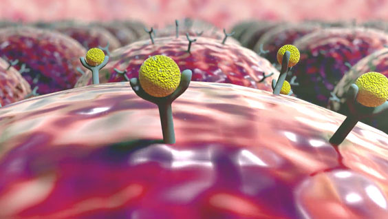
For about 50 percent of HIV-1-infected people, things as simple as buttoning a shirt, remembering the alphabet, and handling money may become compromised by a disorder known as HIV-induced brain injury. The condition occurs when receptors and proteins in an HIV-infected immune system produce toxic substances that lead to brain- and nerve-cell death. There is currently no treatment available for the more than 600,000 affected individuals in the U.S. In a new study by Sanford-Burnham researchers, blocking CCR5—an HIV co-receptor—was found to protect against brain injury and impairment of learning and memory. The findings, reported in TheJournal of Immunology, create a new approach to treating HIV-induced brain injury and may help our understanding of the potential involvement of CCR5 in other diseases of the brain. Continue reading “Research suggests new way to prevent HIV-associated brain injury”
Institute News
Exercise following bariatric surgery provides health benefits
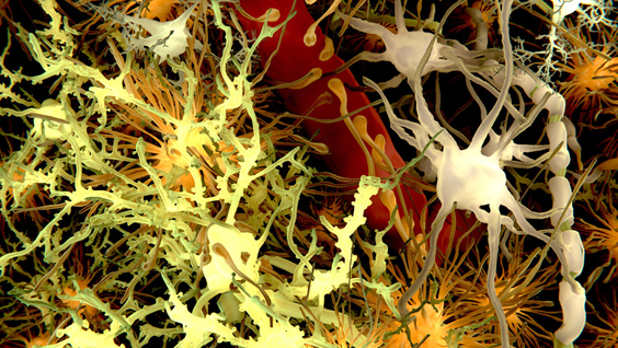
A new study by researchers at Sanford-Burnham reveals the process that leads to changes in the brains of individuals with Down syndrome—the same changes that cause dementia in Alzheimer’s patients. The findings, published in Cell Reports, have important implications for the development of treatments that can prevent damage in neuronal connectivity and brain function in Down syndrome and other neurodevelopmental and neurodegenerative conditions, including Alzheimer’s disease.
Down syndrome is characterized by an extra copy of chromosome 21 and is the most common chromosome abnormality in humans. It occurs in about one per 700 babies in the United States, and is associated with a mild to moderate intellectual disability. Down syndrome is also associated with an increased risk of developing Alzheimer’s disease. By the age of 40, nearly 100 percent of all individuals with Down syndrome develop the changes in the brain associated with Alzheimer’s disease, and approximately 25 percent of people with Down syndrome show signs of Alzheimer’s-type dementia by the age of 35, and 75 percent by age 65. As the life expectancy for people with Down syndrome has increased dramatically in recent years—from 25 in 1983 to 60 today—research aimed to understand the cause of conditions that affect their quality of life are essential.
“Our goal is to understand how the extra copy of chromosome 21 and its genes cause individuals with Down syndrome to have a greatly increased risk of developing dementia,” said Huaxi Xu, PhD, professor in the Degenerative Diseases Program and senior author of the paper. “Our new study reveals how a protein called sorting nexin 27 (SNX27) regulates the generation of beta-amyloid—the main component of the detrimental amyloid plaques found in the brains of people with Down syndrome and Alzheimer’s. The findings are important because they explain how beta-amyloid levels are managed in these individuals.”
Beta-amyloid, plaques, and dementia
Xu’s team found that SNX27 regulates beta-amyloid generation. Beta-amyloid is a sticky protein that’s toxic to neurons. The combination of beta-amyloid and dead neurons form clumps in the brain called plaques. Brain plaques are a pathological hallmark of Alzheimer’s disease and are implicated in the cause of the symptoms of dementia.
“We found that SNX27 reduces beta-amyloid generation through interactions with gamma-secretase—an enzyme that cleaves the beta-amyloid precursor protein to produce beta-amyloid,” said Xin Wang, PhD, a postdoctoral fellow in Xu’s lab and first author of the publication. “When SNX27 interacts with gamma-secretase, the enzyme becomes disabled and cannot produce beta-amyloid. Lower levels of SNX27 lead to increased levels of functional gamma-secretase that in turn lead to increased levels of beta-amyloid.”
SNX27’s role in brain function
Previously, Xu and colleagues found that SNX27-deficient mice shared some characteristics with Down syndrome, and that humans with Down syndrome have significantly lower levels of SNX27. In the brain, SNX27 maintains certain receptors on the cell surface—receptors that are necessary for neurons to fire properly. When levels of SNX27 are reduced, neuron activity is impaired, causing problems with learning and memory. Importantly, the research team found that by adding new copies of the SNX27 gene to the brains of Down syndrome mice, they could repair the memory deficit in the mice.
The researchers went on to reveal how lower levels of SNX27 in Down syndrome are the result of an extra copy of an RNA molecule encoded by chromosome 21 called miRNA-155. miRNA-155 is a small piece of genetic material that doesn’t code for protein, but instead influences the production of SNX27.
With the current study, researchers can piece the entire process together—the extra copy of chromosome 21 causes elevated levels of miRNA-155 that in turn lead to reduced levels of SNX27. Reduced levels of SNX27 lead to an increase in the amount of active gamma-secretase causing an increase in the production of beta-amyloid and the plaques observed in affected individuals.
“We have defined a rather complex mechanism that explains how SNX27 levels indirectly lead to beta-amyloid,” said Xu. “While there may be many factors that contribute to Alzheimer’s characteristics in Down syndrome, our study supports an approach of inhibiting gamma-secretase as a means to prevent the amyloid plaques in the brain found in Down syndrome and Alzheimer’s.”
“Our next step is to develop and implement a screening test to identify molecules that can reduce the levels of miRNA-155 and hence restore the level of SNX27, and find molecules that can enhance the interaction between SNX27 and gamma-secretase. We are working with the Conrad Prebys Center for Chemical Genomics at Sanford-Burnham to achieve this,” added Xu.
A team of scientists led by Sanford-Burnham’s Ranjan J. Perera, PhD, has identified a set of RNA molecules that are detectable in tissue samples and urine of prostate cancer patients, but not in normal healthy individuals. The study sets the stage for the development of more-sensitive and specific non-invasive tests for prostate cancer than those currently available, which could result in fewer unnecessary prostate biopsies with less treatment-related morbidity, according to a new study in The Journal of Molecular Diagnostics.
According to the American Cancer Society, prostate cancer is the second most common type of cancer in American men (behind skin cancer), and the second-leading cause of cancer death in men (after lung cancer). In 2014, more than 230,000 new cases of prostate cancer will be diagnosed. One in seven American men will get prostate cancer during his lifetime, and one in 36 will die from it. Since most men with prostate cancer have indolent (non-aggressive) disease for which conservative therapy or surveillance would be appropriate treatment, the clinical challenge is not only how to identify those with prostate cancer, but also how to distinguish those who would benefit from surgical or other aggressive treatment from those who would not.
Today, prostate cancer is primarily detected and monitored by testing for high concentrations of prostate-specific antigen (PSA) in blood samples. High PSA levels are often followed by a biopsy to confirm the presence of cancer, and whether it’s slow growing or aggressive. “While elevated PSA can be an alert to a lethal cancer, it can also detect less aggressive cancers that may never do any harm,” said Vipul Patel, MD, medical director of the Global Robotics Institute at Florida Hospital in Orlando, and co-author of the study. “Moreover, only 25 percent of men with raised PSA levels that have a biopsy actually have prostate cancer. Prostate cancer needs to be screened for; we just need to find a better marker.”
The researchers believe that they have identified a group of RNA molecules – known as long noncoding RNAs (lncRNAs) – that hold the potential for serving as better prognostic markers for prostate cancer. lncRNAs are noncoding RNA molecules that until recently were dismissed by scientists as non-functional noise in the genome. Now, lncRNAs are thought to regulate normal cellular development and are increasingly reported as contributing to a range of diseases, including cancer.
Detection of lncRNAs in urine
“We have identified a set of lncRNAs that appear to have an important role in prostate cancer diagnostics,” said Perera, associate professor and scientific director of Analytical Genomics and Bioinformatics at our Lake Nona campus. “The findings advance our understanding of the role of lncRNAs in cancer biology and, importantly, broaden the opportunity to use lncRNAs as biomarkers to detect prostate cancer.”
The study profiled the lncRNAs in three distinct groups: (1) human prostate cancer cell lines and normal prostate epithelial cells, (2) prostate adenocarcinoma tissue samples and matched normal tissue samples, (3) urine samples from patients with prostate cancer or benign prostate hypoplasia, and normal healthy individuals. In each case, the lncRNAs were elevated in prostate cancer patient samples, but not in patients with benign prostate hypoplasia or normal healthy individuals.
One advantage of lncRNAs is that the molecules can be detected in urine samples, which are more easily available than blood tests. One lncRNA, PCA3, was recently commercialized as a urine test to identify which men suspected of having prostate cancer should undergo repeat prostate biopsy. However, discrepancies have been found to exist between PCA3 levels and clinicopathologic features, said Perera. In the current study, PCA3 was detected in some, but not all of the study samples, suggesting that reliance on a single biomarker may be insufficient for prostate cancer detection, while combining additional markers may increase the specificity and sensitivity of the test.
“There is a tremendous unmet clinical need for better non-invasive screening tools for early detection of prostate cancer to reduce the overtreatment and morbidity of this disease,” added Patel. “Our findings represent a promising approach to meet this demand.”
Technical details of the study
The goal of the first experiment was to see whether lncRNAs are differentially expressed in prostate cancer by measuring total RNA from prostate cancer cell lines and normal epithelial prostatic cells using NCode human ncRNA array and SurePrint G3 human lncRNA microarrays. Hierarchical clustering revealed distinguishable lncRNA expression profiles. Thirty lncRNAs were up-regulated and the expression levels of three top-ranking candidates [XLOC_007697, LOC100287482, and AK024556 (also known as SPRY4-IT1)] were confirmed in prostate cancer cell lines by quantitative real-time polymerase chain reaction (qPCR) analysis. The SPRY4-IT1 was found to be up-regulated more than 100-fold in PC3 cells compared with prostatic epithelial cells.
In a second experiment, lncRNA expression was compared in pooled prostate cancer tissue samples and matched normal tissues from 10 frozen biopsy specimens. Hierarchical clustering of the differentially expressed lncRNAs was observed and 10 up-regulated lncRNAs were detected using microarrays. An additional set of 18 prostate cancer tissue samples was analyzed by qPCR and five lncRNAs were found to be significantly higher in prostate tumor tissues compared with matched normal tissues.
Researchers used qPCR to analyze total RNA isolated from urine in another experiment. Urine was collected from 13 prostate cancer patients and 14 healthy controls. All six lncRNAs were found to be significantly up-regulated in the urine samples from the prostate cancer patients compared with normal patient controls, while there were no differences between normal and benign prostatic hyperplasia patient samples.
In other studies focused particularly on SPRY4-IT1. Using both qPCR and highly sensitive droplet digital PCR, expression of SPRY-IT1 was found to be increased in 16 of 18 (89 percent) tissue samples from patients with prostatic adenocarcinoma, compared to normal tissue samples. The researchers developed chromogenic in situ hybridization (CISH) techniques to visualize SPRY4-IT1 expression in cancerous and matched normal tissue. Intense staining was seen in all adenocarcinoma samples, but not in normal prostatic tissue. Finally, the investigators showed that reduction of SPRY4-IT1 in prostate cancer cells through the use of small interfering RNA (siRNA) leads to decreased cell viability and cellular invasion as well as increased apoptosis, similar to what is seen in melanoma cells.
About the paper
“Long Noncoding RNAs as Putative Biomarkers for Prostate Cancer Detection,” by Bongyong Lee, Joseph Mazar, Muhammed Nauman Aftab, Feng Qi, John Shelley, Jian-Liang Li, Subramaniam Govindarajan, Felipe Valerio, Inoel Rivera, Tadzia Thurn, Tien Anh Tran, Darian Kameh, Vipul Patel, and Ranjan J. Perera, DOI: http://dx.doi.org/10.1016/j.jmoldx.2014.06.009. Published online ahead of The Journal of Molecular Diagnostics, Volume 16, Issue 6 (November 2014) published by Elsevier.
Medulloblastoma (MB) is the most common malignant brain cancer in children. Children diagnosed with the disease undergo intense therapy, including surgery, radiation, and high-dose chemotherapy. Although current treatment regimens have improved 5-year survival rates, almost a third of MB patients still die from their disease, and children who survive suffer long-term side effects that affect their quality of life. Continue reading ““Survivin” as a new target to treat brain cancer”
Institute News
Unique pathway that homes cancer drugs to tumors is like no other
In a new study published in Nature Communications, Erkki Ruoslahti, MD, Ph.D., and his research team, with Hongbo Pang, PhD, as the lead author, identify the unique pathway that enhances the delivery of anti-cancer drugs to tumors. The pathway, called CendR, is a previously unknown variation of endocytosis—the process by which cells engulf nutrients and extracellular molecules. When activated, the CendR system improves the therapeutic efficacy of existing anti-cancer drugs while minimizing the collateral damage of normal cells and tissue. The findings advance our understanding of the biology of the cell by establishing a new type of trans-tissue transport pathway. Continue reading “Unique pathway that homes cancer drugs to tumors is like no other”
Institute News
A signature for early-stage heart failure could improve diagnosis and prevent disease progression
This is a post by our guest writer Janelle Weaver, PhD
Heart failure affects about five million people in the United States, and about half of these individuals die within five years of diagnosis. This condition occurs when the heart can’t pump enough blood to meet the body’s needs, and evidence suggests that abnormalities in energy metabolism play an important role. However, many past studies addressing the underlying molecular mechanisms have focused on severe, late-stage heart failure, potentially missing out on early events that could guide the development of treatment strategies for early disease stages. Continue reading “A signature for early-stage heart failure could improve diagnosis and prevent disease progression”
Institute News
New insights into how the heart forms may help identify heart defects
This is a post by our guest writer Janelle Weaver, PhD
The formation of the heart during development is a highly complex process that requires precise coordination between cells and molecular signaling pathways. The fruit fly has been widely used for studying the underlying cellular and molecular mechanisms, and a great deal is known about how the fate of heart cells is controlled by signaling pathways and transcription factors—proteins that control gene activity. But beyond that, events that regulate heart formation have not been clear. Continue reading “New insights into how the heart forms may help identify heart defects”