Tumor Initiation and Maintenance Archives - Sanford Burnham Prebys
Institute News

Targeting long-sought EphA2 receptor becomes crystal clear

AuthorMonica May
Date

May 13, 2019

Scientists have long sought to target a cellular receptor called EphA2 because of its known role in many disorders, including cancer, inflammatory conditions, neurological disorders and infectious diseases. However, lack of information about the structure formed when EphA2 links to other molecules—ligands—has hindered drug development. 

Now, scientists at Sanford Burnham Prebys have crystallized EphA2 together with peptide ligands (short proteins) and used the structure to engineer more powerful compounds that activate or inactivate the receptor, paving the way for new therapies. The discovery was published in the Journal of Biological Chemistry.

“EphA2 plays a central role in a plethora of biological and disease processes,” says Elena Pasquale, PhD, professor in the Tumor Initiation and Maintenance Program at Sanford Burnham Prebys. “Our team’s identification of potent, highly selective peptides that regulate the receptor is a key step toward rational design of therapies for the numerous disorders that are driven by EphA2.” 

EphA2 is found in the cells that line the surfaces of our body, including our skin, blood vessels and other organs. The receptor is typically only present at high levels during disease states, making it a promising drug target. Activating the receptor could hinder tumor growth, while inhibiting it could reduce unwanted formation of blood vessels (angiogenesis), treat certain inflammation-driven disorders and block pathogens—such as malaria, chlamydia and the hepatitis C virus—from gaining entry into a cell through the receptor. Because EphA2 travels deep inside of the cell when activated, scientists could also harness it as a Trojan horse by attaching chemotherapies or imaging agents to the peptide ligands, which would subsequently be delivered to the desired cells. 

In the study, the scientists initially crystallized a weakly binding peptide in complex with EphA2, yielding a detailed picture of the binding features and providing clues to the receptor’s “sweet spot” or site of action. The researchers then used this information to repeat this process, engineering increasingly more powerful ligands. This work identified several peptides that strongly clasp the receptor and activate or inactivate it—which can be used to inform drug development.

Further quantitative Förster Resonance Energy Transfer (FRET) microscopy experiments, which measure receptor-receptor interactions, revealed that EphA2 receptors cluster together when activated by a peptide—an effect similar to that caused by its natural ligands—answering an unresolved question in the field. 

“In addition to helping guide therapeutic development paths, these peptides are also valuable research tools for scientists who are working to gain insights into this important receptor,” adds Pasquale. “Our hope is that with this new information, one day we can find targeted therapies to treat cancer, inflammatory disorders and infectious diseases that are regulated by EphA2.”

The co-first authors of the study are Maricel Gomez-Soler, PhD, and Marina Petersen Gehring, PhD, of Sanford Burnham Prebys; and Bernhard C. Lechtenberg, PhD, formerly of Sanford Burnham Prebys and currently of the Walter and Eliza Hall Institute of Medical Research. 

Additional authors include Elmer Zapata-Mercado and Kalina Hristova, PhD, of Johns Hopkins University. The study’s DOI is 10.1074/jbc.RA119.008213. 

This research was supported by the National Institutes of Health (NIH) (R01NS087070, R01GM131374 and P30CA030199). 
 

Interested in keeping up with our latest discoveries, upcoming events and more? Subscribe to our monthly newsletter, Discoveries.

Institute News

Making ERK work as a therapeutic target for colorectal cancer

AuthorJessica Moore
Date

June 3, 2016

Colorectal cancer is the third most common cancer in the US, affecting 1.2 million people. Despite extensive research, the five-year survival rate remains below 15%, underscoring the need for new treatments.

One-third of colorectal cancers are driven by over-activation of extracellular signal-regulated kinases 1 and 2 (ERK1/2), which regulates proliferation, metabolism, and cell movement. However, drugs targeting the ERK1/2 pathway are not widely used to treat colorectal cancer because they don’t appreciably slow cancer growth. New research co-led by Petrus de Jong, MD, PhD, postdoctoral associate at SBP, points to a possible reason for this lack of effect, as well as a solution.

“We genetically deleted the ERK1/2 pathway in the lining of the mouse intestine, and we expected to see less cell proliferation,” said de Jong, a co-first author on the paper. “Instead, the opposite occurred. There was more cell growth and the cells were less organized.” de Jong works in the laboratory of Garth Powis, D.Phil., professor and director of SBP’s NCI-Designated Cancer Center, who also contributed to the investigation.

The new study, published in Nature Communications, shows that the increased cell growth caused by disabling ERK1/2 results from increased activity of a related kinase, ERK5. The team went on to show that inhibiting both pathways suppresses proliferation of human colorectal cancer cell lines and slows growth of tumor-like structures in vitro.

“Therapies aimed at targeting ERK1/2 likely fail because ERK5 compensates,” said Eyal Raz, MD, senior author and professor at the UC San Diego School of Medicine. “Previously, ERK5 didn’t seem important in colorectal cancer. This is an underappreciated escape pathway for tumor cells. Hence, the combination of ERK1/2 and ERK5 inhibitors may lead to more effective treatments for colorectal cancer patients.”

“If you block one pathway, cancer cells usually mutate and find another pathway that ultimately allows for a recurrence of cancer growth,” said Koji Taniguchi, MD, PhD, assistant project scientist at UC San Diego and the other co-first author. “Usually, mutations occur over weeks or months. But other times, as in this case, the tumor does not need to develop mutations to find an escape route from targeted therapy. When you find the compensatory pathway and block both, there is no more escape.”

The scientists suggested that other inhibitors of the ERK1/2 pathway should be tested with ERK5 inhibitors in both human colorectal cancer cells and mouse models to identify the most effective combination that could advance to clinical trials.

This post is a modified version of the press release from UC San Diego. Photo from Ed Uthman via Flickr.

The paper is available online here.

Institute News

Super-oncogenic protein that promotes development of melanoma

AuthorJessica Moore
Date

May 19, 2016

An international collaborative study led by scientists at the Sanford Burnham Prebys Medical Discovery Institute (SBP) has identified a malicious form of a protein that drives the formation of melanoma. The findings, published in Cell Reports, reveal unexpected insight into how this lethal skin cancer develops and progresses, and may help understand and develop novel therapies against these aggressive tumors.

“We found that an inactive version of a protein called activating transcription factor 2 (ATF2) elicits a tumor-promoting effect in a way not seen before,” said Ze’ev Ronai, PhD, chief scientific advisor of SBP and professor of its NCI-designated Cancer Center. “We have known for years that the active version of ATF2 promotes melanoma, but this result was a surprise because we thought ATF2 transcriptional activity was essential to activate cancer-related genes.”

Ronai’s team has been studying ATF2’s role in melanoma for two decades. Their past work led to the view that it’s dangerous when it’s in the nucleus because it controls cancer-enabling genes, but benign when it’s not.

In the current study, researchers looked at the oncogenic potential of a ‘dead’ form of ATF2 in mice with mutations in BRAF, a kinase that transmits signals promoting cell division and is often mutated in pigmented skin cells. The same mutation is found in about half of all human melanomas.

“Inactive ATF2, in mice with mutant BRAF, resulted in the formation of pigmented lesions and later, melanoma tumors,” said Ronai, senior author of the study.

“What makes this discovery relevant to human melanoma is that we identified a structurally similar form of inactive ATF2 in human melanoma samples that has the same effects on cancer cells,” added Ronai. “Inactive ATF2 could be an indicator of tumor aggressiveness in patients with BRAF mutations, and maybe other types of cancer as well.”

“Unlike models with more complex genetic changes, like the inactivation of PTEN and p16 combined with BRAF mutations that result in rapid tumorigenesis (within a few weeks), the inactive ATF2 caused BRAF mutant mice to develop melanoma much slower, more similar to the timescale seen in patients,” commented Ronai. “This improves our ability to monitor the development of melanoma and efficacy of possible interventions.”

“We’re now investigating why inactive ATF2 so potently promotes BRAF-mutant melanoma, and looking for other types of cancer where it acts the same way,” Ronai said. “Our findings may guide precision therapies for tumors with mutant ATF2.”

The paper is available online here.

Institute News

Deeper dive into emerging cancer drugs’ actions

AuthorJessica Moore
Date

April 28, 2016

A major challenge in developing cancer drugs is finding ways to kill tumors without damaging healthy tissue. It’s tough—since cancer cells share the same cellular machinery as normal cells, scientists have to be mindful about the targets they choose. One way to balance these concerns is to target cellular processes—such as protein synthesis and degradation—that tumors frequently overuse to support their rapid and aberrant growth. Continue reading “Deeper dive into emerging cancer drugs’ actions”

Institute News

New drug combination may lead to treatment for childhood brain cancer

AuthorJessica Moore
Date

March 14, 2016

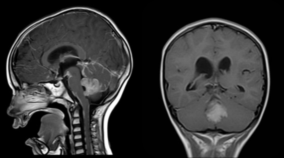
Researchers at SBP have identified a new combination therapy for the most aggressive form of medulloblastoma, a fast growing type of pediatric brain cancer. The study, published  in Cancer Cell, is expected to lead to a clinical trial to confirm the benefits of the novel drug combination. Continue reading “New drug combination may lead to treatment for childhood brain cancer”

Institute News

Molecular “brake” prevents excessive inflammation

AuthorGuest Blogger
Date

February 25, 2016

Inflammation is a catch-22: the body needs it to eliminate invasive organisms and foreign irritants, but excessive inflammation can harm healthy cells, contributing to aging and sometimes leading to organ failure and death. A study published in Cell, co-authored by Jorge Moscat, PhD, and Maria Diaz-Meco, PhD, professors in SBP’s NCI-designated Cancer Center, in collaboration with the laboratory of Michael Karin, PhD, at the University of California, San Diego School of Medicine, shows that a protein known as p62 acts as a molecular brake to keep inflammation in check and avoid collateral damage. Continue reading “Molecular “brake” prevents excessive inflammation”

Institute News

Why new therapies are failing some kids with brain cancer

Authorjmoore
Date

February 1, 2016

The most common type of malignant brain cancer in children is medulloblastoma, a fast growing tumor located in the cerebellum—the lower, rear portion of the brain. Although the standard treatment, an aggressive combination of surgery, radiation, and high-dose chemotherapy, cures more than 70 percent of patients, many survivors are left with profound long-term side effects, including cognitive deficits and increased incidence of other cancers. Continue reading “Why new therapies are failing some kids with brain cancer”

Institute News

Cells on the run: researchers discover a protein required for cell motility

Authorsgammon
Date

April 28, 2015

Your cells move. They need to move for good reasons, such as when white blood cells travel to heal wounds, and for bad reasons, like when cancer cells invade surrounding tissue to metastasize. To move, cells create extensions—like feet—that make contact with a surface and lead the cell to its destination. The abnormal production of these cell extensions is associated with Alzheimer’s disease, epilepsy, and many other neurological disorders. For these reasons, scientists are working to understand the fundamental components of cell movement. What they find may lead to treatments that can promote cell movement when you need it, and prevent it when you don’t. Continue reading “Cells on the run: researchers discover a protein required for cell motility”

Institute News

Expanding the options to treat melanoma

Authorsgammon
Date

March 16, 2015

Melanoma is the most deadly form of skin cancer with approximately 10,000 deaths per year in the U.S. and more than 65,000 worldwide. Although there are more and better treatment options available today than in previous years, there is still an urgent need to develop drugs that target the numerous pathways melanoma cells use to multiply, spread, and kill. Continue reading “Expanding the options to treat melanoma”

Institute News

Study explains control of cell metabolism in patient response to breast cancer drugs

Authorsgammon
Date

March 9, 2015

A new research study has discovered a mechanism that explains why some breast cancer tumors respond to specific chemotherapies and others do not. The findings highlight the level of glutamine, an essential nutrient for cancer development, as a determinant of breast cancer response to select anticancer therapies, and identify a marker associated with glutamine uptake, for potential prognosis and stratification of breast cancer therapy. The study results were published online in Cancer Cell. Continue reading “Study explains control of cell metabolism in patient response to breast cancer drugs”