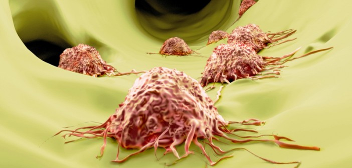
Your cells move. They need to move for good reasons, such as when white blood cells travel to heal wounds, and for bad reasons, like when cancer cells invade surrounding tissue to metastasize. To move, cells create extensions—like feet—that make contact with a surface and lead the cell to its destination. The abnormal production of these cell extensions is associated with Alzheimer’s disease, epilepsy, and many other neurological disorders. For these reasons, scientists are working to understand the fundamental components of cell movement. What they find may lead to treatments that can promote cell movement when you need it, and prevent it when you don’t.
In a recent study led by Rati Fotedar, PhD, adjunct professor in Sanford-Burnham’s NCI-designated Cancer Center, a protein called WISp39 was found to be an essential component of the steering mechanism cells use to drive themselves in the right direction. The steering mechanism, known as “directed motility,” requires a cell to have a front end and a back end (cell polarity), and WISp39 controls this, too. Without cell polarity, cells would be unable to coordinate their movement and reach their destination.
Using a wound-healing assay, the research team examined cells in which WISp39 expression was suppressed, and compared their movement to unmodified WISp39 cells. A wound-healing assay involves culturing cells in vitro and then creating a “scratch” in the cell layer. Normal cells develop a leading edge that moves toward the gap to close the scratch until new cell-cell contacts are established. After the scratch, the effects of cell-matrix and cell-cell interactions are captured in images taken over several hours to two days. It’s a simple method that mimics the migration of cells in vivo.
“We found that without WISp39, cells migrate in a chaotic manner and have multipolar lamellipodia. Lamellipodia are cell projections made of actin that enable cells to move—similar to how we use our feet,” said Fotedar. “Imagine trying to walk with more than one foot at the end of your legs, each foot pulling in a different direction. We observed WISp39 deficient cells trying to migrate in different directions, and this led in some cases to severed cell fragments, and cell death.”
On a molecular level, the study showed that WISp39 binds phosphorylated Coronin 1B and protein chaperone Hsp90, and establishes a complex with Slingshot phosphatase (SSH). The complex plays in important role in regulating Cofilin and localizing the Arp 2/3 complex, which is critical for lamellipodia formation, cell polarity, and directed cell motility.
“Now that we have identified the proteins that interact with WISp39, we will focus our future studies on elucidating the detailed mechanism by which WISp39 coordinates Coronin 1B, Arp2/3 complex, and Cofilin activity in the lamellipodia to achieve directed cell motility,” added Fotedar.
The study was performed in collaboration with The Scripps Research Institute, and the Sanford Consortium for Regenerative Medicine, and published in the The Journal of Cell Biology.
