News
Showing 6 of 1913 Results

Press ReleaseJul 22, 2026
Read MoreBiological sex linked to protein changes in lung adenocarcinoma and other cancers
Study shows lung adenocarcinoma has strong sex-based proteome differences, while five other cancer types have moderate differences.

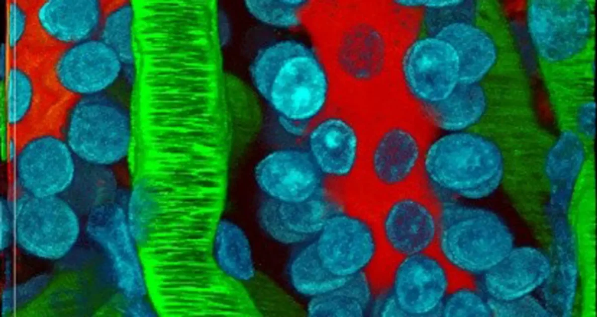
Institute NewsJul 27, 2026
Science in Pictures
A weekly image series featuring selected pictures in science • Leukemia cells apoptosis
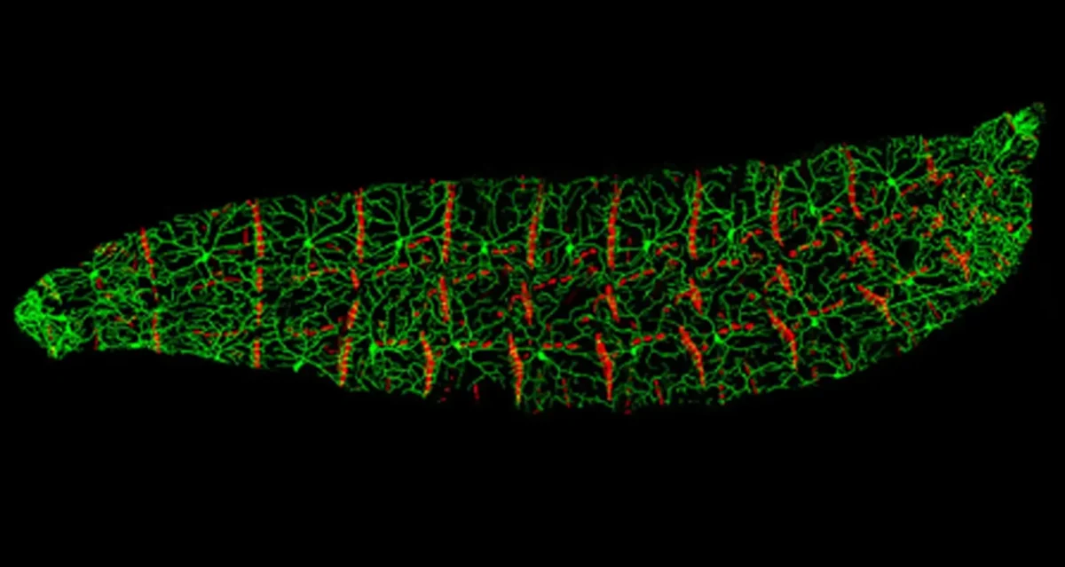
Institute NewsJul 20, 2026
Science in Pictures
A weekly image series featuring selected pictures in science • Drosophila larva

Institute NewsJul 18, 2026
T. Denny Sanford (1935-2026)
Renowned philanthropist and businessman T. Denny Sanford passed away July 18, 2026 in his hometown of Sioux Falls, South Dakota….
Press ReleaseJul 17, 2026
Protective protein reduces tau tangle toxicity linked to dementia
Bolstering SORLA protein has potential as a therapeutic strategy for Alzheimer’s disease and other tauopathies.
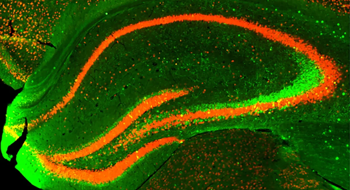
Institute NewsJul 13, 2026
Science in Pictures
A weekly image series featuring selected pictures in science • Kidney microvasculature

Institute NewsJul 6, 2026
LEAP scholars share scientific and professional development achievements
Postbaccalaureate training program participants deliver capstone presentations, thank mentors and colleagues.