Overview
The High-Content Screening (HCS) core facility of the Conrad Prebys Center for Chemical Genomics is part of the Sanford Burnham Prebys screening center. As part of the Prebys Center, the HCS resource has access to the HTS plate and liquid handling infrastructure of the screening center as well as the screening center’s cell culture facility. For details on the other core facilities of the Prebys Center, please refer to the “Related Resources” below.
The HCS core facility provides assay development, screening, and data analysis/mining expertise and services for high content screens, where the readout is based on images obtained using high-throughput microscopy systems. For assay development and screening of non-imaging assays refer to Assay Development and High-Throughput Screening. The HCS team has expertise in all areas of high content image-based screens including sample preparation, image acquisition, image analysis, image data management, and algorithm development.
The HCS core has experience conducting phenotypic assays, ranging from functional assays, such as inhibition of phagocytosis and quantification of cytoskeletal changes, to transcriptional reporter modulation, such as activation of insulin promoter activity. HCS projects range from screening of several hundred chemical compounds to libraries of over 300,000 compounds. Additionally, the Prebys Center screening center is co-located with the Sanford Burnham Prebys Functional Genomics (FGC) core, fostering collaboration between the HCS and FGC resources for RNAi screening projects.
Services
The HCS core facility’s experienced staff is available to help investigators design and execute image-based assays by providing the following services:
- Conceptualization and design of image-based assays for medium- and high-throughput screening.
- For small scale screens:
- Training/use of HCS instruments
- Training/use of analysis software
- Performing selection, development, and/or validation of image analysis methods and/or assay read-outs
- For large scale screens:
- Optimizing assay biology
- Developing algorithms and read-outs
- Performing assay miniaturization/validation
- Screening in collaboration with HTS or Functional Genomics facilities
- For small scale screens:
- Aid in image data management, HC data analysis and visualization
HCS Project Examples
Image-based high content assays are usually employed when an assay utilizes multiplexed fluorescent labels, investigates sub cellular localization or spatial distribution of fluorescence. The technique can also be used when evaluating morphological changes of cells or cellular structures such as shape or size, quantifying co-localization of multiple labels, or monitoring the response of a sub-population of cells. Because of the high content nature of image-based assays, the multi-parametric assay read-outs can provide additional biological or disease relevant information. Some examples of medium and high throughput screening campaigns utilizing image-based high content assays are provided in the table below.
Equipment & Resources
Liquid Handling
Robotic liquid and plate handling equipment and resources are used in common by all groups within the Prebys Center and are described in detail under High Throughput Screening. For information on available screening libraries, refer to Compound Management for chemical libraries and Functional Genomics for RNAi libraries.
HCS imaging systems
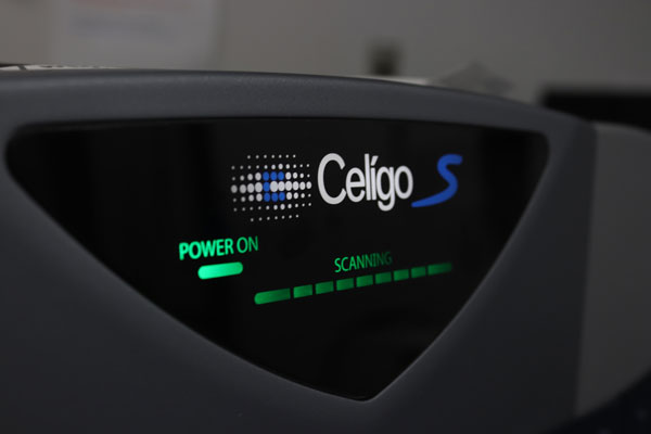
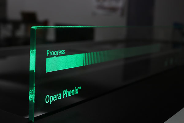
The HCS core facility contains state-of-the-art automated high-throughput microscopy instruments. Depending on the instrument, autofocusing is either image-based (IC200, Eidaq100) or laser-based (IN Cell 1000, Opera QEHS, Celigo). These instruments can be used with different objectives ranging from 4x to 60x magnification and have the ability to image standard clear-bottom multi-well plates (96, 384, 1536), preferably black plates with optical quality glass or plastic bottoms with a bottom thickness of ~200um. The INCell1000 and Opera QEHS in La Jolla have automatic plate loading capabilities (>40 plates). For a summary of each HCS instrument’s capabilities refer to the table below.

HCS image analysis and image data management software
A variety of image processing, data analysis, and visualization tools are available, including InCell Investigator (GE), CytoShop (Q3DM/Beckman Coulter), CyteSeer (Vala Sciences), Acapella (PE), Matlab (The Mathworks), Photoshop (Adobe), ImageJ (NIH), CellProfiler/CellVisualizer (nonprofit open source), and Volocity 3D/time course analysis software (PE). For screen data analysis and mining, CBIS (ChemInnovation) screening database software, as well as Spotfire (Tibco), Genedata Screener, and Prism (Graphpad) data analysis software are available. With these software packages the HCS team can rapidly apply available HCS solutions and algorithms, as well as develop custom HCS solutions, algorithms and tools for the wide variety of HCS projects.
To streamline HCS processing, visualization, and data management, the HCS team implemented a local storage system (LSS) dedicated to HCS based on the Columbus image-data management system (PE). The Columbus system provides ~12TB of fast access storage at the La Jolla site and handles image and data file management for currently ongoing HCS projects. The LSSs is connected via high-speed intranet and provide scientists with web-based access to image visualization, analysis, and data visualization tools. The Columbus image-data management system provides web-based upload capabilities for Opera, INCell 1000, IC200, Eidaq100, and Celigo instruments. It is integrated with the Volocity 3D/time course visualization and analysis package, and is also directly linked to Genedata Screener software for a complete HCS image-data analysis and visualization solution.

Price List
For a Price List, please call (858) 646-3100 ext. 3329 or email us.
Leadership
Michael Jackson, PhD
Vice President, Drug Discovery & Development
Susanne Heynen-Genel, PhD
Facility Director
Contact
Please call (858) 646-3100 ext. 3329 or use the button below to send us an email.
