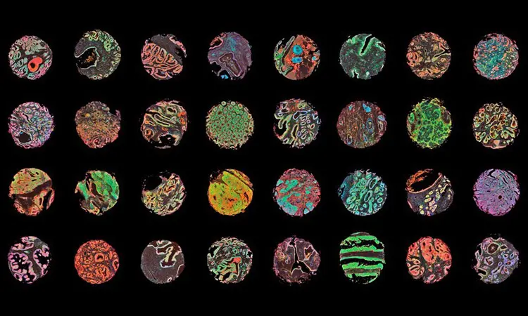
Fluorescence immunohistochemistry and confocal microscopy were used to create this array of normal and cancerous human tissue samples. Human prostate, colon, kidney, intestine and breast tissue sections were stained for the presence of protein biomarkers to improve cancer diagnosis and prognosis.
Image courtesy of Aamir Ahmed, Jane Pendjiky and Michael Millar.
