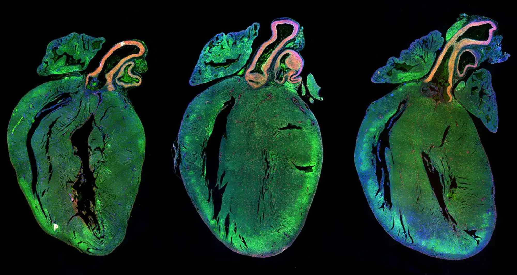
Sections of a rat heart are shown using fluorescence microscopy imaging. The muscle cells of the heart, called cardiomyocytes, are stained in green; cell nuclei are counterstained in blue; and proliferating cells are labeled in green.
Institute News
Science in Pictures