Alzheimer’s is one of the most frightening diseases of our time. Of the top 10 causes of death in the U.S., it is the only disease for which no effective or preventative treatment exists. Recent clinical trial failures have only deepened the pain of patients and their families.
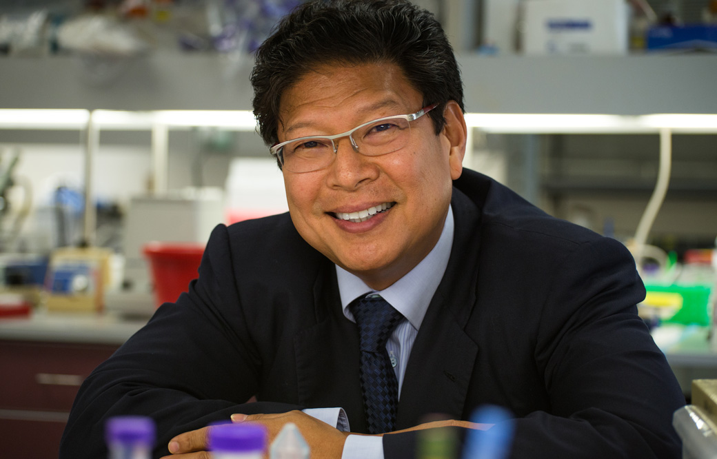
To learn about the state of Alzheimer’s research in the wake of these setbacks—and whether there is hope on the horizon—we caught up with Alzheimer’s expert Jerold Chun, MD, PhD, professor and senior vice president of Neuroscience Drug Discovery at Sanford Burnham Prebys. Chun and his team recently published a Nature study that suggests a potential Alzheimer’s treatment may be closer than we think.
- Why has Alzheimer’s disease become so prevalent? Are we better at diagnosing the disease?
The number of people with Alzheimer’s disease is rising because of the aging Baby Boomers generation—which makes up more than 20% of the U.S. population. As a result, the number of those living with the condition is projected to more than double by 2050 to nearly 14 million people. This will place an incredible economic and social burden on our society—unless a treatment is found. - Are there any treatments that work for Alzheimer’s disease?
No disease-modifying therapies exist. The medicines a patient can receive today just treat symptoms. For example, cholinesterase inhibitors and N-methyl D-aspartate antagonists treat cognitive symptoms, such as memory loss, confusion and problems with thinking and reasoning—but they aren’t able to stop the disease. - Is it possible to prevent Alzheimer’s?
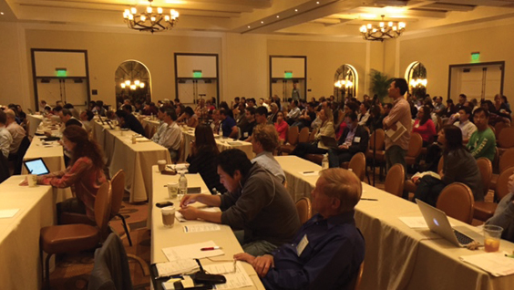
Multiple studies from this year’s Alzheimer’s Association International Conference centered on this topic. Evidence suggests that adopting healthy lifestyle choices such as eating a healthy diet, not smoking, exercising regularly and stimulating the mind may decrease the risk of cognitive decline and dementia.It is encouraging to know that preventing Alzheimer’s may be partially within our control. However, it is undeniable that even individuals who live a healthy lifestyle will still develop Alzheimer’s. We need to remain laser focused on developing effective preventions and treatments.
- Do we know the cause of Alzheimer’s? What are the latest theories?
In short, no. We know that clumps of amyloid-beta and tau proteins in the brain are linked to the disease. We also know that in rare cases genes are involved, because Alzheimer’s can run in families—but this accounts for less than 1% of cases. New research points to unique gene changes within the brain, called somatic gene recombination, as a new potential factor. Some data also implicate aspects of the immune system. It’s most likely that multiple factors lead to disease—and that an effective treatment will tackle Alzheimer’s from several angles. - How would you describe the pipeline of Alzheimer’s treatments in development?
The pipeline of Alzheimer’s treatments is in dire need of expansion. As of February 2019, only 132 drugs were under evaluation in clinical trials. Nearly half of these compounds target beta-amyloid.For comparison, there are nearly 4,000 compounds under development for cancer—which affects almost three times as many Americans each year. We certainly need to continue to invest in cancer treatments—but clearly there is an urgent need to fill the Alzheimer’s pipeline, and an even greater need to find an approach that actually works.
- Tell us more about your research. What did you find? What are the next steps?
In school we learned that all cells have the same DNA. However, in our recent research we found that in the brains of patients, the DNA in the Alzheimer’s-linked APP gene can be “mixed and matched” into many different, new forms, some of which aren’t found in healthy individuals. To create these new gene variants, reverse transcriptase—best known as an enzyme infamously used by HIV—is required. This suggests that existing HIV medications—called reverse transcriptase inhibitors—which halt reverse transcriptase, might be useful as a near-term treatment for Alzheimer’s disease. A doctor can prescribe these medicines now as an “off-label” use for the treatment of Alzheimer’s disease. However, prospective clinical trials are needed to test the efficacy and side-effect profiles of these medicines in actual Alzheimer’s disease patients. - Are humans the only species that get Alzheimer’s disease?
To our knowledge, yes. No other animal has the intellectual and cognitive capacity exhibited by humans. For this reason, scientists have developed animal models that exhibit symptoms and pathologies that approximate the disease. - How far away are we from an effective Alzheimer’s treatment? Years or decades?
What excites me about my team’s findings is that, if true, a partially effective treatment may be available now. Reverse transcriptase inhibitors are medicines currently used to treat HIV and hepatitis B, and have been safely used for 30 years with millions of patient-years of experience. New medicines based on this approach could lead to next-generation drugs with better efficacy and safety.Other agents in the Alzheimer’s pipeline currently in development are many years away from an effective treatment. And, it could take an additional 30 years for such agents to have the same level of proven safety as reverse transcriptase inhibitors. Nevertheless, new therapeutics must be pursued. Hopefully, our adult children will have great medical options in their future.
- What is the biggest hurdle to developing an Alzheimer’s treatment?
A major hurdle is securing funding for early, innovative research. The National Institutes of Health (NIH) is granting more funding than ever before to tackle this disease. However, many people aren’t aware that the NIH overwhelmingly finances projects that are scientifically conservative, which in the case of Alzheimer’s disease has failed to produce effective medicines. Funding that enables scientists to explore new, bold frontiers can be transformational in leading to important advances. This is an area where philanthropic donations can have a major impact—especially now, as the field strives to “think outside of the amyloid box” and explore new approaches. - Are you hopeful for the future? Why or why not?
I am absolutely hopeful for the future. Advances in fundamental brain science will lead to new treatments for Alzheimer’s disease. Our work will hopefully be a start to a world where our children don’t have to live in fear of this disease.
About Jerold Chun
Jerold Chun, MD, PhD, is a world-renowned neuroscientist who seeks to understand the brain and its diseases. His research has discovered genomic mosaicism and somatic gene recombination, surprising phenomena whereby cells in the brain actually have different genomic DNA sequences that can change with disease states. Chun’s research continues to shed light on Alzheimer’s disease, Parkinson’s disease, multiple sclerosis and other neurodegenerative diseases as well as neuropsychiatric disorders and substance abuse.