biomarkers Archives - Sanford Burnham Prebys
Institute News

Protein superfamily crucial to the immune system experiences Broadway-style revival

AuthorGreg Calhoun
Date

November 19, 2024

More than 25 years after targeting a member of this superfamily of proteins led to groundbreaking treatments for several autoimmune diseases including rheumatoid arthritis and Crohn’s disease, San Diego scientists note a resurgence of interest in research to find related new drug candidates.

In 1998, the same year “Footloose” debuted on Broadway, REMICADE® (infliximab) was approved by the FDA for the treatment of Crohn’s disease. This was the first monoclonal antibody ever used to treat a chronic condition, and it upended the treatment of Crohn’s disease.

Research published in February 2024 demonstrated better outcomes for patients receiving infliximab or similar drugs right after diagnosis rather than in a “step up” fashion after trying other more conservative treatments such as steroids.

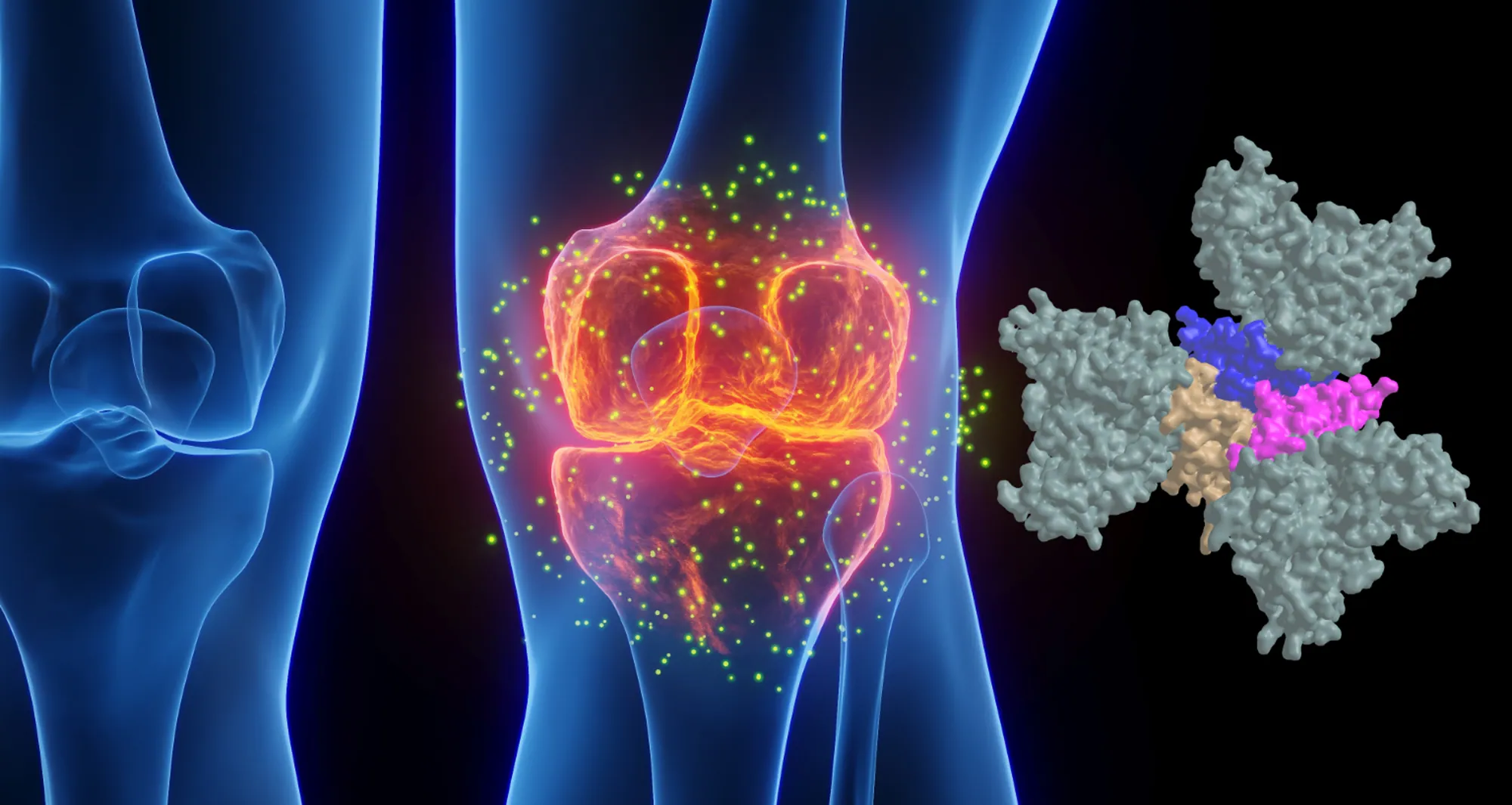
Infliximab and ENBREL® (etanercept) — also approved in 1998 to treat rheumatoid arthritis — were the first FDA-approved tumor necrosis factor-α (TNF) inhibitors. TNF is part of a large family of signaling proteins known to play a key role in developing and coordinating the immune system.

The early success of infliximab and etanercept generated excitement among researchers and within the pharmaceutical industry at the possibility of targeting other members of this protein family. They were interested in finding new protein-based (biologics) drugs to alter inflammation that underlies the destructive processes in autoimmune diseases.

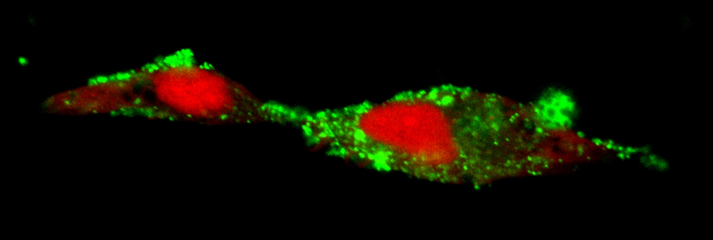
As “Footloose” made it back to Broadway in 2024 for the first time since its initial run, therapies targeting the TNF family are in the midst of their own revival. Carl Ware, PhD, a professor in the Immunity and Pathogenesis Program at Sanford Burnham Prebys, and collaborators at the La Jolla Institute for Immunology and biotechnology company Inhibrx, report in Nature Reviews Drug Discovery that there is a resurgence of interest and investment in these potential treatments.

“Many of these signaling proteins or their associated receptors are now under clinical investigation,” said Ware. “This includes testing the ability to target them to treat autoimmune and inflammatory diseases, as well as cancer.”

Today, there are seven FDA-approved biologics that target TNF family members to treat autoimmune and inflammatory diseases. There also are three biologics and two chimeric antigen receptor (CAR)-T cell-based therapies targeting TNF members for the treatment of cancer. This number is poised to grow as Ware and his colleagues report on the progress of research and many clinical trials to test new drugs in this field and repurpose currently approved drugs for additional diseases.

“The anticipation levels are high as we await the results of the clinical trials of these first-, second- and — in some cases — third-generation biologics,” said Ware.

Ware and his coauthors also weighed in on the challenges that exist as scientists and drug companies develop therapies targeting the TNF family of proteins, as well as opportunities presented by improvements in technology, computational analysis and clinical trial design.

Portrait of Carl Ware

Carl Ware, PhD, is a professor in the Immunity and Pathogenesis Program at Sanford Burnham Prebys.

“There are still many hurdles to get over before we truly realize the potential of these drugs,” noted Ware. “This includes the creation of more complex biologics that can engage several different proteins simultaneously, and the identification of patient subpopulations whose disease is more likely to depend on the respective proteins being targeted.

“It will be important for researchers to use computational analysis of genetics, biomarkers and phenotypic traits, as well as animal models that mimic these variables. This approach will likely lead to a better understanding of disease mechanisms for different subtypes of autoimmune conditions, inflammatory diseases, and cancer, enabling us to design better clinical trials where teams can identify the appropriate patients for each drug.”

Institute News

Helping the brain de-toxify could slow Alzheimer’s

AuthorJessica Moore
Date

February 16, 2017

In Alzheimer’s disease, the space around brain cells becomes clogged with toxic clumps of protein called amyloid. The problem isn’t just that amyloid is generated—that happens in all aging brains—it’s that there’s more of it than the brain’s garbage-scavenging cells, called microglia, can clear out. The lingering amyloid causes neurons to break down, creating a bigger mess for the already overworked microglia to eliminate.

Recent research could lead to a way to turn up the activity of microglia, which should slow the advance of Alzheimer’s. Two related papers with contributions from Huaxi Xu, PhD, the Jeanne and Gary Herberger Chair of Neuroscience and Aging Research at Sanford Burnham Prebys Medical Discovery Institute (SBP), show that a protein called TREM2 helps microglia survive and respond more strongly to damaging material like amyloid and cell debris.

“These results suggest that activating TREM2 could be a viable future treatment strategy for Alzheimer’s,” says Xu. “TREM2 has recently become a hot topic since mutations in the gene have been strongly linked to a greater risk for Alzheimer’s. This work shows that TREM2 is important not just in the relatively small number of patients carrying mutations, but potentially in all of Alzheimer’s.”

TREM2 is a receptor on tissue-resident immune cells, including microglia. TREM2 is activated by fatty molecules released from damaged cells, which stick to amyloid.

The two papers identify the functions of two forms of TREM2—the receptor form that sits on the surface of microglia, and a soluble fragment form that’s released into the space surrounding cells in the brain. Both studies were led by Guojun Bu, PhD, professor at the Mayo Clinic. The first, published in the Journal of Neuroscience, shows that the receptor form of TREM2 is required for microglia to live as long as they normally do. The second, in the Journal of Experimental Medicine, found that the soluble TREM2 fragment also supports microglial survival and turns on their inflammatory response—which in turn supports their ability to remove toxic amyloid clumps.

“The finding that soluble TREM2 has an important function supports its use as a biomarker for Alzheimer’s,” adds Xu. “Levels of soluble TREM2 increase in cerebrospinal fluid before severe symptoms appear, so measuring its levels could aid early diagnosis, which is crucial for effective treatment.”

“We’re now searching for activators of TREM2 in collaboration with drug discovery specialists here at SBP and with support from the Tanz Initiative,” Xu comments. “We’re also looking at whether enhancing TREM2 function lowers levels of amyloid in models of Alzheimer’s.” 

Institute News

Un-blurring the lines in diabetes

AuthorJessica Moore
Date

September 14, 2016

You’re probably familiar with two types of diabetes—juvenile or type 1, which affects kids and young adults, and type 2, in older adults who are generally overweight and inactive. But the lines are less clear than they used to be. Now kids are developing type 2 diabetes, and some adult cases are actually more like a slowly progressing type 1. These fuzzy boundaries make it difficult to accurately diagnose all patients.

“Incorrect classification of diabetes is a major problem,” said Richard Pratley, MD, adjunct professor in the Integrative Metabolism Program, director of the Florida Hospital Diabetes Institute, and senior investigator at the Florida Hospital-SBP Translational Research Institute for Metabolism and Diabetes (TRI-MD). “Some patients can go months before their need for insulin is recognized, which allows damage to the pancreas to continue and increases the likelihood that they’ll develop complications.”

To make it easier to determine which kind of diabetes a patient has, Pratley’s lab looked at an emerging class of biomarkers called microRNAs—small RNAs that regulate the translation of other RNAs into proteins. These molecules are ideal indicators of disease because they are easily measured and remain intact after samples are collected.

“We observed that each subtype of diabetes has its own pattern of microRNAs that are increased or decreased,” Pratley explained. “With further validation in more patients, these results could lead to better diagnostics that, in combination with other standard lab tests, help distinguish the various forms.”

There are three conditions in which the body doesn’t produce enough insulin:

  • In type 1 diabetes, the immune system attacks the cells that make insulin—the beta cells of the pancreas.
  • In type 2, the body becomes resistant to insulin, so beta cells have to work extra hard to make more and more, which eventually wears them out.
  • Latent autoimmune diabetes of adults (LADA), like type 1, is caused by immune destruction of beta cells, but progresses much more slowly, and may also involve diet-related insulin resistance.

In the new study, published in Scientific Reports, Pratley’s team compared levels of microRNAs in the blood of patients with each diabetic condition. A total of eight microRNAs were significantly altered in the diabetic population compared to healthy controls.

“MicroRNA measurements alone weren’t enough to differentiate the three subtypes,” added Pratley. “But since each signature was unique, that information may improve our ability to diagnose each type of diabetic disease. Since microRNAs can be assayed noninvasively and cheaply, these tests may one day become commonplace in diabetes care.”

The paper is available online here.