Dorit Hanein Archives - Sanford Burnham Prebys
Institute News

Super advanced microscopy reveals how cellular anchors are activated

Authorjmoore
Date

March 15, 2016

Integrins are indispensable cell-surface proteins that form bridges with the surrounding protein matrix and other cells. They also transmit the biochemical and mechanical signals that regulate essential cellular processes, such as proliferation, differentiation, migration and cell death. Understanding how integrins become activated is important for developing ways to modulate their function, which is relevant to many disease processes, including cancer, autoimmunity, and fibrosis (hardening of tissue).

The laboratory of Dorit Hanein, PhD, in collaboration with Niels Volkmann, PhD, both professors in SBP’s Bioinformatics and Structural Biology Program, have made an important contribution in this area. Their recent study fundamentally changes our understanding of how integrins are activated—instead of a binary one-step process, it turns out to be a much more nuanced process involving multiple states.

The details

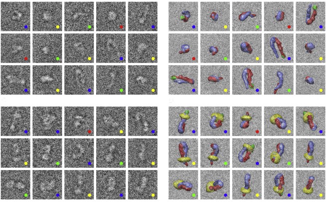
Publishing in the Biophysical Journal, the team used cryo electron microscopy (cryoEM) and advanced computational methods to determine the three-dimensional structures of full-length human integrin, αIIbβ3, which mediates blood clotting. CryoEM allows scientists to look at how a protein works in a near-physiological environment—surrounded by water and interacting with other proteins.

“Because cryoEM captures multiple states, we were able to show that the activation of integrins is much more complex than we thought—they exist in an equilibrium of conformations between bent and upright, and binding to their partners makes them more likely to straighten. Also, the matrix-binding domain is accessible even in the bent form, making it clear how integrins can be initially activated by extracellular signals,” explained Hanein.

Until now, the model for integrin activation was based on structures generated using other techniques that have value, but significant drawbacks as well. For example, X-ray crystallography constrained the protein to a highly compact and bent conformation, which implied an inactive configuration. Another type of electron microscopy revealed an upright conformation, assumed to be the active form that’s triggered by a one-step switch.

What the results mean

The new model aligns better with integrins’ ability to integrate multiple signals to control key cell processes. Instead of one binding event turning the integrin all the way on, each additional signal shifts it towards the upright form, which transmits mechanical signals more easily.

This refinement of the model is critical for designing and interpreting experiments involving integrins. According to Volkmann, “To do translational research, you have to understand the language, and in this case the language is structure.”

The paper is available online here.

Institute News

Sanford-Burnham hosts the Titan Microscope Inauguration Symposium and Reception

Authorrbruni
Date

August 22, 2014

Dorit Hanein, PhD, professor in the Bioinformatics and Structural Program, hosted the Titan Microscope Inauguration Symposium and reception on August 21 at our La Jolla, Calif., campus. The Titan Krios, a state-of-the-art electron microscope, will help our scientists visualize cells, viruses, and bacteria at the atomic level.

The symposium was held to inaugurate the new Titan Krios Transmission Electron Microscope (FEI Company) in honor of our Institute founders Dr. William and Lillian Fishman, who acquired the Institute’s first microscope over 35 years ago and began a legacy of cutting-edge technology that is continued today.

The symposium’s distinguished speakers included more than 14 presenters from peer research institutes, including UC San Diego, The Scripps Research Institute, Caltech, Stanford, and the National University of Singapore.

Guests enjoyed a full day of presentations focused on cutting-edge research in the fields of biophotonics and bioinformatics. Hot topics included the challenges of data processing, connecting cell structures with functions, specimen preparation to maximize results, and real-time analysis of pathogens.

Following the symposium, guests and donors gathered in Chairmen’s Hall for a cocktail reception and tour of the new Titan Krios suite. Kristiina Vuori, MD, Ph.D., president of Sanford-Burnham, led a short program describing the importance of having access to such an advanced instrument for our researchers and the quality of their work.

Ze’ev Ronai, scientific director of Sanford-Burnham in La Jolla, also said a few words, specifically sharing how Jonas Salk of the Salk Institute for Biological Studies generously gifted the first electron microscope to Dr. William Fishman over 35 years ago.

Finally, Nina Fishman, Dr. William and Lillian Fishman’s daughter, joined the reception and praised the Institute’s continued commitment to her parent’s vision for the Institute. A beautiful brushed metal plaque bearing our founder’s image was unveiled shortly after the remarks concluded.

The plaque is permanently mounted on the wall directly outside of the new Titan Microscope suite. It is placed there to honor the Institute’s outstanding commitment to employing the latest and most advanced technology available to accelerate and improve the quantity and quality of our researchers’ discoveries.

About the Titan Krios Microscope

The Titan microscope is a rare state-of-the-art electron microscope specifically designed and developed for life science and medical research applications. It is not a traditional electron microscope, but rather a cryo-electron microscope, meaning that samples within it are frozen at the temperature of liquid nitrogen (between -346°F and -320°F)—the microscope’s operating temperature—and they are never exposed to any form of dehydration. This technique produces the most-accurate imaging results.

Produced by the FEI Company of Hillsboro, Ore., the Titan Microscope is a very exceptional instrument. There are only a handful of them in the U.S. and fewer than 45 total worldwide. The purchase of this $5.5-million instrument was made possible through an NIH Shared Instrumentation Grant.