extracellular matrix Archives - Sanford Burnham Prebys
Institute News

This enzyme is one of the hardest working proteins in the body

AuthorMiles Martin
Date

October 21, 2021

Researchers from Sanford Burnham Prebys have shown that a protein they identified plays a major role in the breakdown of hyaluronic acid, a compound found in the scaffolding between our cells. The findings, published recently in the Journal of Biological Chemistry, could have implications for epilepsy, cancer and other human diseases associated with hyaluronic acid and similar compounds.

They also shed light on one of the most active biochemical processes in the body. 

“Our body turns over hyaluronic acid at an extremely rapid rate, far faster than the other compounds surrounding our cells,” says senior author Yu Yamaguchi, MD, PhD, a professor in the Human Genetics Program at Sanford Burnham Prebys.

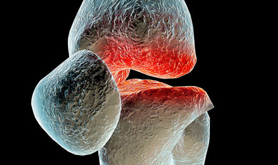
Hyaluronic acid, a common ingredient in cosmetic anti-aging products, is a one of several large sugar molecules known as glycosaminoglycans (GAGs). These are found naturally in the extracellular matrix, the complex network of organic compounds surrounding our cells that gives structure to our tissues. In addition to its structural role, the extracellular matrix is involved in regulating the immune system and is critical in the early development of connective tissues like cartilage, bone and skin.

“The extracellular matrix is found in every organ and tissue of the body, and malfunctions in its biochemistry can trigger or contribute to a variety of diseases, some of which we don’t even know about yet,” says Yamaguchi. His team studies how GAGs affect childhood diseases including congenital deafness, epilepsy and multiple hereditary exostoses, a rare genetic disorder that causes debilitating cartilage growths on the skeleton.

Hyaluronic acid is also known to be correlated with several health conditions, depending on its concentration in certain tissues. Reduced levels of hyaluronic acid in the skin caused by aging contribute to loss of skin elasticity and reduced capacity to heal without scarring. Levels of hyaluronic acid in the blood dramatically increase in alcoholic liver disease, fatty liver and liver fibrosis. In addition, hyaluronic acid levels have been correlated with increased tumor growth in certain cancers.

“These compounds are literally everywhere in the body, and we continue to learn about how GAG’s influence disease, but there’s also a lot we still don’t know about how these molecules are processed,” says Yamaguchi, “Research like this is about understanding what’s happening at the molecular level so we can later translate that into treatments for disease.” 

For this study, the team focused on a protein called TMEM2, which they had previously found to break down hyaluronic acid by cutting the longer molecule into manageable pieces for other enzymes to process further. Using mice as a research model, they selectively shut off the gene that codes for TMEM2 and were able to successfully measure precisely how much the absence of TMEM2 affects the overall levels of hyaluronic acid.

The answer: a lot.

“We saw up to a 40-fold increase in the amount of hyaluronic acid in the study mice compared to our controls,” says Yamaguchi. “This tells us that TMEM2 is one of the key players in the process of degrading this compound, and its dysfunction may be a key player in driving human diseases.” 

The team further confirmed this role of the TMEM2 protein by using fluorescent compounds that detect hyaluronic acid to determine where the TMEM2 protein is most active. They found the most activity on the surface of cells lining blood vessels in the liver and lymph nodes, which are known to be the main sites of hyaluronic acid degradation. 

“These findings refine our understanding of this critical biochemical process and set us up to explore it further in the interest of developing treatments for human diseases,” says Yamaguchi. “Hyaluronic acid is so much a part of our tissues that there could be any number of diseases out there waiting to benefit from discoveries like these.”

Institute News

Research may explain congenital deafness

AuthorJessica Moore
Date

March 15, 2017

If you’ve heard of hyaluronic acid (HA), it’s probably as an ingredient in cosmetic products meant to help keep skin moisturized. But HA—a polysaccharide, or long chain of sugars—is also a major component of the material that surrounds cells in almost every tissue. It’s particularly important in joints, where it’s part of the fluid that lubricates the cartilage ends of bones as they move against each other, and in the eyes, where it helps maintain the shape of the eyeball.

HA is broken down and replaced much faster than other molecules that make up the structure of tissues—one-third of all the HA in your body (about 15 grams in an average-sized human) is turned over each day. But despite its ubiquity and rapid turnover, how HA is degraded remained a mystery. The only enzymes known to cut it up are located inside cells, but HA is far too large to be taken into a cell whole.

That conundrum has now been untangled. Research from the lab of Yu Yamaguchi, MD, PhD, professor at Sanford Burnham Prebys Medical Discovery Institute (SBP), has identified the enzyme that chops extracellular HA into pieces, a protein called TMEM2 (transmembrane protein 2).

“Until now, the function of TMEM2 was unknown,” says Yamaguchi. “We show that it specifically cleaves HA and none of the other polysaccharides that surround cells.”

The discovery, published in the Journal of Biological Chemistry, could help explain the seemingly unrelated effects of altering or inactivating the TMEM2 gene. Zebrafish lacking TMEM2 die as embryos because their hearts don’t develop properly, and mutations in TMEM2 have been linked to severe inherited deafness. That those presentations are so different is less perplexing now that we know that TMEM2 helps control levels of HA. HA is an important ingredient in cardiac jelly, the gelatinous tissue that fosters proper heart formation, and in the fluid in the inner ear that conducts sound waves.

“Our findings open the door to better understanding of an unknown number of disease states,” Yamaguchi adds. “The fact that eliminating the TMEM2 gene causes mortality suggests that it’s really important, but keeps us from seeing how it supports the function of various tissues later in life. To figure that out, we’re making mice that lack TMEM2 only in certain organs.”

Institute News

A new approach to treating osteoarthritis

AuthorSusan Gammon
Date

January 20, 2015

In a recent collaborative research study between two brothers—one a rheumatologist and the other a medical engineer—novel shaped nanoparticles were able to deliver anti-osteoarthritis drugs directly to the cells that drive the onset and progression of osteoarthritis (OA). The findings show promise to improve the treatment options for the nearly 21 million Americans, 25 years of age and older, that suffer from this chronic, often debilitating disease.

“We are excited to have developed nanoparticles which can efficiently and safely bring anti-OA drugs into the cells called chondrocytes that cause OA,” said Massimo Bottini, Ph.D., adjunct assistant professor in the Bioinformatics and Structural Biology Program at Sanford-Burnham. “Our method not only delivered the drug effectively, but stayed in the joint for a prolonged time without causing side effects. This is a significant improvement over previous attempts to deliver anti-OA drugs to affected joints.”

About osteoarthritis Under normal conditions, the extracellular matrix of the joint is maintained through a continual remodeling process in which low levels of different enzymes that produce and degrade cartilage are maintained. However, with increasing age and general wear and tear on joints, OA can occur when the enzymes that degrade cartilage are overproduced, creating an imbalance that leans toward the loss of collagen and joint impairment.

“For advanced OA, joint-replacement surgery is the only option for patients to regain comfortable and normal joint functions. For less severe cases, there is currently no medical therapy that can slow down or halt progression of the disease. This makes OA one of the largest unmet clinical needs in the field of rheumatology,” said Nunzio Bottini, MD, Ph.D., an associate professor in the Division of Cellular Biology at the La Jolla Institute for Allergy and Immunology (LIAI), who is also a practicing rheumatologist—and Massimo’s brother.

“The goal of treating OA is to restore the balance of the enzymes that control the matrix environment. Since there is no blood supply to the joint, drugs to treat the disease must be injected directly into the joint,” said Nunzio.

“Until now, scientists have tried using spherical nanoparticles to deliver anti-OA drugs. But the physical shape and size of the spheres predisposes them to diffuse into the synovial fluid and be flushed out of the joint before they can be effective,” said Massimo. “We have designed a one-dimensional linear nanoparticle made of graphite that is 100,000 times thinner than a human hair. This unique nanoparticle is engineered to travel through the negatively charged extracellular matrix and carry molecules to the nucleus of chondrocytes to turn off the genes that cause the disease.”

The study Using a mouse model for OA, the brothers injected the novel nanoparticle loaded with a gene inhibitor into the knees of affected mice. The nanoparticle delivered large amounts of the gene inhibitor to the cytoplasm and the nucleus of chondrocytes. Importantly, particles remained in the joint for two weeks compared to only few days for spherical nanoparticles.

“This is a significant improvement over previous attempts to deliver drugs to OA joints,” said Massimo. “Our next step is to further optimize the nanoparticle, see how long it remains in the body, and move to clinical studies in humans,” said Massimo.

Arthritis is a complex disease and integrated work between technologists—such as my brother Massimo—and biologists like me significantly increases the chance to make major treatment advances. Our next objective is to secure NIH funding to continue applying our complementary expertise to the quest to improve the lives of those suffering from arthritis,” added Nunzio.

The collaborative study was published in ACS Nano and performed at both Sanford-Burnham and LIAI. Cristiano Sacchetti, PhD, a shared postdoctoral fellow in Massimo and Nunzio Bottini’s laboratories was lead author on the paper.

A link to the paper can be found at: http://pubs.acs.org/doi/abs/10.1021/nn504537b.