hypoxia Archives - Sanford Burnham Prebys
Institute News

“Flying” high to understand what happens when hearts don’t get enough oxygen

AuthorSusan Gammon
Date

October 23, 2017

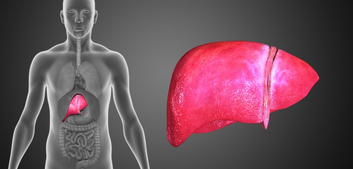
A good supply of oxygen is important for the survival of tissue, but it’s especially critical for organs with high-energy demands, such as the heart. Lack of oxygen (hypoxia) can occur under a variety of conditions, including high altitude, inflammation and cardiopulmonary disorders such as heart attacks and blood clots. Understanding how the heart compensates—or doesn’t compensate—under hypoxic conditions can open avenues to find treatments for hypoxia-related cardiac diseases.

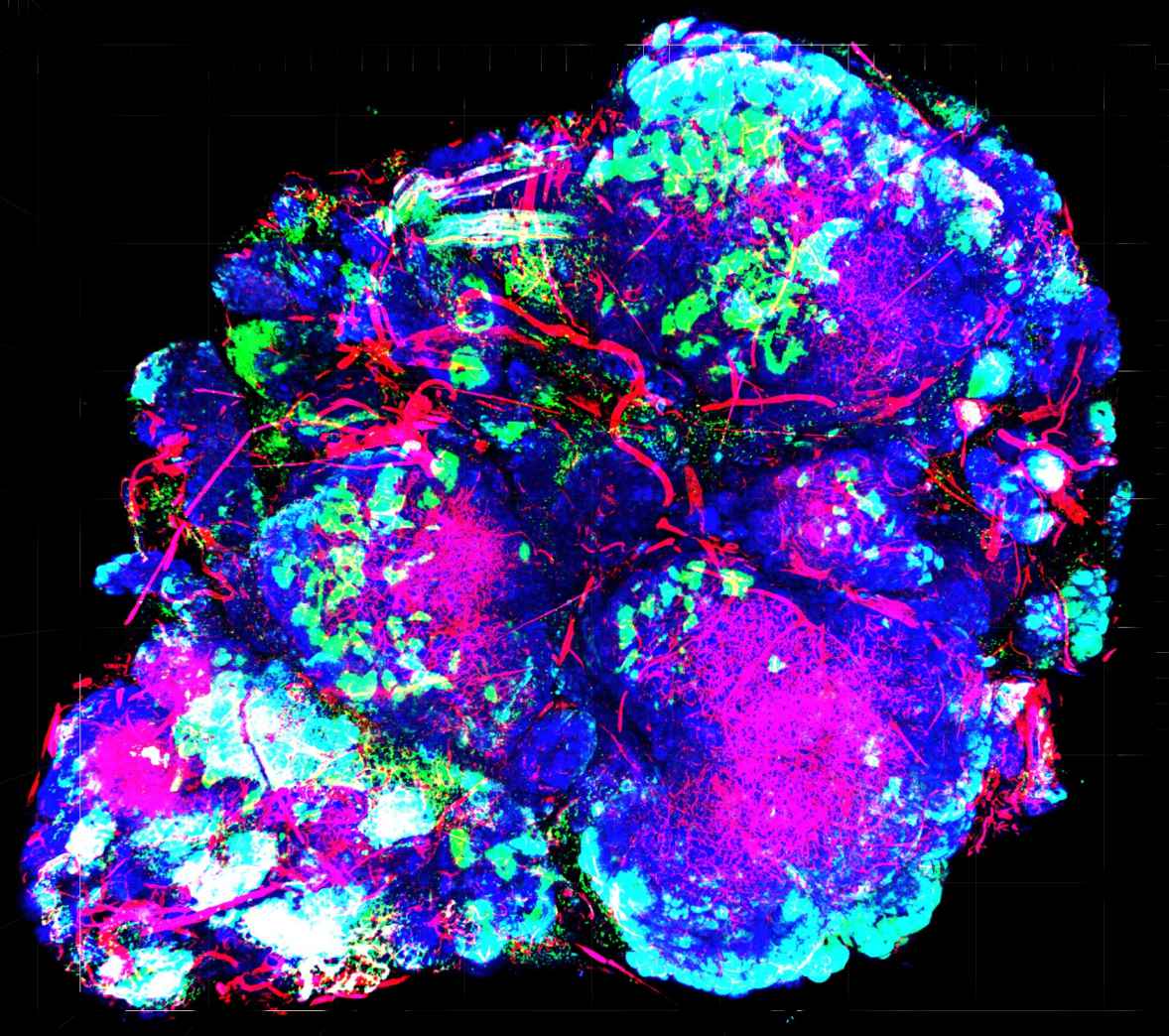
Rolf Bodmer, PhD, director and professor of the Development, Aging and Regeneration Program, and Karen Ocorr, PhD, assistant professor at SBP, study hypoxia in the Drosophila model. Drosophila, a common fruit fly about 3mm long has a heart that doesn’t look much like a human’s—it’s a long tube—but it has many of the same components and genes as a human heart, making it a very useful model to study how genes and environmental conditions affect heart function.

Bodmer and Ocorr’s new study, published in the journal Circulation Cardiovascular Genetics, looked into how hypoxia can lead to long-term heart defects in Drosophila. Their research team studied two sets of flies that underwent different hypoxia treatments: Set (1) flies were subjected to chronic hypoxia for three weeks (hypoxia-treated flies), which is about half of a fly’s life, and Set (2) flies were selected for survival in hypoxic conditions over 250 generations (hypoxia-selected) flies.

While there were some significant differences discovered in the hearts of the two sets of flies, one thing was the same—the expression profile of calcineurin genes were much lower under both conditions.

“Calcineurin is actually an enzyme that promotes the enlargement of the heart (hypertrophy) under some prolonged stress conditions,” says Bodmer. “In mammals, we knew that inhibiting calcineurin reduces the pathological condition of an enlarged heart, but we didn’t know how calcineruin worked in long-term hypoxia, where hypertrophy is a defining feature of diseases linked to chronic hypoxia, most notably known as chronic mountain sickness, which is notorious for affecting high altitude dwellers in the Andes, but surprisingly not as much in Tibet.

Using calcineurin knockdown flies, the team found that without the enzyme, hearts were impaired in normal oxygen conditions. In hypoxic conditions, the damage was even worse, suggesting a careful balance of pro- and antigrowth signaling is necessary for a well-functioning and responsive heart.

“Our study in Drosophila shows that reduced cardiac calcineurin levels cause heart defects that mimic some characteristics we see during long-term hypoxia,” explains Bodmer. “Since calcineurin genes are very similar between Drosophila and human—approximately 75% identical—we believe that reduced levels of calcineurin in mammals—including humans—may play a crucial role in the progression of heart disease during long-term hypoxia exposure, and help understand cardiac complications associated with hypoxia, including population living at high altitude.”

Institute News

To treat breast cancer, give it a lifeline

AuthorJessica Moore
Date

October 17, 2016

In honor of Breast Cancer Awareness Month, we’re highlighting the work our scientists are doing towards the next generation of breast cancer therapies.

Providing more oxygen to a tumor might seem like exactly the wrong way to treat cancer. But Masanobu Komatsu, PhD, associate professor in the Cardiovascular Metabolism Program and the NCI-designated Cancer Center, is trying to find treatments that do exactly that. Enhancing a tumor’s blood supply, which carries oxygen to cancer cells, actually lowers the chance that the cancer will spread.

“We’re aiming to minimize one of the most challenging and devastating aspects of breast cancer—metastasis,” said Komatsu. ”Mortality rates for metastatic breast cancers are still incredibly high. Of the patients with cancer that has spread and led to tumors in other organs, almost 80% will survive less than five years.”

Cancer cells become more likely to move into other tissues as they adapt to a low-oxygen environment due to the tumor’s defective vasculature. Because these blood vessels grow abnormally fast, they form improperly—oxygen and nutrients leak out before reaching the tumor’s interior. However, the cancer cells buried within continue to divide and mutate, so some can survive the lack of oxygen. The master switch that enables cancer cells to generate energy by alternate means also triggers changes that let them enter the circulation and find new homes.

Strengthening the blood supply could also help make the cancer more vulnerable to therapeutic attack, Komatsu added. “Improving the circulation inside a tumor would help anticancer drugs—and the body’s own T cells, which also help eliminate cancer—reach all the tumor cells, and increasing oxygen levels helps sensitize them to radiation and immunotherapy.”

Animal studies suggest that normalizing tumor blood vessels confers such benefits, but existing drugs known to stabilize the vasculature have shown limited benefit. Komatsu and his lab are looking for better therapies by screening microRNAs, small pieces of genetic material that regulate gene activity.

With funding from the Florida Breast Cancer Foundation, the scientific team is testing each of hundreds of microRNAs to look for those that affect signaling pathways controlling the stability of tumor blood vessels. The microRNAs that come up positive could either be developed as drugs (to be used in combination with other cancer-killing treatments), or studied further to find new drug targets.

“This strategy is relevant not only to breast cancer, but to any solid tumor,” commented Komatsu. “The therapies we hope to find could help a huge number of patients.”

Institute News

Unveiling a tumor survival strategy points to new drug target

AuthorJessica Moore
Date

June 20, 2016

One of the reasons tumors can grow out of control is that they survive harsh conditions that normal cells can’t. For example, many can thrive even when supplies of oxygen are low, which happens when tumor growth outpaces the formation of oxygen-supplying blood vessels. Garth Powis, D.Phil., professor and director of SBP’s NCI-designated Cancer Center, has been studying how tumors adapt to this condition, called hypoxia, in hopes of finding ways to block it, which would kill certain cancers.

Surviving hypoxia requires a protein called hypoxia inducible factor-1 (HIF-1), which controls genes involved in switching tumor metabolism to oxygen-independent pathways and promotes the growth of new blood vessels. Though blocking HIF-1 would kill hypoxic tumors, finding drugs that achieve this has so far proven difficult.

A new study from the Powis lab published in Cancer Research may have found another way to overcome cancers’ hypoxia resistance.

The research team found that eliminating or blocking an enzyme called aldolase A lowers activity of HIF-1 and inhibits growth of breast cancer tumors in mice. Aldolase A is responsible for one of the steps in glycolysis, a metabolic process crucial for tumor survival, as cancer cells use it to generate energy more than normal cells.

“Our findings suggest that HIF-1 and glycolysis are a self-perpetuating cycle,” commented Petrus R. de Jong, MD, PhD, postdoctoral associate in Powis’ lab and co-lead author of the study.

“Turning off aldolase A breaks the cycle, decreasing both glycolysis and HIF-1 activity,” Geoffrey Grandjean, PhD, co-first author, explained. “This treatment strategy is a double whammy— it keeps tumors from generating energy without oxygen and it keeps them from becoming better vascularized to get more oxygen.”

To show that aldolase A can be blocked by a drug, Powis teamed with medicinal chemists at the University of Texas at Austin led by Kevin Dalby, PhD, professor of chemical biology, to develop an inhibitor, which slowed proliferation in cultured cancer cells

“The inhibitor we used hasn’t been optimized for use as an anticancer drug,” de Jong said. “However, it could inform future drug design— aldolase A is a very promising target.”

The paper is available online here.


Institute News

Blocking RANTES may slow growth of liver cancer

AuthorJessica Moore
Date

May 16, 2016

Liver cancer is often deadly—less than 20% of patients survive five years—and it’s the leading cause of cancer-related deaths worldwide. And things aren’t getting better. Between the prevalence of hepatitis C and an escalating rate of obesity that leads to fatty liver disease and potentially cancer, new treatments are desperately needed for these cancer patients. Continue reading “Blocking RANTES may slow growth of liver cancer”

Institute News

New STRIVE awards announced

Authorsgammon
Date

October 8, 2015

“The STRIVE award is providing funds that allow us to accelerate the testing of a new hypothesis that connects the microbiome, epigenetics, and colorectal cancer.”        

          –Alex Strongin, PhD professor in the Bioinformatics and Structural Biology Program at SBP. Continue reading “New STRIVE awards announced”

Institute News

Scientists solve structure of important protein for tumor growth

Authorsgammon
Date

August 5, 2015

In a collaborative study between SBP and the Argonne National Laboratory, scientists have used a highly specialized X-ray crystallography technique to solve the protein structure of hypoxia-inducible factors (HIFs), important regulators of a tumor’s response to low oxygen (hyopoxia). The findings, published today in the journal Nature, open the door to search for new drugs to treat tumors by cutting off their supply of oxygen and nutrients. Continue reading “Scientists solve structure of important protein for tumor growth”

Institute News

The switch that promotes kidney cancer progression and metastasis

Authorsgammon
Date

December 12, 2014

Kidney cancer is the sixth most common cancer in adults in the United States. Clear-cell kidney cancer, or clear-cell renal cell carcinoma (CRCC)—the cells appear pale and clear under a microscope—accounts for seven out of 10 people with kidney cancer, and is the most aggressive form of the disease. Because kidney cancer does not cause symptoms until the tumor has already grown, and is very difficult to detect in a routine physical examination, approximately 25 to 30 percent of patients aren’t diagnosed until the disease is metastatic. Unfortunately, metastatic clear-cell kidney cancer is currently incurable. Continue reading “The switch that promotes kidney cancer progression and metastasis”