kinases Archives - Sanford Burnham Prebys
Institute News

What SBP Scientists are Researching to Battle Skin Cancer

AuthorHelen I. Hwang
Date

May 16, 2017

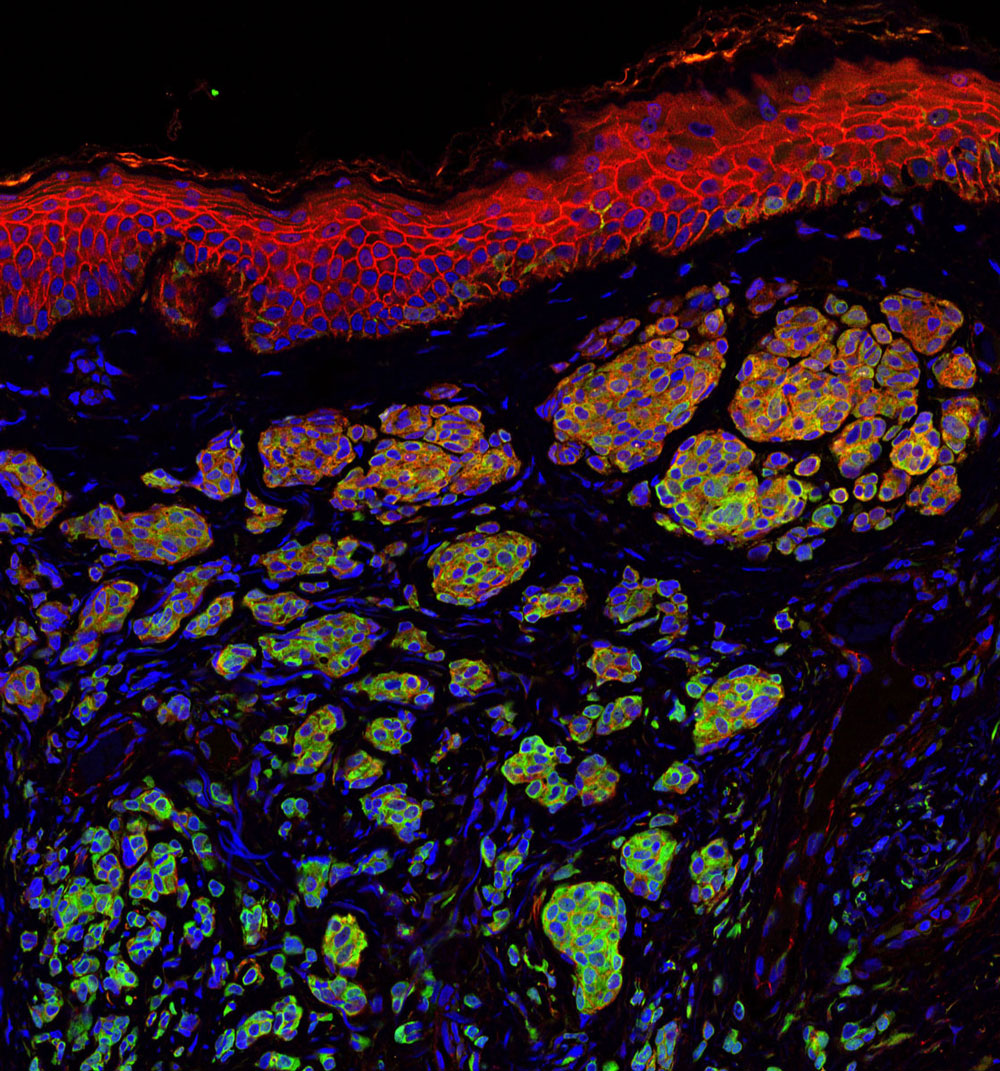
Skin cancer is one of the most common of all cancers, and melanoma accounts for about 1 percent of skin cancers. However, melanoma causes a large majority of deaths from that particular type of cancer. Alarmingly, rates of skin cancer have been on the rise in the last 30 years. Here in Southern California, our everlasting summer comes with a price. Exposure to sun increases our risk to melanoma.

Melanoma occurs when the pigment-producing cells that give color to the skin become cancerous. Symptoms might include a new, unusual growth or a change in an existing mole. Melanomas can occur anywhere on the body.

At Sanford Burnham Prebys Medical Discovery Institute (SBP), we have several researchers working on the causes of melanoma and discovering new ways to treat this deadly disease.

Here is a roundup of SBP’s latest research:

Key findings show how melanoma develops in order to identify potential therapeutic targets

Ze’ev Ronai, PhD
Professor and SBP Chief Scientific Advisor

Ronai’s laboratory has been studying how rewired signaling networks can underlie melanoma development, including resistance to therapy and metastatic propensity. One player in that rewiring is a protein called ATF-2, which can switch from its usual tumor-preventive function to become a tumor promoter when combined with a mutation in the human gene called BRAF.

Ronai’s work on a protein, ubiquitin ligases, led to the identification of RNF125 as an important regulator of melanoma resistance to a common chemotherapy drug. RNF125 impacts melanoma resistance by its regulation of JAK2, an important protein kinase which could play an important role in melanoma resistance to therapy.

Work on the ubiquitin ligase Siah2 identified its important role in melanoma growth and metastasis, and its contribution to melanomagenesis. Melanoma is believed to be a multi-step process (melanomagenesis) of genetic mutations that increase cell proliferation, differentiation, and death.

Work in the lab also concern novel metabolic pathways that are exploited by melanoma for their survival, with the goal of identifying combination drug therapies to combat the spread of melanoma. Earlier work on the enzyme PDK1 showed how it can be a potential therapeutic target for melanoma treatment.

Immunotherapy discovery has led to partnership with Eli Lilly

Linda Bradley, PhD
Professor, Immunity and Pathogenesis Program, Infectious and Inflammatory Diseases Center

Bradley’s group is focused on understanding how anti-tumor T cells can be optimized to kill melanoma tumors. They discovered an important molecule (PSGL-1) that puts the “break” on killer T cells, allowing melanoma tumors to survive and grow. Using animal models, they removed this “break” and T cells were able to destroy melanoma tumors. They have extended their studies and found that in melanoma tumors from patients, T cells also have this PSGL-1 “break”. Bradley’s lab has partnered with Eli Lilly to discover drugs that can modulate PSGL-1 activity in human disease that may offer new therapies for patients.

Knocking out a specific protein can slow melanoma growth 

William Stallcup, PhD
Professor, Tumor Microenvironment and Cancer Immunology Program

The danger of melanomas is their metastasis to organs, such as the brain, in which surgical removal is not effective. By injecting melanoma cells into the brains of mice, we have shown that the NG2 protein found in host tissues makes the brain a much “friendlier” environment for melanoma growth.

Specifically, NG2 is found on blood vessel cells called pericytes and on immune cells called macrophages. The presence of NG2 on both cell types improves the formation of blood vessels in brain melanomas, contributing to delivery of nutrients and thus to accelerated tumor growth. Genetically knocking out NG2 in either pericytes or macrophages greatly impairs blood vessel development and slows melanoma growth.

Mysterious molecule’s function in skin cancer identified

Ranjan Perera, PhD
Associate Professor, Integrative Metabolism Program

Ranjan’s research uncovered the workings of a mysterious molecule called SPRIGHTLY that has been previously implicated in colorectal cancer, breast cancer and melanoma. These findings bolster the case for exploring SPRIGHTLY as a potential therapeutic target or a biological marker that identifies cancer or predicts disease prognosis.

 Drug discovery to help babies has led to a clinical trial at a children’s hospital

Peter D. Adams, PhD
Professor, Tumor Initiation and Maintenance Program

Approximately 1 in 4 cases of melanoma begins with a mole, or nevus. Genetic mutations can cause cells to grow uncontrollably. By investigating how this occurs, we can understand why melanoma develops from some moles, but not others.

Babies born with a giant nevus that covers a large part of the body have especially high risk of melanoma, and the nevus cells can spread into their spine and brain. Adams’ research identified a drug that deters the cells from growing. The drug identified will be used in a clinical trial at Great Ormond Street Children’s Hospital in London, England that may help babies with this debilitating disease.

Discovery of a receptor mutation correlates with longer patient survival

Elena Pasquale, PhD
Professor, Tumor Initiation and Maintenance Program

Pasquale’s work has included whether mutations in the Eph receptor, tyrosine kinases, play a role in melanoma malignancy. Eph receptor mutations occur in approximately half of metastatic melanomas. We found that some melanoma mutations can drastically affect the signaling ability of Eph receptors, but could not detect any obvious effects of the mutations on melanoma cell malignancy.

Bioinformatic analysis of metastatic melanoma samples showed that Eph receptor mutations correlate with longer overall patient survival. In contrast, high expression of some Eph receptors correlates with decreased overall patient survival, suggesting that Eph receptor signaling can promote malignancy.
Institute News

Measuring heart toxicity of cancer drugs in a dish

AuthorJessica Moore
Date

February 22, 2017

A class of cancer drugs known as tyrosine kinase inhibitors (TKIs) are often damaging to the heart, sometimes to the degree that they can’t be used in patients. These toxic effects are not always predictable using current preclinical methods, so they may not be discovered until they make it to clinical trials.

New research could make it possible to tell which TKIs cause heart toxicity without putting any humans at risk. The collaborative study, involving Wesley McKeithan, a PhD student in the Sanford Burnham Prebys Medical Discovery Institute (SBP) graduate program and Mark Mercola, PhD, adjunct professor at SBP and a professor at Stanford University, used lab-grown heart muscle cells to assess the drugs’ potential to cause damaging effects.

“This new method of screening for cardiotoxicity should help pharma companies focus their efforts on TKIs that will be safe,” says Mercola, who collaborated with Joseph Wu, MD, PhD, also a professor at Stanford, on the study published in Science Translational Medicine. “That could mean better new TKIs will make it to the market, since we will be able to predict whether or not they cause heart problems early in the development process.”

TKIs with tolerable cardiac side effects, which include imatinib (Gleevec) and erlotinib (Tarceva), are widely used to treat multiple types of cancer. Because tumors often become resistant to these drugs, new compounds in this class continue to be developed to provide replacement treatments. Other TKIs can harm the heart in a variety of ways, from altering electrical patterns to causing arrhythmias, reducing pumping capacity, or even increasing risk of heart attacks.

Mercola and Wu’s team used heart muscle cells derived from induced pluripotent stem cells (iPSCs), which can be generated from adult skin or blood cells. After treating heart muscle cells with one of 21 TKIs, they assessed their survival, electrical activity, contractions (beating) and communication with adjacent cells. They used a new method for measuring heart cell contraction developed by the lab of Juan Carlos del Álamo, Ph.D., at UC San Diego to create a ‘cardiac safety index’, which correlates in vitro assay results with the drugs’ serum concentrations in humans. Importantly, the safety index values matched nicely with clinical reports on the cardiotoxicity of currently used TKIs.

The study also identified a possible way to protect heart muscle cells from impairment caused by TKIs—treating them with insulin or insulin-like growth factor. Although more research is needed, the findings suggest that it may be possible to alleviate some of the heart damage in patients receiving these chemotherapies.

Mercola adds, “By using cells derived from a broader group of individuals, this screening strategy could easily be adopted by the pharma industry to predict cardiotoxicity.”

This story is based in part on a press release from Stanford University School of Medicine.

Institute News

New therapeutic target for Crohn’s disease

AuthorJessica Moore
Date

September 20, 2016

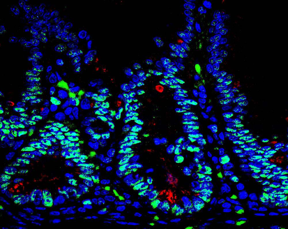
Research from the Sanford Burnham Prebys Medical Discovery Institute (SBP) identifies a promising new target for future drugs to treat inflammatory bowel disease (IBD). The study, published in Cell Reports, also indicates that another protein, protein kinase C (PKC) λ/ι, may serve as a biomarker of IBD severity.

“The intestine is protected by specialized cells, called Paneth cells, that secrete antimicrobial peptides,” said Jorge Moscat, PhD, deputy director and professor in the NCI-designated Cancer Center and senior author of the paper. “We found that maintaining normal numbers of Paneth cells requires PKC λ/ι, and that the amount of PKC λ/ι decreases as IBD gets worse. We also discovered a way to prevent Paneth cell loss—inhibiting a protein called EZH2, which could be a new therapeutic strategy for IBD.”

IBD, which includes Crohn’s disease and ulcerative colitis, affects 1.4 million people in the U.S. These chronic conditions are often debilitating, as they cause unpredictable abdominal pain and diarrhea. Because current medications only help control symptoms and not the underlying disease, 70% of Crohn’s patients and 30% of those with colitis must eventually undergo surgery. In addition, IBD increases risk of intestinal cancer by as much as 60%.

“We also examined the effect of PKC λ/ι on tumor formation,” said Maria Diaz-Meco, PhD, also a professor in the Cancer Center and co-author of the paper. “In contrast to some previous studies indicating that it might promote cancer development, we demonstrate that in the intestine, PKC λ/ι is protective.”

“We inactivated the PKC λ/ι gene in the intestine of mice, which caused them to have very few Paneth cells,” added Diaz-Meco. “Without Paneth cells, the intestine is more susceptible to bacterial infiltration, which leads to inflammation. Since inflammation favors cancer, it makes sense that PKC λ/ι is a tumor suppressor in this setting.”

To find a way to boost Paneth cell numbers and possibly treat IBD, the team looked for what drives the deficit in these protector cells. The key link was overactive EZH2, which turns off genes needed to generate Paneth cells.

“We used an in vitro model—‘mini guts’ in a dish—to show that blocking EZH2 helps return the number of Paneth cells to normal,” said Yuki Nakanishi, MD, a postdoctoral fellow in the Moscat/ Diaz-Meco lab and lead author of the work. “This demonstrates that inhibiting EZH2 could be a new way to slow the progression of IBD.”

Importantly, the team verified the relevance of their findings in intestinal biopsy samples from 30 patients with Crohn’s disease. Disease progression correlated with lower levels of PKC λ/ι.

“EZH2 inhibitors are currently being developed by the pharmaceutical industry to treat other cancers, so they could be tested for IBD relatively soon,” said Moscat. “But first, we need to do preclinical studies to test whether they block progression of the disease.”

The paper is available online here.

Institute News

Fine-tuning cellular energy increases longevity

AuthorJessica Moore
Date

February 25, 2016

New research from SBP has identified a protein that can extend the natural lifespan of C. elegans, a microscopic roundworm commonly used for research on aging and longevity. The findings, published in Cell Reports, expand what we know about the aging process and may lead to new ways to delay the onset of human age-related diseases such as cancer and neurodegenerative diseases. Continue reading “Fine-tuning cellular energy increases longevity”

Institute News

Is there a type 3 diabetes?

AuthorGuest Blogger
Date

November 10, 2015

This article was written by guest blogger Jessica Frisch-Daiello, PhD

People with type 2 diabetes are twice as likely to develop Alzheimer’s disease—a type of dementia affecting behavior, memory, and cognitive functions. According to the Centers for Disease Control and Prevention, in 2013 Alzheimer’s ranked sixth and diabetes was seventh as the leading causes of death in the United States. Recent studies are suggesting a link between insulin resistance in the brain and Alzheimer’s disease, prompting some researchers to consider a new classification for the disease: type 3 diabetes.

People with diabetes can’t effectively break down blood sugar. Either their bodies don’t produce enough insulin (type 1 diabetes) or their bodies become desensitized to insulin (type 2 diabetes).

The exact mechanisms between insulin resistance and Alzheimer’s disease are not well understood and research is on-going. However, studies suggest that insulin resistance in the brain leads to the formation of two pathological hallmarks of Alzheimer’s disease—the formation of tau tangles and the build-up of clusters of beta amyloid peptides called plaques in the brain. The degree of insulin resistance is correlated with the amount of plaques deposited between nerve cells. Plaques create a blockade that inhibits cell-to-cell signaling in the brain. Additionally, insulin dysfunction has also been shown to affect the formation of tau tangles by mediating the activity of an important enzyme in the body, GSK-3β (glycogen synthase kinase 3).

Juan Pablo Palavicini, PhD, an SBP postdoctoral fellow in the lab of Xianlin Han, PhD, is studying the role of a particular class of molecules found in the body that might give more clues to the mechanisms connecting these two seemingly disparate diseases. According to Palavicini, “We have found that a specific lipid class called sulfatide is severely deficient in the brains of both Alzheimer’s disease patients and type 2 diabetics. Moreover, our research shows that when sulfatide is removed, there is a dramatic change in insulin levels, beta amyloid peptides, and tau tangles. We are currently exploring therapeutic techniques to restore sulfatide content as a treatment for both diseases.”

Sulfatide serves many functions in the body, including aiding neural plasticity and memory. It also plays a role in insulin secretion. A change in the expression of sulfatide has been associated with a number of conditions, including Alzheimer’s disease, Parkinson’s disease, and diabetes.

Given the association between Alzheimer’s disease and diabetes, it is important for people to incorporate healthy habits in everyday life. Both the American Diabetes Association and the Alzheimer’s Association say that daily exercise, social interaction, and a diet emphasizing fruits, vegetables, and whole grains may reduce the risk of developing, or slowing the progression of, these diseases.

Dr. Palavicini and Dr. Han are pursuing this research as part of a mentor-based postdoctoral fellowship awarded by the American Diabetes Association. This article was written by Dr. Jessica Frisch-Daiello, a postdoctoral associate in Dr. Han’s laboratory at SBP.

Institute News

Expanding the options to treat melanoma

Authorsgammon
Date

March 16, 2015

Melanoma is the most deadly form of skin cancer with approximately 10,000 deaths per year in the U.S. and more than 65,000 worldwide. Although there are more and better treatment options available today than in previous years, there is still an urgent need to develop drugs that target the numerous pathways melanoma cells use to multiply, spread, and kill. Continue reading “Expanding the options to treat melanoma”