macrophages Archives - Sanford Burnham Prebys
Institute News

Study reveals how immune cells manage cholesterol levels

AuthorLindsay Ward-Kavanagh
Date

March 12, 2018

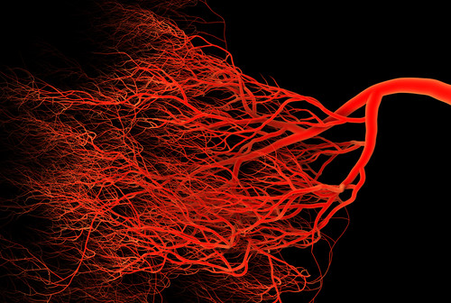
Atherosclerosis, the buildup of plaques inside arteries, is a key step in the development of cardiovascular disease, the leading cause of death in the United States. Elevated cholesterol levels are a risk factor for atherosclerosis, as the molecule is one of the building blocks of these plaques. However, since cholesterol is also essential in healthy cells, scientists are researching how cholesterol biology is controlled to better understand the changes that lead to disease.

Laszlo Nagy, MD, PhD, professor and director of the Genomic Control of Metabolism Program, recently collaborated with Peter Tontonoz, MD, PhD, professor, the leading senior scientist of the study and Francis and Albert Piansky Endowed Chair in Pathology and Laboratory Medicine at the UCLA David Geffen School of Medicine, to assess a network of molecules that control cholesterol transport out of macrophages, immune cells normally associated with inflammation.

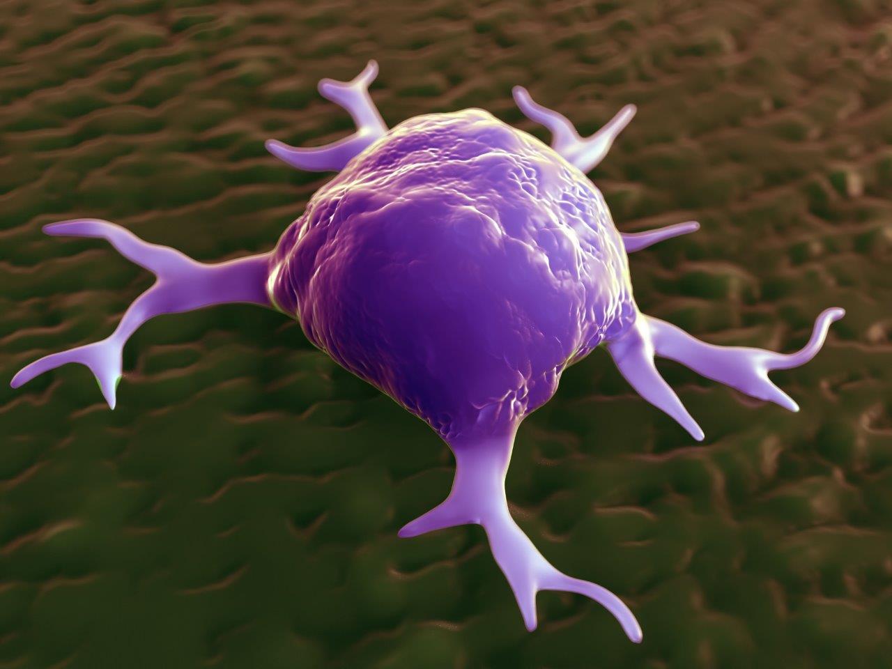
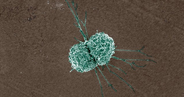
“Although macrophages are usually thought of as the white blood cells that ingest invading bacteria and cleaning up cell debris after injury or infection, they can also enter a ‘alternatively activated’ state to help tissue repair and remodeling,” Nagy says. “In blood vessels, these repair state macrophages protect the body by removing cholesterol from the bloodstream. However, the accumulation of excess cholesterol in macrophages is a key event in the development of atherosclerosis. How macrophages control cholesterol transport is not well understood, but needs to be explored to better understand atherosclerosis.”

“We were interested in how macrophages are able to switch on the gene Abca1, the gene that encodes the protein that pumps cholesterol out of these cells,” Nagy explained. “We used our expertise in epigenomics to define regions of the genome that controlled the amount of Abca1 RNA produced.”

By examining long non-coding RNA strands that regulate gene expression, the study identified an RNA called MeXis that increases the expression of Abca1. Although MeXis cannot start transcription of Abca1 by itself, it does impact the ability of other proteins to transcribe the gene.

“Using our molecular tools, we were able to show that MeXis recruited another protein that helps to start transcription to Abca1, says Nagy. “Without MeXis, this protein did not interact with Abca1 and transcription was dramatically reduced, even when the cells received signals to start the process of ridding themselves of cholesterol.

“The more we understand about the biological processes that control cholesterol metabolism the better informed we are to develop strategies to prevent and treat atherosclerosis, says Nagy.  “This study reveals key insights on the regulation of Abca1, which could ultimately lead to new therapeutic approaches.”

The study was published in Nature Medicine.

Institute News

How tumors trick immune cells

AuthorSusan Gammon
Date

September 19, 2017

The aggressiveness of a tumor is determined partly by the properties of the actual cancer cells, but also to a surprisingly large degree by the surrounding environment in which the tumor grows, including blood vessels, fat cells, fibroblasts and immune cells.

Of these elements, the roles of immune cells are arguably the most baffling. Immune cells are recruited to tumors to attack and destroy cancer cells, but their properties can change in response to specific signals they receive from the tumor. Macrophages are a good example of immune cells that play complex roles in tumor progression.

Laszlo Nagy, MD, PhD, professor and director of the Genomic Control of Metabolism Program at SBP’s Florida campus, is well-versed in many aspects of macrophage biology. Nagy comments that, “Our lab is studying the way the retinoid X receptor (RXR) controls how macrophages direct the aggressiveness of a tumor—specifically whether it spreads to other parts of the body (metastasizes). Scientists have known for some time that RXR is a regulatory protein found in the cell nucleus, but how it effects tumor metastasis has not been studied.”

Nagy’s lab recently published a study in PNAS that compared RXR macrophage knockout mice with normal mice to assess the how the protein affects the metastasis of subcutaneous tumors. Surprisingly, metastasis was increased in RXR macrophage knockout mice, even though growth of the primary tumors was not affected. Further research showed that the loss of RXR resulted in increased macrophage production of several factors that promote tumor cell colonization of sites in the lungs.

“This is a really tricky concept to grasp,” says Nagy. “The idea being developed by several research groups is that macrophages promote tumor metastasis by producing factors that modify sites in distant organs (known as pre-metastatic niches). This “conditioning” makes these sites more attractive as new homes for migrating tumor cells.

“Our contribution to understanding this phenomenon is discovering the involvement of RXR in suppressing macrophage production of metastasis-enhancing factors. The importance of RXR loss in our mouse model is underscored by finding that macrophage RXR activity is also low in patients with metastatic cancer. Since metastasis is the most dangerous aspect of cancer, we are toying with ideas for how to boost RXR activity in macrophages as a means of suppressing the metastatic phase of the disease.”

Institute News

Immune scavenger cells: Good guys or bad guys?

AuthorSusan Gammon
Date

July 9, 2017

The immune system is our main defense against foreign invaders (bacteria and viruses) and also against mutant cells that develop into cancer. Some of the first immune responders to these threats are scavenger cells called macrophages that destroy targets by internalizing and degrading them.

But macrophages can be tricked. When they are continually activated in a chronic disease like cancer, they can be “instructed” by cancer cells to perform functions that benefit the growing tumor instead of destroying it.

Recently, William Stallcup, PhD, professor at Sanford Burnham Prebys Medical Discovery Institute (SBP) published a study in Trends in Cell and Molecular Biology describing one way that cancer cells transform macrophages from tumor fighters into tumor helpers.

“We found that a protein called NG2 on macrophages is important for the ability of these scavengers to exit from blood vessels and migrate into tumors,” explains Stallcup. “When we knocked out NG2 on macrophages in mice, instead of allowing mouse brain tumors to grow faster (in the absence of the scavenging macrophages), the tumors actually grew much more slowly. This was somewhat surprising since we expect macrophages to help fight cancer.”

“It turns out that tumor cells can modify macrophage function, making them produce factors that stimulate the formation of tumor-nourishing blood vessels, says Pilar Cejudo Martin, PhD, a postdoc in Stallcup’s lab and first author of the paper. “By knocking out NG2, we blocked macrophage entry into tumors so that there were fewer macrophages for the tumors to use to their own advantage. So due to poor blood vessel development, tumor growth was slowed.”

So does this mean that blocking NG2 could be a general means of improving treatment of diseases involving chronic macrophage recruitment?

“It depends,” says Stallcup. “When we used a mouse model of multiple sclerosis (MS) to show that knockout of NG2 impaired macrophage entry into the damaged spinal cord, we found that recovery was hindered. That’s because macrophages are needed to produce factors that stimulate production of the cells responsible for damage repair.

“So while blocking macrophage NG2 might be useful for cancer therapy, it looks like this approach would be counter-productive for treating MS. Even though macrophages seem like attractive targets for therapy, they have such a large bag of tricks that we will probably have to deal with blocking their effects on a case-by-case basis,” adds Stallcup.

Read the paper here.

Institute News

Research suggests new way to prevent HIV-associated brain injury

Authorsgammon
Date

December 5, 2014

For about 50 percent of HIV-1-infected people, things as simple as buttoning a shirt, remembering the alphabet, and handling money may become compromised by a disorder known as HIV-induced brain injury. The condition occurs when receptors and proteins in an HIV-infected immune system produce toxic substances that lead to brain- and nerve-cell death. There is currently no treatment available for the more than 600,000 affected individuals in the U.S. In a new study by Sanford-Burnham researchers, blocking CCR5—an HIV co-receptor—was found to protect against brain injury and impairment of learning and memory. The findings, reported in The Journal of Immunology, create a new approach to treating HIV-induced brain injury and may help our understanding of the potential involvement of CCR5 in other diseases of the brain. Continue reading “Research suggests new way to prevent HIV-associated brain injury”

Institute News

A nuclear receptor that binds more than 5,000 sites in the genome—and promotes angiogenesis.

Authorsgammon
Date

July 17, 2014

The retinoid X receptor (RXR) is a nuclear hormone receptor—meaning that it sits on various parts of the genome—and turns genes “on” and “off.” RXR is known to play an important role in many fundamental biological processes such as reproduction, cellular differentiation, bone development, and hematopoiesis. Continue reading “A nuclear receptor that binds more than 5,000 sites in the genome—and promotes angiogenesis.”