microRNA Archives - Sanford Burnham Prebys
Institute News

Taking out a microRNA to thwart melanoma

AuthorSusan Gammon
Date

August 6, 2018

Melanoma is a deadly disease with limited treatment options. However, even when those therapies are initially successful, the cancer often comes back. Researchers continue to hunt for new approaches to make the disease more vulnerable.

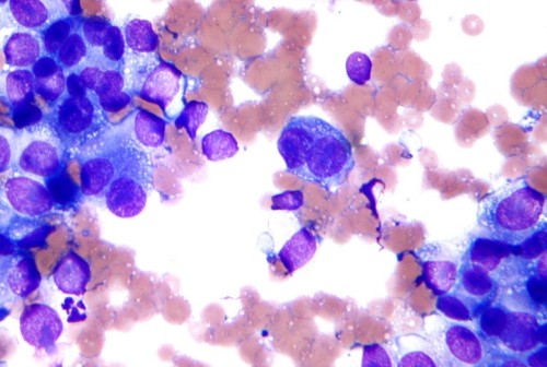
Ranjan Perera, PhD, an adjunct professor at SBP’s Lake Nona campus, has been studying melanoma for many years, looking for mechanisms that can help control the disease. These efforts helped his lab discover miR-211, a molecule found in melanocytes, the cells that sometimes go awry and become cancerous. Not surprisingly, miR-211 is sometimes overexpressed in melanoma. 

Ranjan Perara, PhD
     Ranjan Perera, PhD 

Because miR-211 is a microRNA—a small molecule that interferes with the cellular machinery that produces proteins—it can have a big impact on gene expression, and the gene it impacts is pretty interesting.

“MicroRNA-211 is targeted to a gene called PDK4, which is important for mitochondrial energy metabolism,” says Perera. 

Scientists have known for more than 100 years that tumors restructure their metabolisms to compensate for their out-of-control growth. If miR-211 is part of that process, taking it out could make cancer cells more treatable.

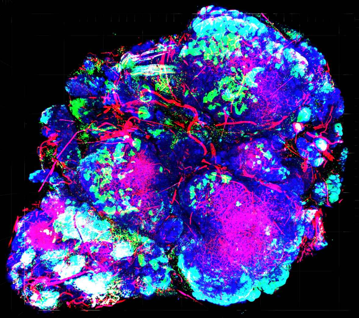
To better understand what miR-211 is doing, researchers in Perera’s lab used CRISPR/Cas9 gene editing tools to eliminate it from cancer cell lines. They found that removing the molecule impacted mitochondria, the cells’ energy plants, and made them metabolically vulnerable. In addition, miR-211 loss dampened pathways that drive melanoma growth—so there was a double benefit. In animal models, cells without miR-211 had trouble forming tumors. These results were recently published in the Journal of Investigative Dermatology.

Perera and colleagues were also curious whether removing the microRNA might affect how cancer cells respond to the drug Vemurafenib—a therapy used for the treatment of late-stage melanoma. While the drug is effective in certain patients, tumors often develop resistance after several months. Further study showed that eliminating miR-211 made the melanoma cells much more sensitive to Vemurafenib.

These findings add to the body of evidence that helped Perera and SBP get a patent covering approaches using miR-211 to detect and treat melanoma. Perera’s team will continue to study this molecule, as well as the genes it impacts, to gain more insights and potentially transform these findings into new melanoma diagnostics and treatments.

Though it’s still early, these findings make miR-211 an interesting potential drug target, and Perera believes further investigation is definitely warranted.

“Given that miR-211 loss has a dual anti-cancer effect, by inhibiting both critical growth-promoting cell signaling pathways and rendering cells metabolically vulnerable, it is an extremely attractive candidate for combinatorial therapeutics,” says Perera. “This is especially true if, like here, miR-211 is upregulated in Vemurafenib-resistant melanomas in the clinic, since it provides both a highly specific target.”

Interested in keeping up with SBP’s latest discoveries, upcoming events and more? Subscribe to our monthly newsletter, Discoveries.

Institute News

To treat breast cancer, give it a lifeline

AuthorJessica Moore
Date

October 17, 2016

In honor of Breast Cancer Awareness Month, we’re highlighting the work our scientists are doing towards the next generation of breast cancer therapies.

Providing more oxygen to a tumor might seem like exactly the wrong way to treat cancer. But Masanobu Komatsu, PhD, associate professor in the Cardiovascular Metabolism Program and the NCI-designated Cancer Center, is trying to find treatments that do exactly that. Enhancing a tumor’s blood supply, which carries oxygen to cancer cells, actually lowers the chance that the cancer will spread.

“We’re aiming to minimize one of the most challenging and devastating aspects of breast cancer—metastasis,” said Komatsu. ”Mortality rates for metastatic breast cancers are still incredibly high. Of the patients with cancer that has spread and led to tumors in other organs, almost 80% will survive less than five years.”

Cancer cells become more likely to move into other tissues as they adapt to a low-oxygen environment due to the tumor’s defective vasculature. Because these blood vessels grow abnormally fast, they form improperly—oxygen and nutrients leak out before reaching the tumor’s interior. However, the cancer cells buried within continue to divide and mutate, so some can survive the lack of oxygen. The master switch that enables cancer cells to generate energy by alternate means also triggers changes that let them enter the circulation and find new homes.

Strengthening the blood supply could also help make the cancer more vulnerable to therapeutic attack, Komatsu added. “Improving the circulation inside a tumor would help anticancer drugs—and the body’s own T cells, which also help eliminate cancer—reach all the tumor cells, and increasing oxygen levels helps sensitize them to radiation and immunotherapy.”

Animal studies suggest that normalizing tumor blood vessels confers such benefits, but existing drugs known to stabilize the vasculature have shown limited benefit. Komatsu and his lab are looking for better therapies by screening microRNAs, small pieces of genetic material that regulate gene activity.

With funding from the Florida Breast Cancer Foundation, the scientific team is testing each of hundreds of microRNAs to look for those that affect signaling pathways controlling the stability of tumor blood vessels. The microRNAs that come up positive could either be developed as drugs (to be used in combination with other cancer-killing treatments), or studied further to find new drug targets.

“This strategy is relevant not only to breast cancer, but to any solid tumor,” commented Komatsu. “The therapies we hope to find could help a huge number of patients.”

Institute News

Un-blurring the lines in diabetes

AuthorJessica Moore
Date

September 14, 2016

You’re probably familiar with two types of diabetes—juvenile or type 1, which affects kids and young adults, and type 2, in older adults who are generally overweight and inactive. But the lines are less clear than they used to be. Now kids are developing type 2 diabetes, and some adult cases are actually more like a slowly progressing type 1. These fuzzy boundaries make it difficult to accurately diagnose all patients.

“Incorrect classification of diabetes is a major problem,” said Richard Pratley, MD, adjunct professor in the Integrative Metabolism Program, director of the Florida Hospital Diabetes Institute, and senior investigator at the Florida Hospital-SBP Translational Research Institute for Metabolism and Diabetes (TRI-MD). “Some patients can go months before their need for insulin is recognized, which allows damage to the pancreas to continue and increases the likelihood that they’ll develop complications.”

To make it easier to determine which kind of diabetes a patient has, Pratley’s lab looked at an emerging class of biomarkers called microRNAs—small RNAs that regulate the translation of other RNAs into proteins. These molecules are ideal indicators of disease because they are easily measured and remain intact after samples are collected.

“We observed that each subtype of diabetes has its own pattern of microRNAs that are increased or decreased,” Pratley explained. “With further validation in more patients, these results could lead to better diagnostics that, in combination with other standard lab tests, help distinguish the various forms.”

There are three conditions in which the body doesn’t produce enough insulin:

  • In type 1 diabetes, the immune system attacks the cells that make insulin—the beta cells of the pancreas.
  • In type 2, the body becomes resistant to insulin, so beta cells have to work extra hard to make more and more, which eventually wears them out.
  • Latent autoimmune diabetes of adults (LADA), like type 1, is caused by immune destruction of beta cells, but progresses much more slowly, and may also involve diet-related insulin resistance.

In the new study, published in Scientific Reports, Pratley’s team compared levels of microRNAs in the blood of patients with each diabetic condition. A total of eight microRNAs were significantly altered in the diabetic population compared to healthy controls.

“MicroRNA measurements alone weren’t enough to differentiate the three subtypes,” added Pratley. “But since each signature was unique, that information may improve our ability to diagnose each type of diabetic disease. Since microRNAs can be assayed noninvasively and cheaply, these tests may one day become commonplace in diabetes care.”

The paper is available online here.