neurodegeneration Archives - Sanford Burnham Prebys
Institute News

Mutations in protein receptor gene linked to Alzheimer’s disease

AuthorGreg Calhoun
Date

January 7, 2025

New research on four variants in the EPHA1 gene reveals how its genetic typos may contribute to risk of dementia

Upon inspecting the DNA sequences in patients suffering from Alzheimer’s disease, scientists have found evidence of an inconspicuous conspirator.

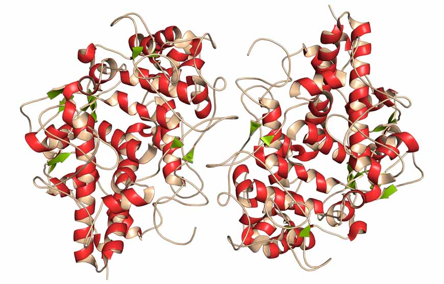
The EPHA1 gene contains the blueprint for the EPHA1 receptor protein, one of 14 such receptor proteins in the Eph receptor family. Relatively little is known about EPHA1 when compared to many of its siblings, making it difficult for researchers to ascertain why changes in its source code would contribute to such a debilitating disease.

Scientists at Sanford Burnham Prebys published results on December 18, 2024, in the Journal of Biological Chemistry, detailing the effects of four miniature mutations of just a single typo each in the sequence of nucleotides forming the EPHA1 gene.

These seemingly minor mutations are known as single nucleotide polymorphisms (SNPs), and they can lead to larger issues depending on where the typos fall in the sequence of a gene. The Sanford Burnham Prebys team focused on four missense mutations that are caused when SNPs result in different amino acids being used to build the EPHA1 receptor protein.

“Our data show that all four Alzheimer’s mutations we have characterized disrupt EPHA1 physiological signaling, and that the specific effects depend on the particular mutation,” said Elena Pasquale, PhD, professor in the Cancer Metabolism and Microenvironment Program at Sanford Burnham Prebys.

The team reported that the functional consequences of EPHA1 missense mutations identified in patients suffering from Alzheimer’s disease included misplacement of EPHA1 within cells, decreased protein stability and dysregulated signaling.

“To continue advancing knowledge on this topic, more work is needed to uncover the physiological role of the different EPHA1 signaling features and how their disruption may lead to neurodegeneration,” said Pasquale.


Additional authors on the study from Sanford Burnham Prebys include Mike Matsumoto and Sara Lombardi, PhD. Maricel Gomez-Soler, PhD, now works at Crinetics Pharmaceuticals in San Diego. Bernhard C. Lechtenberg, PhD, now works at the Walter and Eliza Hall Institute of Medical Research in Parkville, Australia.

Institute News

New roles for autophagy genes in cellular waste management and aging

AuthorCommunications
Date

January 3, 2024

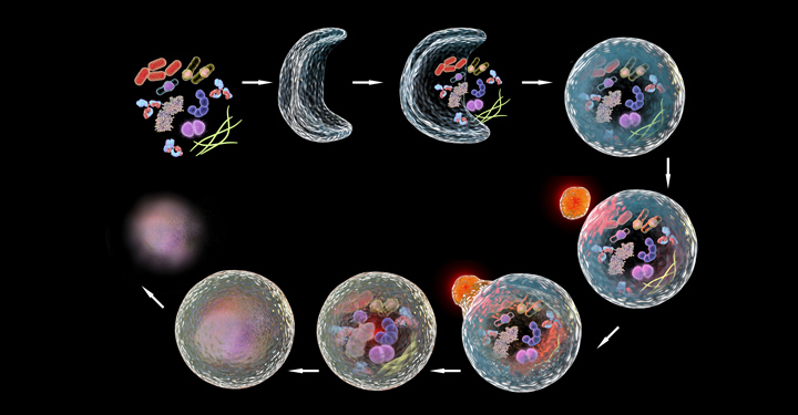
Autophagy genes help extrude protein aggregates from neurons in the nematode C. elegans.

Autophagy, which declines with age, may hold more mysteries than researchers previously suspected. In the January 4 issue of Nature Aging, it was noted that scientists from the Buck Institute, Sanford Burnham Prebys and Rutgers University have uncovered possible novel functions for various autophagy genes, which may control different forms of disposal including misfolded proteins—and ultimately affect aging.

“While this is very basic research, this work is a reminder that it is critical for us to understand whether we have the whole story about the different genes that have been related to aging or age-related diseases,” said Professor Malene Hansen, PhD, Buck’s chief scientific officer, who is also the study’s co-senior author. “If the mechanism we found is conserved in other organisms, we speculate that it may play a broader role in aging than has been previously appreciated and may provide a method to improve life span.”

These new observations provide another perspective to what was traditionally thought to be occurring during autophagy.

Autophagy is a cellular “housekeeping” process that promotes health by recycling or disposing of damaged DNA and RNA and other cellular components in a multi-step degradative process. It has been shown to be a key player in preventing aging and diseases of aging, including cancer, cardiovascular disease, diabetes and neurodegeneration. Notably, research has shown that autophagy genes are responsible for prolonged life span in a variety of long-lived organisms.

The classical explanation of how autophagy works is that the cellular “garbage” to be dealt with is sequestered in a membrane-surrounded vesicle, and ultimately delivered to lysosomes for degradation. However, Hansen, who has studied the role of autophagy in aging for most of her career, was intrigued by an accumulation of evidence that indicated that this was not the only process in which autophagy genes can function.

“There had been this growing notion over the last few years that genes in the early steps of autophagy were ‘moonlighting’ in processes outside of this classical lysosomal degradation,” she said. “Additionally, while it is known that multiple autophagy genes are required for increased life span, the tissue-specific roles of specific autophagy genes are not well defined.”

To comprehensively investigate the role that autophagy genes play in neurons—a key cell type for neurodegenerative diseases—the team analyzed Caenorhabditis elegans, a tiny worm that is frequently used to model the genetics of aging and which has a very well-studied nervous system. The researchers specifically inhibited autophagy genes functioning at each step of the process in the neurons of the animals, and found that neuronal inhibition of early-acting, but not late-acting, autophagy genes, extended life span.

An unexpected aspect was that this life span extension was accompanied by a reduction in aggregated protein in the neurons (an increase is associated with Huntington’s disease, for example), and an increase in the formation of so-called exophers. These giant vesicles extruded from neurons were identified in 2017 by Monica Driscoll, PhD, a collaborator and professor at Rutgers University.

“Exophers are thought to be essentially another cellular garbage disposal method, a mega-bag of trash,” said Caroline Kumsta, PhD, co-senior author and assistant professor at Sanford Burnham Prebys “When there is either too much trash accumulating in neurons, or when the normal ‘in-house’ garbage disposal system is broken, the cellular waste is then being thrown out in these exophers.

“Interestingly, worms that formed exophers had reduced protein aggregation and lived significantly longer. This finding suggests a link between this process of this massive disposal event to overall health,” said Kumsta. The team found that this process was dependent on a protein called ATG-16.2.

The study identified several new functions for the autophagy protein ATG-16.2, including in exopher formation and life span determination, which led the team to speculate that this protein plays a nontraditional and unexpected role in the aging process. If this same mechanism is operating in other organisms, it may provide a method of manipulating autophagy genes to improve neuronal health and increase life span.

“But first we have to learn more—especially how ATG-16.2 is regulated and whether it is relevant in a broader sense, in other tissues and other species,” Hansen said. The Hansen and Kumsta teams are planning on following up with a number of longevity models, including nematodes, mammalian cell cultures, human blood and mice.

“Learning if there are multiple functions around autophagy genes like ATG-16.2 is going to be super important in developing potential therapies,” Kumsta said. “It is currently very basic biology, but that is where we are in terms of knowing what those genes do.”

The traditional explanation that aging and autophagy are linked because of lysosomal degradation may need to expand to include additional pathways, which would have to be targeted differently to address the diseases and the problems that are associated with that. “It will be important to know either way,” Hansen said. “The implications of such additional functions may hold a potential paradigm shift.” 
 
DOI: 10.1038/s43587-023-00548-1

Institute News

Emily Wu awarded Melvin and Phyllis McCardle Clause Scholarship

AuthorSusan Gammon
Date

September 28, 2023

A scholarship program enabled by the Clause family’s generous donations to the Institute has been awarded to Jiaqian (Emily) Wu, a graduate student in the lab of Nicholas Cosford, PhD, co-director and professor of the Cancer Molecular Therapeutics Program.

“This award is special to me because it’s more than a scholarship—it’s inspiring and encouraging for early-stage scholars,” says Wu. “My research goal is to discover innovative treatments of Alzheimer’s disease and enhance our understanding of the disease. Receiving this honor from a family who was affected by this devastating disease makes me even more motivated to advance my research. I sincerely appreciate the support.”

The McCardle Clause Scholarship was established in honor of Phyllis McCardle Clause who passed away after a long struggle with Alzheimer’s disease in 2008, in San Diego, California. The award supports graduate student education in neurodegeneration and aging within the Graduate Program for Biomedical Sciences.

Wu’s research focuses on a brain-specific enzyme called STEP, whose levels are increased in the human prefrontal cortex of AD patients. Genetic and pharmacological evidence from mouse studies suggest that targeting STEP, a signaling molecule involved in the initial synaptic dysfunction that occurs prior to the loss of neurons, may provide an early treatment option for Alzheimer’s disease.

“We are using a bold approach to screen for potential drugs that modulate STEP,” says Wu. “The strategy holds great potential in overcoming the historical challenges of drug potency, selectivity and blood-brain barrier penetration efficacy for Alzheimer’s disease.”

“More approaches to stemming Alzheimer’s disease are desperately needed. I’m hopeful that our research will contribute to the field and help people suffering this disease.”

Institute News

Antimicrobial protein implicated in Parkinson’s disease

AuthorMonica May
Date

July 17, 2019

An immune system protein that usually protects the body from pathogens is abnormally produced in the brain during Parkinson’s disease, scientists from Sanford Burnham Prebys report. The discovery, published in Free Radical Biology & Medicine, indicates that developing a drug that blocks this protein, called myeloperoxidase (MPO), may help people with Parkinson’s disease.

“Prior to this study we knew that MPO was a powerful oxidizing enzyme found in white blood cells used to protect us from microbial infections,” says Wanda Reynolds, PhD, senior author of the study and adjunct associate professor at Sanford Burnham Prebys. “This is the first time that scientists have found that MPO is produced by neurons in the Parkinson’s disease brain, which opens important new directions for drug development.

Parkinson’s disease occurs when the neurons that control movement are impaired or destroyed. Over time, people with the disease lose mobility. The disorder affects men more than women; most people develop the disease around age 60. Currently available medicines address the disease’s symptoms, not the root cause. There is no cure.

“For this research we compared brain samples from people who had succumbed to Parkinson’s disease to those from normally aged brains,” says Reynolds. “We found that MPO was only expressed in neurons in people who succumbed to Parkinson’s disease—and not the healthy samples. 

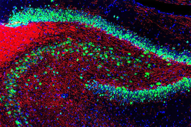
“We then created unique mice that modeled Parkinson’s disease and expressed MPO. These mice accumulated toxic, misfolded proteins in the brain. Additionally, the MPO produced in the brain had an altered shape. As a result, instead of being stored inside neurons, MPO is capable of being ejected from the cell and cause further brain damage. We also found that MPO was located preferentially in the memory-associated regions of the brain—the cortex and hippocampus—indicating it plays a role in memory disruption.” 

Reynolds and her team are already working to develop an MPO inhibitor, which they hope will slow the progression of Parkinson’s disease. Based on Reynold’s previous research showing that MPO is abnormally expressed in the Alzheimer’s disease brain, an MPO inhibitor may also hold potential as an Alzheimer’s disease treatment. 


The first author of the study is Richard A. Maki, PhD, of Sanford Burnham Prebys. Additional authors include Michael Holzer, PhD, Gunther Marsche, PhD, and Ernst Malle, PhD, of the Medical University of Graz; Khatereh Motamedchaboki of Sanford Burnham Prebys; and Eliezer Masliah, MD, of the National Institutes of Health (NIH) and University of California, San Diego.

This work was supported by the NIH (ROINS074303, ROIAG017879, and ROI AG040623) and the Austrian National Bank (17600). The content is solely the responsibility of the authors and does not necessarily represent the official views of the NIH.

Institute News

High-throughput screening against a new target for Alzheimer’s drugs

AuthorJessica Moore
Date

March 27, 2017

More than 5 million people in the U.S. have Alzheimer’s, and by 2050 that number could rise as high as 16 million. There are currently no treatments that slow the advance of this cruel disease that slowly destroys a person’s memory and reason. Given the overwhelming need for drugs that prevent or limit the brain degeneration caused by Alzheimer’s, scientists are attacking the problem from all angles.

A new strategy for finding possible Alzheimer’s therapeutics has recently been developed by Nicholas Cosford, PhD, professor at Sanford Burnham Prebys Medical Discovery Institute (SBP), in collaboration with Varghese John, PhD, associate professor at UCLA. The high-throughput screening method, reported in Frontiers in Pharmacology, identified several compounds worthy of further investigation.

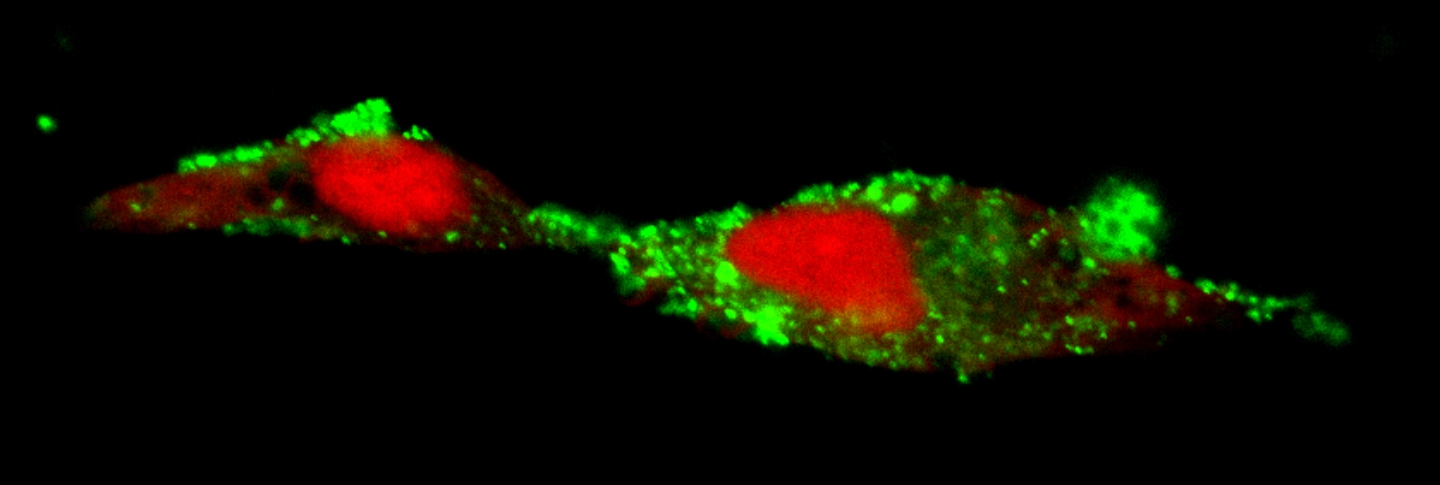
“We looked for inhibitors of a process that isn’t the standard target in drug screening for Alzheimer’s,” says Cosford. “This process—generation of a toxic peptide called APP (amyloid precursor protein) delta C31, or the 31 amino acids at the end of APP—may be especially important at early stages of Alzheimer’s, before neurons start to die.”

APP delta C31, which kills neurons, is made when APP is first cut by an enzyme called gamma secretase to form amyloid beta (the best-studied contributor to brain deterioration in Alzheimer’s) and the remaining portion of APP is then further cut by an intracellular caspase enzyme. Amyloid beta—also toxic—sticks together in clumps that clog up the spaces between brain cells. Most of the drugs that have recently been tested in Alzheimer’s patients are intended to eliminate amyloid beta or prevent it from being created, but so far none have been successful in clinical studies.

Cosford and John’s team found several compounds that inhibit production of APP delta C31. “A few of these compounds block signaling pathways not previously implicated in Alzheimer’s—so this is really a new avenue of research that we are pursuing,” adds Cosford.

Worryingly, generation of APP delta C31 is enhanced by statins, drugs that are widely used to lower blood cholesterol. In fact, statins were used in this study to bump up production of APP delta C31 and make reductions in levels of the peptide easier to detect. However, statins also protect against other damaging processes in Alzheimer’s.

“We speculate that whether statins are good or bad in the context of Alzheimer’s may depend on the stage of the disease,” notes Cosford.

“The next step for us is to test the most promising compounds in animal models of Alzheimer’s,” Cosford adds. “We also plan to use the same approach to screen another library of molecules to identify more potential drugs.”

Institute News

Helping the brain de-toxify could slow Alzheimer’s

AuthorJessica Moore
Date

February 16, 2017

In Alzheimer’s disease, the space around brain cells becomes clogged with toxic clumps of protein called amyloid. The problem isn’t just that amyloid is generated—that happens in all aging brains—it’s that there’s more of it than the brain’s garbage-scavenging cells, called microglia, can clear out. The lingering amyloid causes neurons to break down, creating a bigger mess for the already overworked microglia to eliminate.

Recent research could lead to a way to turn up the activity of microglia, which should slow the advance of Alzheimer’s. Two related papers with contributions from Huaxi Xu, PhD, the Jeanne and Gary Herberger Chair of Neuroscience and Aging Research at Sanford Burnham Prebys Medical Discovery Institute (SBP), show that a protein called TREM2 helps microglia survive and respond more strongly to damaging material like amyloid and cell debris.

“These results suggest that activating TREM2 could be a viable future treatment strategy for Alzheimer’s,” says Xu. “TREM2 has recently become a hot topic since mutations in the gene have been strongly linked to a greater risk for Alzheimer’s. This work shows that TREM2 is important not just in the relatively small number of patients carrying mutations, but potentially in all of Alzheimer’s.”

TREM2 is a receptor on tissue-resident immune cells, including microglia. TREM2 is activated by fatty molecules released from damaged cells, which stick to amyloid.

The two papers identify the functions of two forms of TREM2—the receptor form that sits on the surface of microglia, and a soluble fragment form that’s released into the space surrounding cells in the brain. Both studies were led by Guojun Bu, PhD, professor at the Mayo Clinic. The first, published in the Journal of Neuroscience, shows that the receptor form of TREM2 is required for microglia to live as long as they normally do. The second, in the Journal of Experimental Medicine, found that the soluble TREM2 fragment also supports microglial survival and turns on their inflammatory response—which in turn supports their ability to remove toxic amyloid clumps.

“The finding that soluble TREM2 has an important function supports its use as a biomarker for Alzheimer’s,” adds Xu. “Levels of soluble TREM2 increase in cerebrospinal fluid before severe symptoms appear, so measuring its levels could aid early diagnosis, which is crucial for effective treatment.”

“We’re now searching for activators of TREM2 in collaboration with drug discovery specialists here at SBP and with support from the Tanz Initiative,” Xu comments. “We’re also looking at whether enhancing TREM2 function lowers levels of amyloid in models of Alzheimer’s.” 

Institute News

Research points to new way to prevent optic nerve degeneration in glaucoma

AuthorJessica Moore
Date

June 2, 2016

Over 3 million Americans have glaucoma, the group of eye diseases that damage the nerve that carries information from the eye to the brain. This damage slowly degrades patients’ vision, even with treatment. Current glaucoma drugs lower the pressure in the eye, which lessens the injury to the nerve, although it is not eliminated. Finding ways to protect the optic nerve could lead to treatments that are much more effective in preserving or restoring sight in glaucoma patients. Continue reading “Research points to new way to prevent optic nerve degeneration in glaucoma”

Institute News

Closing in on the causes of Alzheimer’s disease

AuthorGuest Blogger
Date

April 5, 2016

This post was written by Nicole Le, a guest blogger.

Imagine if we could clear the brain of plaque that accumulates and causes Alzheimer’s disease (AD) as simply as having the plaque removed from our teeth? The body has a natural clearing mechanism in place to rid the brain of these deposits, but if this mechanism gets overwhelmed or disrupted, the plaques can accumulate and lead to neurodegeneration. Continue reading “Closing in on the causes of Alzheimer’s disease”