Atherosclerosis, the buildup of plaques inside arteries, is a key step in the development of cardiovascular disease, the leading cause of death in the United States. Elevated cholesterol levels are a risk factor for atherosclerosis, as the molecule is one of the building blocks of these plaques. However, since cholesterol is also essential in healthy cells, scientists are researching how cholesterol biology is controlled to better understand the changes that lead to disease.
Laszlo Nagy, MD, PhD, professor and director of the Genomic Control of Metabolism Program, recently collaborated with Peter Tontonoz, MD, PhD, professor, the leading senior scientist of the study and Francis and Albert Piansky Endowed Chair in Pathology and Laboratory Medicine at the UCLA David Geffen School of Medicine, to assess a network of molecules that control cholesterol transport out of macrophages, immune cells normally associated with inflammation.
“Although macrophages are usually thought of as the white blood cells that ingest invading bacteria and cleaning up cell debris after injury or infection, they can also enter a ‘alternatively activated’ state to help tissue repair and remodeling,” Nagy says. “In blood vessels, these repair state macrophages protect the body by removing cholesterol from the bloodstream. However, the accumulation of excess cholesterol in macrophages is a key event in the development of atherosclerosis. How macrophages control cholesterol transport is not well understood, but needs to be explored to better understand atherosclerosis.”
“We were interested in how macrophages are able to switch on the gene Abca1, the gene that encodes the protein that pumps cholesterol out of these cells,” Nagy explained. “We used our expertise in epigenomics to define regions of the genome that controlled the amount of Abca1 RNA produced.”
By examining long non-coding RNA strands that regulate gene expression, the study identified an RNA called MeXis that increases the expression of Abca1. Although MeXis cannot start transcription of Abca1 by itself, it does impact the ability of other proteins to transcribe the gene.
“Using our molecular tools, we were able to show that MeXis recruited another protein that helps to start transcription to Abca1, says Nagy. “Without MeXis, this protein did not interact with Abca1 and transcription was dramatically reduced, even when the cells received signals to start the process of ridding themselves of cholesterol.
“The more we understand about the biological processes that control cholesterol metabolism the better informed we are to develop strategies to prevent and treat atherosclerosis, says Nagy.“This study reveals key insights on the regulation of Abca1, which could ultimately lead to new therapeutic approaches.”
The Zika virus has been relatively quiet lately, but that doesn’t mean the danger is over. Zika can come roaring back at any time, generating severe birth defects and other health issues.
That makes it a race against time. Can scientists and clinicians develop effective Zika therapies before the virus rebounds? The normal drug discovery process takes 10 to 20 years, and we clearly don’t have that much time. But there’s another option: Retasking drugs that have already been approved for other conditions could dramatically shorten the approval process.
“It’s important to find drugs that are immediately available that you can put in the pipeline to treat Zika,” says Alexey Terskikh, PhD, associate professor at SBP.
A recent paper from Terskikh and UC San Diego’s Alysson Muotri, PhD, highlighted this possible solution. Published in Nature Scientific Reports, the study showed sofosbuvir (Sovaldi), a drug made by Gilead to treat hepatitis C, could also be effective against Zika.
Sovaldi neutralizes an RNA polymerase – an enzyme that transcribes RNA from DNA. By disrupting this pathway, Sovaldi prevents hepatitis C from replicating. Because Zika has a similar RNA polymerase, a number of labs have been looking at Sovaldi’s possible impact on the virus. The collaborative work conducted by the Terskikh and Muotri labs showed the drug decreased viral levels, reduced neural cell death and limited the amount of virus transmitted to babies.
This work reinforces a previous study, published in Nature Scientific Reports last November, that shows the anti-malaria drug chloroquine reduces Zika transmission between mothers and babies in a mouse model.
Chloroquine offers a number of advantages. It’s inexpensive, generally available in the countries most affected by Zika and poses no risk to pregnancy.
“Chloroquine has been tested in pregnant women for the past 50 years in high doses and has been found safe,” says Terskikh.
The researchers believe a drug cocktail, including Sovaldi, chloroquine and perhaps other drugs, could be effective against Zika.
“It’s possible we could use both drugs,” says Terskikh. “We are working on a grant proposal to test different combinations. Most antiviral therapies rely on a cocktail—for example many HIV treatments are a mix of three or four drugs.”
But most importantly for Zika, these drugs are already being made in significant quantities and, should another epidemic arise, could potentially help stem the outbreak and protect vulnerable people.
Institute News
Brain map connects brain diseases to specific cell types
Researchers have developed new single-cell sequencing methods that could be used to map the cell origins of various brain disorders, including Alzheimer’s, Parkinson’s, schizophrenia and bipolar disorder.
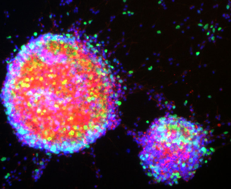
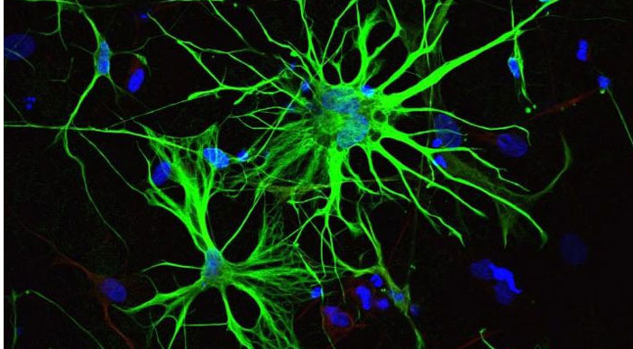
By analyzing individual nuclei of cells from adult human brains, Jerold Chun, MD, PhD, professor at SBP, in collaboration with researchers at UC San Diego and Harvard Medical School, have identified 35 different subtypes of neurons and glial cells and discovered which of these subtypes are most susceptible to common risk factors for different brain diseases.
There are multiple theories regarding the roots of brain diseases. The new study, published in Nature Biotechnology, allows scientists to narrow down and rank the cell types in the brain that carry the most genetic risk for developing brain disorders. The information can guide researchers to pick the best drug-targets for future therapies.
The work builds off of a previous study by the authors that identified 16 subtypes of neurons in the cerebral cortex. That study was the first large-scale mapping of gene activity in the human brain and provided a basis for understanding the diversity of individual brain cells.
In the new study, researchers developed a new generation of single-cell sequencing methods that enabled them to identify additional neuronal subtypes in the cerebral cortex as well as the cerebellum, and even further divide previously identified neuronal subtypes into different classes. The new methods also enabled researchers to identify different subtypes of glial cells, which wasn’t possible in the previous study due to the smaller size of glial cells.
“These data confirm and significantly expand our prior work, further highlighting the enormous transcriptional diversity among brain cell types, especially neurons,” says Chun. “This diversity, which continues to emerge from our single-cell analytical approach, will provide a foundation for better understanding the normal and diseased brain.” The advance was made possible by combining next-generation RNA sequencing with chromatin mapping—mapping of DNA and proteins in the nucleus that combine to form chromosomes—for more than 60,000 individual neurons and glial cells.
“While the analysis of RNA can tell us how cell types differ in their activity, the chromatin accessibility can reveal the regulatory mechanisms driving the distinctions between different cells”, notes Peter Kharchenko, PhD, an assistant professor of biomedical informatics at Harvard Medical School who co-led the study.
Using the information from RNA sequencing and chromatin mapping methods, researchers were able to map which cell types in the brain were affected by common risk alleles—snippets in DNA that occur more often in people with common genetic diseases. Researchers could then rank which subtypes of neurons or glial cells are more genetically susceptible to different brain diseases. For example, they found that two subtypes of glial cells, microglia and oligodendrocytes, were the first and second most at risk, respectively, for Alzheimer’s disease. They also identified microglia as most at risk for bipolar disorder, and a subtype of excitatory neurons as most at risk for schizophrenia.
“Now we can locate where the disease likely starts,” says Kun Zhang, PhD, professor of bioengineering at the UC San Diego Jacobs School of Engineering and co-senior author of the study. “However, we are only mapping the genetic risk. We don’t know the precise mechanism of how these specific cells actually trigger the disease.”
One caveat of this study, explains Zhang, is that it primarily analyzed data from adult brains (ages 20 to 50), so the findings do not represent younger or older populations. In order to better understand brain disorders that manifest early on, for example in infants, like autism spectrum disorder, the study would need to analyze cells from younger brains, he said.
The team also plans to expand their studies to map additional regions of the brain.
Authors of the study are Blue B. Lake*, Song Chen*, Brandon C. Sos*, Thu E. Duong, Derek Gao and Kun Zhang of UC San Diego; Jean Fan* and Peter V. Kharchenko of Harvard Medical School; and Gwendolyn Kaeser, Yun C. Yung and Jerold Chun of Sanford Burnham Prebys Medical Discovery Institute.
*These authors contributed equally to this work.
This story is based on a UC San Diego press release written by Liezel Labios.
Cancer, which is one of the leading causes of death worldwide, arises from the disruption of essential mechanisms of the normal cell life cycle, such as replication control, DNA repair and cell death. Thanks to the advances in genome sequencing techniques, biomedical researchers have been able to identify many of the genetic alterations that occur in patients that are common among and between tumor types. But until recently, only mutations in DNA were thought to cause cancer. In a new study published in the journal Cell Reports, researchers show that alterations in a process known as alternative splicing may also trigger the disease.
Although DNA is the instruction manual for cell growth, maturation, division, and even death, it’s proteins that actually carry out the work. The production of proteins is a highly regulated and complex mechanism: cellular machinery reads the DNA fragment that makes up a gene, transcribes it into RNA and, from the RNA, makes proteins. However, each gene can lead to several RNA molecules through alternative splicing, an essential mechanism for multiple biological processes that can be altered in disease conditions.
Using data for more than 4,000 cancer patients from The Cancer Genome Atlas (TCGA project), an international team of scientists that included Adam Godzik, PhD, professor at Sanford Burnham Prebys Medical Discovery Institute (SBP), has analyzed the changes in alternative splicing that occur in each tumor patient and studied how these changes could impact the function of genes. The results of the study show that alternative splicing changes lead to a general loss of functional protein domains, and particularly those domains related to functions that are also affected by genetic mutations in cancer patients.
From previous work, the research team learned that tumor type and stage can be predicted by observing alterations in alternative splicing. With this new study, the team discovered that changes in alternative splicing that occur in cancer impact protein functions in a way that is similar to that previously described for genetic mutations.
All of these alterations in protein functions would cause changes in cells morphology and function, giving them the characteristics of tumor cells, such as a high proliferative potential or the ability to avoid programmed cell death.
According to Godzik, “These changes potentially have oncogenic power in cells, which means, the ability to turn a healthy cell into a cancer cell.” A novel aspect of the study is that these changes tend to occur in genes other than those often mutated in cancer, and in patients with a low number of mutated genes.
“Changes in alternative splicing provide cancer with new ways in which it can escape fine cellular regulation. Therefore, the study of alternative splicing opens new doors in the research to cure cancer and may provide new alternatives to the treatment of this disease.
About one in seven men will be diagnosed with prostate cancer during his lifetime. Though it has one of the highest survival rates of any type of cancer—95% make it through the first ten years—diagnosis and treatment could still be improved. Since November is Prostate Cancer Awareness Month, we’re highlighting the work our scientists are doing to address key challenges and unresolved questions in prostate cancer.
Early, accurate detection—The current method of screening for prostate cancer is a blood test for prostate specific antigen, or PSA, which detects cancer early, but isn’t very specific—only one in four men with high PSA levels actually has cancer. In general, high levels of PSA mean the next step is a biopsy. A more specific test would avoid unnecessary biopsies, which are invasive and stressful for patients.
Ranjan Perera, PhD, associate professor in the Integrative Metabolism Program, is looking for biomarkers that would enable just such a test. His lab is making progress—they identified five long noncoding RNAs (RNAs that, instead of carrying genetic information to be translated, regulate the translation of other RNAs) found at higher-than-normal levels in the urine of prostate cancer patients. An RNA-based test is on the market, but could be improved—measuring multiple markers would be more sensitive and specific.
Better therapies—For advanced cases, current treatments are either insufficient or overly toxic. Prostate cancer is usually first treated with drugs that block the actions of androgens, the hormones that drive its growth. If the tumor recurs later, as happens for cancers that are already at a late stage before treatment, it forms from cells that are resistant to those drugs. Then, the only option is chemotherapy or radiation.
Towards therapies that cause less collateral damage, Nicholas Cosford, PhD, associate director of Translational Research in the NCI-designated Cancer Center, is designing new drugs against targets found to be important in prostate cancer. These drugs are intended to block two strategies by which cancer cells survive—avoiding cell death, and generating extra energy by recycling the cell’s own parts.
Understanding why obesity correlates with aggressiveness—Obese men are no more likely than others to get prostate cancer, but if they do, it’s more likely to become advanced quickly. To figure out why this happens, Jorge Moscat, PhD, director, and Maria Diaz-Meco, PhD, professor in the Cancer Metabolism and Signaling Networks Program, are looking at how fat adjacent to the prostate interacts with tumor cells. A detailed picture of how fat cells and tumor cells interact could reveal new ways to treat prostate cancer in overweight men.
Institute News
New molecular markers for prostate cancer identified
A team of scientists led by Sanford-Burnham’s Ranjan J. Perera, PhD, has identified a set of RNA molecules that are detectable in tissue samples and urine of prostate cancer patients, but not in normal healthy individuals. The study sets the stage for the development of more-sensitive and specific non-invasive tests for prostate cancer than those currently available, which could result in fewer unnecessary prostate biopsies with less treatment-related morbidity, according to a new study in The Journal of Molecular Diagnostics.
According to the American Cancer Society, prostate cancer is the second most common type of cancer in American men (behind skin cancer), and the second-leading cause of cancer death in men (after lung cancer). In 2014, more than 230,000 new cases of prostate cancer will be diagnosed. One in seven American men will get prostate cancer during his lifetime, and one in 36 will die from it. Since most men with prostate cancer have indolent (non-aggressive) disease for which conservative therapy or surveillance would be appropriate treatment, the clinical challenge is not only how to identify those with prostate cancer, but also how to distinguish those who would benefit from surgical or other aggressive treatment from those who would not.
Today, prostate cancer is primarily detected and monitored by testing for high concentrations of prostate-specific antigen (PSA) in blood samples. High PSA levels are often followed by a biopsy to confirm the presence of cancer, and whether it’s slow growing or aggressive. “While elevated PSA can be an alert to a lethal cancer, it can also detect less aggressive cancers that may never do any harm,” said Vipul Patel, MD, medical director of the Global Robotics Institute at Florida Hospital in Orlando, and co-author of the study. “Moreover, only 25 percent of men with raised PSA levels that have a biopsy actually have prostate cancer. Prostate cancer needs to be screened for; we just need to find a better marker.”
The researchers believe that they have identified a group of RNA molecules – known as long noncoding RNAs (lncRNAs) – that hold the potential for serving as better prognostic markers for prostate cancer. lncRNAs are noncoding RNA molecules that until recently were dismissed by scientists as non-functional noise in the genome. Now, lncRNAs are thought to regulate normal cellular development and are increasingly reported as contributing to a range of diseases, including cancer.
Detection of lncRNAs in urine
“We have identified a set of lncRNAs that appear to have an important role in prostate cancer diagnostics,” said Perera, associate professor and scientific director of Analytical Genomics and Bioinformatics at our Lake Nona campus. “The findings advance our understanding of the role of lncRNAs in cancer biology and, importantly, broaden the opportunity to use lncRNAs as biomarkers to detect prostate cancer.”
The study profiled the lncRNAs in three distinct groups: (1) human prostate cancer cell lines and normal prostate epithelial cells, (2) prostate adenocarcinoma tissue samples and matched normal tissue samples, (3) urine samples from patients with prostate cancer or benign prostate hypoplasia, and normal healthy individuals. In each case, the lncRNAs were elevated in prostate cancer patient samples, but not in patients with benign prostate hypoplasia or normal healthy individuals.
One advantage of lncRNAs is that the molecules can be detected in urine samples, which are more easily available than blood tests. One lncRNA, PCA3, was recently commercialized as a urine test to identify which men suspected of having prostate cancer should undergo repeat prostate biopsy. However, discrepancies have been found to exist between PCA3 levels and clinicopathologic features, said Perera. In the current study, PCA3 was detected in some, but not all of the study samples, suggesting that reliance on a single biomarker may be insufficient for prostate cancer detection, while combining additional markers may increase the specificity and sensitivity of the test.
“There is a tremendous unmet clinical need for better non-invasive screening tools for early detection of prostate cancer to reduce the overtreatment and morbidity of this disease,” added Patel. “Our findings represent a promising approach to meet this demand.”
Technical details of the study
The goal of the first experiment was to see whether lncRNAs are differentially expressed in prostate cancer by measuring total RNA from prostate cancer cell lines and normal epithelial prostatic cells using NCode human ncRNA array and SurePrint G3 human lncRNA microarrays. Hierarchical clustering revealed distinguishable lncRNA expression profiles. Thirty lncRNAs were up-regulated and the expression levels of three top-ranking candidates [XLOC_007697, LOC100287482, and AK024556 (also known as SPRY4-IT1)] were confirmed in prostate cancer cell lines by quantitative real-time polymerase chain reaction (qPCR) analysis. The SPRY4-IT1 was found to be up-regulated more than 100-fold in PC3 cells compared with prostatic epithelial cells.
In a second experiment, lncRNA expression was compared in pooled prostate cancer tissue samples and matched normal tissues from 10 frozen biopsy specimens. Hierarchical clustering of the differentially expressed lncRNAs was observed and 10 up-regulated lncRNAs were detected using microarrays. An additional set of 18 prostate cancer tissue samples was analyzed by qPCR and five lncRNAs were found to be significantly higher in prostate tumor tissues compared with matched normal tissues.
Researchers used qPCR to analyze total RNA isolated from urine in another experiment. Urine was collected from 13 prostate cancer patients and 14 healthy controls. All six lncRNAs were found to be significantly up-regulated in the urine samples from the prostate cancer patients compared with normal patient controls, while there were no differences between normal and benign prostatic hyperplasia patient samples.
In other studies focused particularly on SPRY4-IT1. Using both qPCR and highly sensitive droplet digital PCR, expression of SPRY-IT1 was found to be increased in 16 of 18 (89 percent) tissue samples from patients with prostatic adenocarcinoma, compared to normal tissue samples. The researchers developed chromogenic in situ hybridization (CISH) techniques to visualize SPRY4-IT1 expression in cancerous and matched normal tissue. Intense staining was seen in all adenocarcinoma samples, but not in normal prostatic tissue. Finally, the investigators showed that reduction of SPRY4-IT1 in prostate cancer cells through the use of small interfering RNA (siRNA) leads to decreased cell viability and cellular invasion as well as increased apoptosis, similar to what is seen in melanoma cells.
About the paper
“Long Noncoding RNAs as Putative Biomarkers for Prostate Cancer Detection,” by Bongyong Lee, Joseph Mazar, Muhammed Nauman Aftab, Feng Qi, John Shelley, Jian-Liang Li, Subramaniam Govindarajan, Felipe Valerio, Inoel Rivera, Tadzia Thurn, Tien Anh Tran, Darian Kameh, Vipul Patel, and Ranjan J. Perera, DOI: http://dx.doi.org/10.1016/j.jmoldx.2014.06.009. Published online ahead of The Journal of Molecular Diagnostics, Volume 16, Issue 6 (November 2014) published by Elsevier.