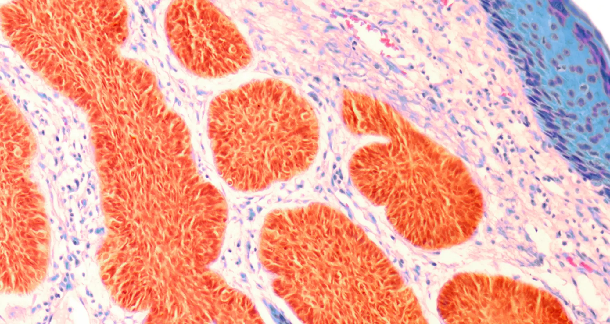
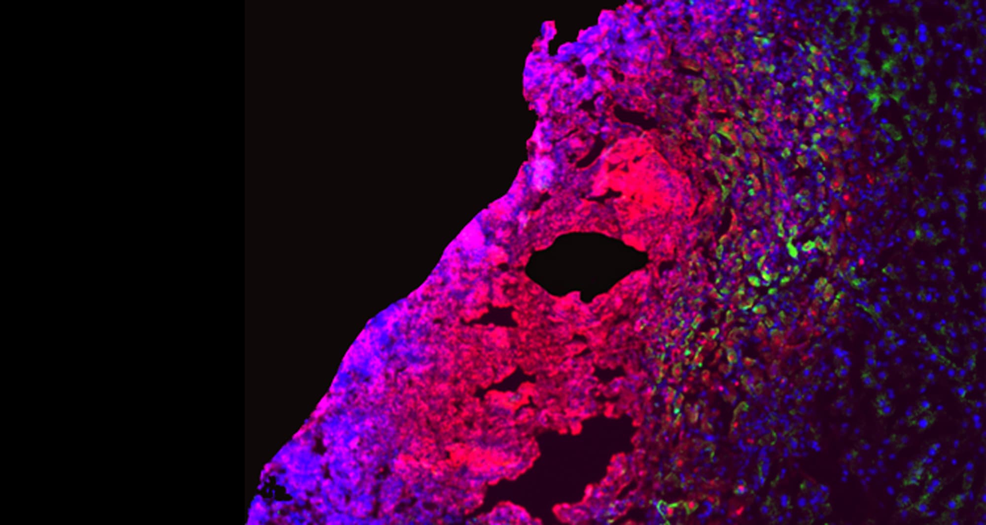
Science in Pictures Archives - Page 4 of 7 - Sanford Burnham Prebys
Institute News

Science in Pictures

AuthorScott LaFee
Date

October 7, 2024

A quantum dot fluorescence image depicts a section of mouse small intestine. Different fluorescent markers indicate different cellular constituents. Actin, a protein that forms the contractile filaments of muscle cells, is shown in red. Lamin (green) are fibrous proteins that provide structural function. Cell nuclei are depicted in blue.

Image courtesy of Thomas J. Deerinck, UC San Diego National Center for Microscopy and Imaging Research.

Institute News

Science in Pictures

AuthorScott LaFee
Date

September 30, 2024

A confocal micrograph of the mammalian inner ear. The inner ear or auris internal is the innermost part of the vertebrate ear. It consists of a bony labyrinth—a hollow cavity in the temporal bone of the skull with a system of passages comprising two main functional parts. The cochlea is dedicated to hearing, converting sound pressure patterns from the outer ear into electrochemical impulses passed to the brain for processing. The vestibular system is dedicated to maintaining balance.

Image courtesy of Glen MacDonald, University of Washington/Bioscapes.

Institute News

Science in Pictures

AuthorScott LaFee
Date

September 23, 2024

 A colorized scanning electron micrograph depicts T regulatory cells (red) interacting with antigen-presenting cells (blue). T regulatory cells can suppress responses by T cells to maintain homeostasis in the immune system

Image courtesy of NIAID.

Institute News

Science in Pictures

AuthorScott LaFee
Date

September 16, 2024

 A colorized scanning electron micrograph of a human proximal tubule. These tubules make up a significant portion of the kidneys and carry out diverse regulatory and endocrine functions. For example, under normal circumstances more than two-thirds of filtered salt and water is reabsorbed in proximal tubules.

Image courtesy of David Gergory and Debbie Marshall, Wellcome Collection.

Institute News

Science in Pictures

AuthorScott LaFee
Date

September 9, 2024

A polarized light micrograph captures the colorful character of caffeine crystals. Caffeine is a central nervous system stimulant that works by blocking the binding of adenosine to its receptor, which enhances release of the neurotransmitter acetylcholine, which in the brain supports cognitive functions and boosts muscle activity in the body.

Image courtesy of Stefan Eberhard, University of Georgia and Nikon Small World.

Institute News

Science in Pictures

AuthorScott LaFee
Date

September 3, 2024

A false color scanning electron micrograph depicts an axon terminal broken open to reveal internal neurotransmitter vesicles. These vesicles are released at neural synapses to carry electrical nerve impulses from one neuron to another.

Image courtesy of Tina Carvalho/NIH-NIGMS.

Institute News

Science in Pictures

AuthorScott LaFee
Date

August 26, 2024

The distribution of metabolites (small molecules produced while chemical reactions take place in a cell to provide energy) in cells in a mouse kidney. Three small molecules (the amino acids aspartate and glutamine and the antioxidant glutathione) were visualized using a technique called computational molecular phenotyping (CMP). Grayscale light micrographs were taken of silvered antibodies against aspartate, glutamine and glutathione and these were then digitally converted into red, blue and green respectively.

Image courtesy of Jefferson R. Brown, Robert E. Marc, Bryan W. Jones, Glen Prusky and Nazia Alam.

Institute News

Science in Pictures

AuthorScott LaFee
Date

August 19, 2024

Fluorescence immunohistochemistry and confocal microscopy were used to create this array of normal and cancerous human tissue samples. Human prostate, colon, kidney, intestine and breast tissue sections were stained for the presence of protein biomarkers to improve cancer diagnosis and prognosis.

Image courtesy of Aamir Ahmed, Jane Pendjiky and Michael Millar.

Institute News

Science in Pictures

AuthorScott LaFee
Date

August 12, 2024

A polarized light micrograph of a section of cat skin showing hairs, whiskers and their blood supply, created from a vintage prepared slide dating before 1900.

Blood vessels were injected with dye (carmine; black) before fixing and sectioning the tissue in order to visualize the capillaries in the tissue. This was a newly developed technique at that time. The capillary bed seen here supplies the hairs and whisker. The fine hairs, thicker whisker, blood vessels and underlying muscle are all visible.

Image courtesy of David Linstead, Wellcome Images.

Institute News

Science in Pictures

AuthorScott LaFee
Date

August 5, 2024

A large-scale, high-resolution montage of the entire surface of a mouse retina using antibody labeling and laser scanning confocal imaging. Complete networks of both astrocytes and blood vessels on the retinal surface are shown: astrocyte cell bodies are red, the star-shaped extensions from the astrocyte cell bodies are green and blood vessels are blue.

Image courtesy of Gabriel Luna, UC Santa Barbara.