Science in Pictures Archives - Page 7 of 7 - Sanford Burnham Prebys
Institute News

Science in Pictures

AuthorScott LaFee
Date

March 11, 2024

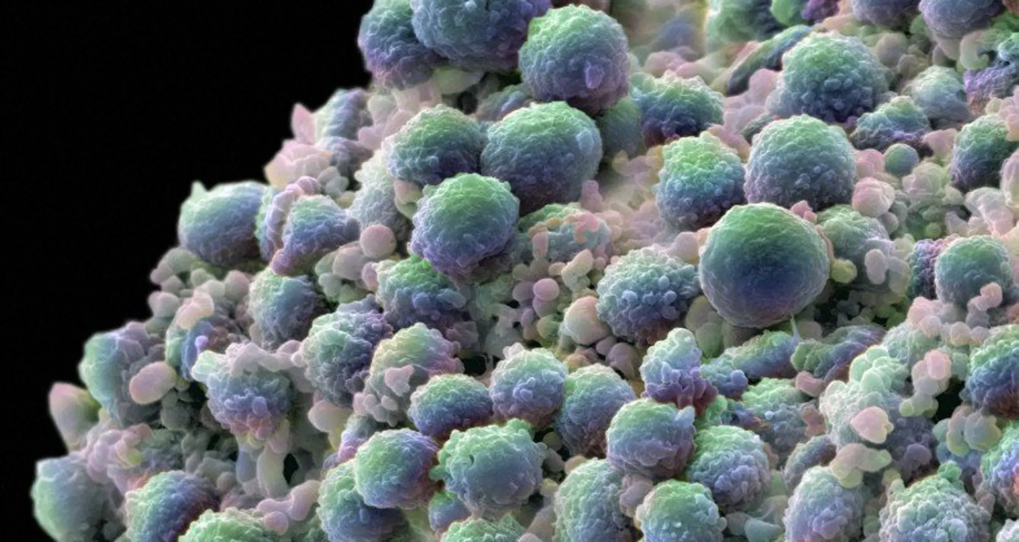
A colorized scanning electron micrograph of a natural killer cell. These immune system cells provide rapid response to cells infected by viruses and other intracellular pathogens. They are increasingly being investigated for their potential in anticancer therapies.

Image courtesy of NIAID.

Institute News

Science in Pictures

AuthorScott LaFee
Date

March 4, 2024

A fluorescent image depicts a section of the tube-like mosquito heart magnified 100 times. The triangular-shaped bundles perpendicular to the heart are alary muscles which hold the organ up against the mosquito’s back. The insect’s body consists of a series of segments; the broad strips of muscle that run parallel to the heart are intersegmental muscles that hold the segments together

Image courtesy of Jonas King, Vanderbilt University.

Institute News

Science in Pictures

AuthorScott LaFee
Date

February 26, 2024

Metastatic chordomas, like the one pictured in this brightfield image in a pulmonary artery, are rare, slow-growing malignant tumors derived from notochord remnants that can arise anywhere along the neuronal axis.

Image courtesy of Steffan Albrecht, Baylor College of Medicine.

Institute News

Science in Pictures

AuthorScott LaFee
Date

February 19, 2024

In this confocal image of a breast organoid, contractile myoepithelial cells (blue) are shown crawling on secretory breast cells. Myoepithelial cells are present in mammary, sweat, lacrimal and salivary glands where they help regulate flow of fluids.

Image courtesy of Jakup Sumbal, Masaryk University, Czech Republic.

Institute News

Science in Pictures

AuthorScott LaFee
Date

February 12, 2024

Zebrafish (Danio rerio) make excellent model organisms. Their genome is fully sequenced and easy to manipulate. They are fecund, have short generation times (a few months) and rapid embryonic development. In this confocal image, the embryo head of a Zebrafish is depicted just 72 hours after fertilization. 

Image courtesy of Layra G. Cintron-Rivera, Brown University.

Institute News

Science in Pictures

AuthorScott LaFee
Date

February 5, 2024

HeLa cells is the oldest immortalized cell line and widely used in research, derived from cancer cells taken from Henrietta Lacks in February 1951. In this scanning electron micrograph, colorized HeLa cells are undergoing apoptosis or programmed cell death. The string-like microvilli are used by the cells to move around and metastasize.

Image courtesy of Thomas Deerinck, UC San Diego, NIGMS and NIH.

Institute News

Science in Pictures

AuthorScott LaFee
Date

January 29, 2024

In 1965, Sydney Brenner settled on Caenorhabditis elegans, a nematode or worm, as a model organism to study animal development and behavior. The anatomy of C. elegans is simple, but complex enough to model nearly every aspect of biology, including aging.

Image courtesy of Jim McCarter and Tim Schedl, WormBook.

Institute News

Science in Pictures

AuthorScott LaFee
Date

January 22, 2024

A colorized scanning electron micrograph of a cell (purple) infected with SARS-CoV-2 virus particles (yellow), isolated from a patient sample.

Image courtesy of NIAID Integrated Research Facility in Fort Detrick, Maryland. National Institute of Allergy and Infectious Diseases.

Institute News

Science in Pictures

AuthorScott LaFee
Date

January 15, 2024

A color-enhanced scanning electron micrograph depicts a clump of prostate cancer cells. prostate cancer cells. Other than skin cancer, prostate cancer is the most common cancer in American men, with approximately 288,300 new cases annually and 34,700 deaths.

Image courtesy of Annie Cavanagh, Wellcome Collection.

Institute News

Science in Pictures

AuthorScott LaFee
Date

January 8, 2024

An Alzheimer’s disease brain cell is depicted in this transmission electron micrograph. Abnormal tangles of the protein tau (red) lie in the cytoplasm (yellow) of the cell body, adjacent to the nucleus (green).  These tangles, with beta-amyloid plaques, are hallmarks of the degenerative neurological condition.

Image courtesy of Thomas Deerinck, National Center for Microscopy and Imaging Research, UC San Diego.