Technique and Technology: Biochemistry
Guy Salvesen earned his PhD in biochemistry from Cambridge University in 1980. He conducted postdoctoral research at Strangeways Laboratory and MRC Laboratory of Molecular Biology in Cambridge, followed by further post-doctoral training at the University of Georgia. In 1991 he was appointed Assistant Professor at Duke University. Dr. Salvesen was recruited to Sanford-Burnham Medical Research Institute in 1996, where he is professor and director of the Apoptosis and Cell Death Research Program and dean of the Graduate School of Biomedical Sciences. He also holds an adjunct position as professor in the Department of Pathology at the University of California, San Diego.
Education
1981: PhD, Cambridge University, England, Biology
1977: B. Sc., London University, London, England, Microbiology
Other Appointments
Adjunct Professor, Department of Pathology, University of California, San Diego
Honors and Recognition
2014: Organizer, Keystone Meeting on Cell Death, February
2013: IUBMB Gold Medal Recipient, October
2010: Keynote Speaker, European Cell Death Organization Conference,
2010: Keynote Speaker, Gordon Research Conference on Cell Death
2009: Lifetime Achievement Award of the International Proteolysis Society
2008: Keynote Speaker, Queenstown Molecular Biology Conference
2008: Chair, Gordon Research Conference on Cell Death
2005: Helmut Holzer Memorial Prize
1999: International Proteolysis Society, Elected Secretary
1999: Keynote Speaker, Gordon Research Conference on Matrix Metalloproteinases
1988: American Association for the Study of Liver Diseases, State of the Art Lecture
1996: Chair, Gordon Research Conference on Proteolytic Enzymes and Their Inhibitors
Related Disease
Cancer, Inflammatory/Autoimmune Disease, Neurodegenerative and Neuromuscular Diseases, Pancreatic Cancer, Skin Cancer and Melanoma, Structural Biology
Phenomena or Processes
Apoptosis and Cell Death, Caspase Family, Cytokines, Inflammation, Protein Structure-Function Relationships, Proteolytic Pathways, Ubiquitin, Ubiquitin Protease System and Ubiquitin-like Proteins
Research Models
Bacteria, Human Cell Lines, Mouse, Mouse Cell Lines, Primary Human Cells
Techniques and Technologies
Biochemistry, Cellular and Molecular Imaging, Chemical Biology, Fluorescence Microscopy, Mass Spectrometry, Protein Engineering, Protein Structure Prediction, Protein-Protein Interactions, Proteomics
The human body contains cells with different life expectancies. Some (white blood cells or skin, for example) are programmed to rapidly die and be replaced. Others (such as nerve cells) are programmed to survive the lifetime of the individual and are seldom replaced. Dr. Salvesen’s research focuses on the central role enzyme pathways play in the life and death of cells. When death pathways slow down in cells that are normally programmed to die, cancer results. Conversely, when death pathways become overactive in cells that are programmed to survive, degenerative disease occurs. Dr. Salvesen’s laboratory focuses on understanding the fundamental molecular interactions that occur within these enzyme pathways. This knowledge is used to engineer synthetic compounds to stimulate cell destruction in cancer cells, or delay cell destruction in neurodegenerative diseases and stroke.
Guy Salvesen’s Research Report
Structure and Function of Proteases and Their Natural Inhibitors
Our research seeks to delineate the structure –> activity –> function algorithm as it applies to proteases and their inhibitors. Our laboratory has very broad interests in principles of proteolysis in humans, and we take multi-pronged approaches to research on proteases and their inhibitors.
Apoptosis
In one approach we apply basic biochemical knowledge to investigate newly emerging principles of proteolysis in human systems. This research is currently dissecting the proteolytic components of the intracellular pathway that lead to apoptotic cell death. Programmed cell death monitors the growth of new cells and the elimination of old ones. This program contains a number of proteolytic steps that are essential for efficient execution of the death pathway. Thus the proteases of the pathway – the caspases – are involved in the normal maintenance of correct cell number, and are therefore implicated in a number of pathologic and physiologic conditions. Using the techniques of protein chemistry, enzymology, crystallography, and recombinant DNA methodologies, we analyze the basic mechanism utilized by caspases to promote cell death pathways, and the mechanisms and specificity of the natural inhibitors that control them.
Cell Signaling
Modification of proteins by the small ubiquitin-like modifier SUMO is a dynamic and reversible process. The SUMO cycle begins when SUMO precursors are processed to remove short C-terminal extensions, thereby uncapping the C-terminal Gly-Gly motif that is essential for conjugation. SUMO ligases conjugate the protein, via its C-terminal carboxylate, to the side-chain lysine of target proteins to generate an isopeptide linkage. Eventually, SUMO is removed intact from its substrate SUMOylated proteins, and so the SUMOylation/deSUMOylation cycle regulates SUMOs function. A group of proteases known as SENPs are involved in both the activation of SUMO precursors (endopeptidase cleavage) and deconjugation of the targets (isopeptidase cleavage). Our laboratory is currently involved in projects to define the mechanisms that regulate SENP activity and access to their natural substrates.
Technology Development
The principle of proteolysis in vivo is to instigate irreversible changes to a set of protein substrates that alters their function and generates the required biological event. The sum total of the proteases and their target substrates operating in a physiologic pathway therefore defines the global event. Consequently, the identity of the substrate cleavages defines the proteases acting on them. We are developing proteomics-based methodologies, including selective protein labeling, multi-dimensional electrophoresis, and mass spectrometry techniques, to identify the products of proteolysis in vivo.
Related Disease
HIV/AIDS, Infectious Diseases, Molecular Biology
Phenomena or Processes
Host-Pathogen Interactions, Infectious Disease Processes, Inflammation, Innate Immunity
Anatomical Systems and Sites
Immune System and Inflammation
Research Models
Clinical and Transitional Research, Computational Modeling, Human, Human Cell Lines, Mouse, Mouse Cell Lines, Primary Cells, Primary Human Cells
Techniques and Technologies
Biochemistry, Bioinformatics, Cellular and Molecular Imaging, Drug Discovery, Drug Efficacy, Gene Expression, Gene Knockout (Complete and Conditional), Gene Silencing, High-Throughput/Robotic Screening, RNA Interference (RNAi), Systems Biology
Select Publications
Showing 3 of 3HIV-1 Vpu is a potent transcriptional suppressor of NF-κB-elicited antiviral immune responses.
Langer S, Hammer C, Hopfensperger K, Klein L, Hotter D, De Jesus PD, Herbert KM, Pache L, Smith N, van der Merwe JA, Chanda SK, Fellay J, Kirchhoff F, Sauter D
Elife 2019 Feb 5 ;8():Transcription Elongation Can Affect Genome 3D Structure.
Heinz S, Texari L, Hayes MGB, Urbanowski M, Chang MW, Givarkes N, Rialdi A, White KM, Albrecht RA, Pache L, Marazzi I, García-Sastre A, Shaw ML, Benner C
Cell 2018 Sep 6 ;174(6):1522-1536.e22Large-Scale Arrayed Analysis of Protein Degradation Reveals Cellular Targets for HIV-1 Vpu.
Jain P, Boso G, Langer S, Soonthornvacharin S, De Jesus PD, Nguyen Q, Olivieri KC, Portillo AJ, Yoh SM, Pache L, Chanda SK
Cell Rep 2018 Feb 27 ;22(9):2493-2503Xueqin (Sherine) Sun seeks to better understand the genetic and epigenetic underpinnings of cancers, using genome editing technologies, animal and patient-derived models, and other tools to develop more effective cancer therapies.
“My lab is interested in studying how DNA or the machinery that interprets it leads to the transformation of normal cells into cancerous cells and concurrently, their specific vulnerabilities. Identifying these intrinsic vulnerabilities and targeting them properly is profoundly important to developing effective cancer therapies.”
Another aspect of Sun’s work is understanding how cancer cells and tumors change their circumstances and environment to improve survival, including hiding from or repressing the immune system.
“Changes to DNA itself and the way how DNA is interpreted by cells can transform normal cells into cancer cells. And transformed cells propagate by enhancing the misinterpreted DNA information, which in turn becomes the Achilles’ heel of cancer cells. Our goal is to find out how DNA information is misinterpreted in different ways and how to correct it to halt cancer.”
At Sanford Burnham Prebys, Sun and colleagues will employ a host of leading-edge tools and approaches, including functional genomics, artificial intelligence, structural biology, large-scale drug screening, and advanced imaging/spatial technologies.
Sun conducted her postdoctoral fellowship at Cold Spring Harbor Laboratory under the guidance of Alea Mills, PhD, a professor at the National Cancer Institute-designated cancer center at Cold Spring Harbor.
She received her PhD from Wuhan University in China.
Related Disease
Aging-Related Diseases, Brain Cancer, Cancer, Childhood Diseases, Immune Disorders, Inflammatory/Autoimmune Disease, Leukemia/Lymphoma
Phenomena or Processes
Adapter Proteins, Adult/Multipotent Stem Cells, Aging, Angiogenesis, Apoptosis and Cell Death, Bcl-2 Family, Cancer Biology, Cancer Epigenetics, Cell Adhesion and Migration, Cell Biology, Cell Cycle Progression, Cell Differentiation, Cell Motility, Cell Proliferation, Cell Signaling, Cell Surface Receptors, Cellular Senescence, Chromosome Dynamics, Combinatorial Therapies, Cytokines, Development and Differentiation, Disease Therapies, DNA Damage Checkpoint Function, Embryonic/Pluripotent Stem Cells, Epigenetics, Gene Regulation, Genomic Instability, Growth Factors, Hematopoiesis, Host Defense, Host-Pathogen Interactions, Inflammation, Innate Immunity, Kinase Inhibitors, Metastasis, Neurogenesis, Oncogenes, Phosphorylation, Posttranslational Modification, Receptor Tyrosine Kinases, Serine/Threonine Kinases, Signal Transduction, TNF-Family, Transcription Factors, Transcriptional Regulation, Tumor Microenvironment, Tumorigenesis, Tyrosine Kinases, Ubiquitin, Ubiquitin Protease System and Ubiquitin-like Proteins
Anatomical Systems and Sites
Brain, General Cell Biology, Hematopoietic System, Immune System and Inflammation, Nervous System
Research Models
Bacteria, Cultured Cell Lines, Human Adult/Somatic Stem Cells, Human Cell Lines, Mouse, Mouse Cell Lines, Mouse Embryonic Stem Cells, Mouse Somatic Stem Cells, Primary Cells, Primary Human Cells
Techniques and Technologies
3D Image Analysis, 3D Reconstructions, Biochemistry, Bioinformatics, Cell Biology, Cellular and Molecular Imaging, Chemical Biology, Computational Biology, Confocal Microscopy, Correlative Light and Electron Microscopy, Drug Delivery, Drug Discovery, Drug Efficacy, Electron Microscopy, Fluorescence Microscopy, Fragment-Based Drug Design, Gene Expression, Gene Knockout (Complete and Conditional), Gene Silencing, Genetics, Genomics, High Content Imaging, High-Throughput/Robotic Screening, In vivo Modeling, Live Cell Imaging, Live Imaging, Mass Spectrometry, Microscopy and Imaging, Molecular Biology, Molecular Genetics, Nucleic Acid Synthesis, Protein-Protein Interactions, Protein-Small Molecule Interactions, Proteomics, Rational Drug Design, RNA Interference (RNAi), Scanning Cytometry, Small Molecule Compounds, Transgenic Organisms, Transplantation
We seek to understand why cancer occurs and what is the Achille’s heel of cancer, and to develop effective therapeutic interventions.
The successful treatment of any disease requires a good understanding of the mechanisms at work. Cancer is fundamentally caused by aberrant gene expression, which reflects the misinterpretation of DNA information at both genetic and epigenetic levels. We are interested in uncovering DNA-related alterations that drive cancer-favored transcriptional programs, identifying cancer-specific vulnerabilities, and developing effective therapeutic interventions for cancer treatment.
Xueqin Sun’s Research Report
Precise gene expression (the interpretation of DNA) is essential for almost all biological processes, and understanding gene regulation is one of the most pivotal frontiers in biological research under both health and disease circumstances. Gene expression is mainly regulated at genetic (with changes of DNA sequence) and epigenetic (without changing DNA sequence) levels. And gene dysregulation can lead to various health conditions and diseases, including developmental disorders, aging, and cancer. The overarching goal of Sun Lab is to uncover driving genetic and epigenetic alterations involved in cancer, to understand how developmental pathways and aging process impact cancer progression, and to identify mechanisms of action for developing more effective therapeutic strategies.
We are an interdisciplinary lab particularly focused on the following research directions:
- The EP400 chromatin remodeling complex
The EP400 complex is an evolutionarily conserved SWR1-class ATP-dependent chromatin remodeling complex encompassing ~17 components, with a total molecular mass of ~1.5 mega-dalton. The EP400 complex plays critical roles in diverse cellular processes, including chromosome stability, transcription, DNA recombination, DNA damage repair, embryonic stem cell renewal/development, and oncogenesis. The EP400 complex can incorporate histone variants, such as H2AZ and H3.3, into the genome to regulate gene expression. Our recent work discovers BRD8—one of the core subunits of the EP400 complex—as a unique vulnerability of p53 wildtype glioblastoma (GBM), the most prevalent and devastating type of brain cancer. BRD8-driven EP400 complex highjacks H2AZ at p53 target loci to block p53-mediated transactivation and tumor suppression (Nature, 2023). The bromodomain of BRD8 plays the key role in this process. Bromodomain is a druggable domain as evidenced by a number of successful small molecules targeting diverse bromodomains encoded by the human genome across multiple cancer types. Furthermore, findings from others and us suggest that the EP400 complex is involved in different cancers. Thus, we seek to unravel the roles of the EP400 complex in health and disease, and to better understand how to target the EP400 complex for developing effective therapeutic interventions. - The NuRD chromatin remodeling complex
The NuRD complex is also a highly conserved class of ~ 1 MDa multi-subunit chromatin remodeling complexes that consume energy derived from ATP hydrolysis to remodel the configuration of chromatin to control gene transcription programs, with a primary role in gene silencing. Chromatin remodeling is vital for efficiently framing the cellular response to both intrinsic and extrinsic signals and has enormous implications for determining cellular states. NuRD complex is unique in combining ATP-dependent chromatin remodeling, protein deacetylase activity, and recognition of methylated DNA and histone modifications, and has multifarious roles in chromatin organization, transcription regulation, and genome maintenance; thereby, largely impacts health and disease. The NuRD complex has been in the central stage of brain development studies, and is significantly related to brain disorders/diseases. Interestingly, NuRD complex re-assembles by exchanging the chromatin remodeling subunits CHD3/4/5 to achieve specific regulation of an array of genes required for generating distinct cell types in a highly organized manner, especially over brain development. Amongst the genes encoding NuRD complex components, CHD5 is located in human chromosome 1 short arm (1p36), a region that is frequently hemizygously deleted in diverse cancers. Besides genetic deletion, CHD5 is also often silenced in cancer cells due to epigenetic mechanisms, such as promoter hypermethylation, aberrant expression of other chromatin regulators, and microRNAs-mediated translational repression and/or mRNA instability. Our current work seeks to determine whether and how CHD5-driven NuRD complex is involved in tumorigenesis (In preparation, 2024). We will further understand how NuRD complex is involved in both development and tumorigenesis, and identify mechanism of action to develop rational therapeutic strategies. - Novel genetic and epigenetic underpinnings in GBM
GBM is notorious for being a highly complex and plastic cancer type. However, at the genetic level, GBM harbors a relatively low genetic alteration burden compared to the majority of other cancers from pan-cancer profiling studies. This indicates the largely undocumented epigenetic mechanisms that interplay with genetic alterations and co-reprogram transcriptional networks essential for GBM development. Epigenetic changes are usually reversible by nature, as evidenced by numerous successes in targeting epigenetic regulators using small chemical compounds. As actionable therapeutic targets for GBM have been scarce, we are keen to uncover novel epigenetic pathways underlying gliomagenesis under different genetic backgrounds, which will potentially provide promising therapeutic opportunities for GBM treatment. - Novel GBM mouse models
Despite decades of effort, our knowledge about GBM biology is still very limited. GBM harbors a number of genetic alterations. However, among these recurrent genetic lesions, only several have been implicated in gliomagenesis, with most being undocumented. Moreover, the mechanisms by which these genetic alterations are involved in establishing GBM-favored epigenetic landscapes and transcription programs during GBM progression are still largely elusive. The lack of efficient approach to establish mouse models for investigating gene function in gliomagenesis and the limit of current mouse models to recapitulate clinical GBM features in brain is the prime reason that hinders GBM biological research. To this end, we have developed an engineered neural stem cells (NSCs)-based strategy to rapidly generate highly aggressive GBM with desired genetic lesions (genotypes) in mouse brain. Therefore, we will further optimize this strategy to establish a series of novel mouse models possessing recurrent combinations of genetic alterations (genotypes) in GBM, in order to systematically study whether and how these genetic lesions are involved in gliomagenesis and identify genotype-specific dependencies. - Crosstalk between GBM cells and tumor microenvironment
GBM exhibits highly diffuse and infiltrative nature, which contributes to therapeutic resistance and tumor relapse after surgical removal, resulting in dismal prognosis. A better understanding of gliomagenesis involving not only malignant cells themselves, but also the holistic bidirectional interactions of malignant cells with a variety of proximal and distal cells within the organism, is profound for developing novel effective therapies to improve GBM prognosis. Individual invasive GBM cells intermingle with normal brain cells and often cause relapse in brain areas essential for patient survival. Emerging evidence indicates that glioma cells highjack normal brain cells to thrive, and even transform them. However, how gliomagenesis reshapes ecological composition/landscape in host brain and how brain microenvironment affects gliomagenesis are still largely unclear. By using our novel highly invasive mouse models that recapitulate the multiforme diffuse topographies of GBM in brain, we seek to understand the interactions between GBM cells and brain microenvironment, and identify extrinsic pathways that are essential for GBM progression and migration.
Our lab is focused on both fundamental questions in cancer biology and translation of promising therapeutic strategies.
To achieve these, we work together with many fantastic collaborators to develop and leverage cutting-edge technologies, including but not limited to, high-throughput functional genomics (CRISPR/Cas9 screens, exon tiling scan, targeted mutagenesis, etc.), cell and molecular biology, genomics, epigenomics, proteomics, biochemistry, microscopy (2D/3D, time-lapse, two-photon, light sheet, etc.), automated large-scale drug synthesis/screening, structural biology, single cell and spatial multi-omics, artificial intelligence, and bioinformatics. We also establish novel patient-derived models and novel mouse models to facilitate our research programs. Our ultimate goals are to better understand fundamental genetic and epigenetic apparatuses involved in cancer-specific transcriptional networks, provide more effective therapeutic opportunities, and contribute to shifting the paradigms in cancer treatment and precision medicine.
- Aug 19, 2024
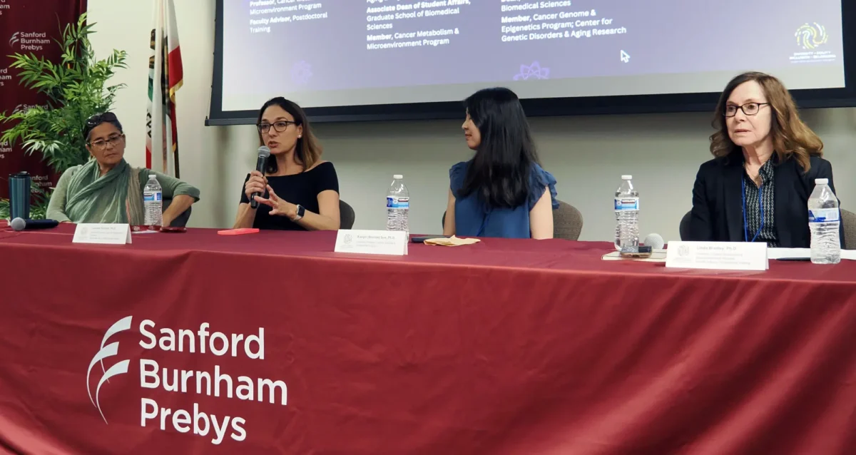
Women in Science event at Sanford Burnham Prebys examines how female faculty members navigate research careers
Aug 19, 2024Topics at the event included work/life balance, caregiving and family obligations, and gender disparities in academic rank at research and…
- Mar 13, 2024
Xueqin Sun seeks to illuminate the underlying causes of cancer
Mar 13, 2024New Sanford Burnham Prebys scientist investigates the mutational powers of cancer cells — and their vulnerabilities
- Dec 19, 2023
Sanford Burnham Prebys continues unprecedented recruitment of early-career scientists
Dec 19, 2023Continuing its rapid and dramatic recruitment of emerging, top-tier researchers, Sanford Burnham Prebys has hired two more highly regarded early-career…
Select Publications
Showing 3 of 3BRD8 maintains glioblastoma by epigenetic reprogramming of the p53 network.
Sun X, Klingbeil O, Lu B, Wu C, Ballon C, Ouyang M, Wu XS, Jin Y, Hwangbo Y, Huang YH, Somerville TDD, Chang K, Park J, Chung T, Lyons SK, Shi J, Vogel H, Schulder M, Vakoc CR, Mills AA
Nature 2023 Jan ;613(7942):195-202ANP32A regulates histone H3 acetylation and promotes leukemogenesis.
Yang X, Lu B, Sun X, Han C, Fu C, Xu K, Wang M, Li D, Chen Z, Opal P, Wen Q, Crispino JD, Wang QF, Huang Z
Leukemia 2018 Jul ;32(7):1587-1597Induction of INKIT by Viral Infection Negatively Regulates Antiviral Responses through Inhibiting Phosphorylation of p65 and IRF3.
Lu B, Ren Y, Sun X, Han C, Wang H, Chen Y, Peng Q, Cheng Y, Cheng X, Zhu Q, Li W, Li HL, Du HN, Zhong B, Huang Z
Cell Host Microbe 2017 Jul 12 ;22(1):86-98.e4Timothy Huang completed his PhD at the University of Calgary (Canada) under Dr. Dallan Young, studying kinase pathways involved in mediating cell polarity in yeast. He studied mechanisms underlying actin cytoskeletal dysfunction in Alzheimer’s disease at Scripps with Dr. Gary Bokoch (La Jolla), before joining Dr. Huaxi Xu’s laboratory in 2012/2013.
Related Disease
Alzheimer’s Disease, Molecular Biology
Phenomena or Processes
Cell Biology, Cell Signaling, Neurobiology, Neurodegeneration, Neurogenesis, Neuron-Glia Interactions in Myelin, Proteolytic Pathways, Tyrosine Kinases
Anatomical Systems and Sites
Brain
Research Models
Human, Human Cell Lines, Mouse, Mouse Cell Lines
Techniques and Technologies
Biochemistry, Cellular and Molecular Imaging, Confocal Microscopy, Electrophysiology, Mass Spectrometry, Protein Engineering, Protein-Protein Interactions, Protein-Small Molecule Interactions, Proteomics
My research is focused on identifying and characterizing mechanisms of neurodegeneration in Alzheimer’s disease (AD) and other related neurodegenerative disorders, and identifying neuroprotective pathways that may be involved in slowing disease progression. Currently, my research is focused on the genetic AD risk factors SORLA (SORL1, LR11) and TREM2, which may be involved in attenuating pathogenic effects associated with cognitive decline. By implementing methods to enhance these neuroprotective pathways, we may be able to reverse neuronal and cognitive damage in AD, and possibly other associated disorders.
Timothy Huang’s Research Report
There are two projects that comprise my main focus: 1) SORLA: Sortilin-related receptor with LDLR class A repeats (SORLA, SORL1, or LR11) is a genetic risk factor associated with Alzheimer’s disease (AD). Although SORLA is known to regulate trafficking of the amyloid β (Aβ) precursor protein to decrease levels of proteotoxic Aβ oligomers, whether SORLA can counteract synaptic dysfunction induced by Aβ oligomers remains unclear. Our work indicates that SORLA interacts with the EphA4 receptor tyrosine kinase and attenuates ephrinA1 ligand–induced EphA4 clustering and activation to limit downstream effects of EphA4 signaling in neurons. Consistent with these findings, SORLA transgenic mice, compared with WT mice, exhibit decreased EphA4 activation and redistribution to postsynaptic densities, with milder deficits in long-term potentiation and memory induced by Aβ oligomers. Importantly, we detected elevated levels of active EphA4 in human AD brains, where EphA4 activation is inversely correlated with SORLA/EphA4 association. These results demonstrate a novel role for SORLA as a physiological and pathological EphA4 modulator, which attenuates synaptotoxic EphA4 activation and cognitive impairment associated with Aβ-induced neurodegeneration in AD. 2) TREM2: Although ligands for TREM2 such as ApoE have been previously identified, a definitive mechanism for TREM2 in AD has not been established. We recently determined that TREM2 directly binds Aβ oligomers with nanomolar affinity and R47H mutations attenuate TREM2/Aβ interaction. TREM2 deletion impairs Aβ turnover in primary microglia, and abrogates Aβ clearance in vivo. Aβ also triggers changes in microglial membrane potential which is impaired with TREM2 deletion. Moreover, TREM2 deletion attenuates Aβ-induced microglial morphogenic changes associated with activation, and inhibits Aβ-mediated induction of proinflammatory cytokine expression. Together, these results indicate that TREM2 may have opposing neuroprotective roles in mediating microglial Aβ clearance and turnover, while concurrently transducing potentially neurotoxic Aβ-induced inflammatory signals. To further define a role for TREM2 in Aβ clearance and Aβ-mediated microglial activation/cytokine expression, we plan to exploit use of TREM2 R47H knock-in and TREM2 WT and R47H overexpression mouse models currently housed in our laboratory to determine whether impaired TREM2/Aβ interactions can impair microglial response in the presence of Aβ. This provides essential groundwork in future strategies to optimize neuroprotective TREM2 Aβ clearance while limiting Aβ-induced microglial inflammation.
- Mar 3, 2025
Investigating individual immune responses to COVID-19 vaccination and infection
Mar 3, 2025Study analyzes changes in blood plasma proteins following vaccination and may contribute to improving vaccine development.
- Dec 20, 2023
Tim Huang awarded $4.3 million NIH grant to study the protective role of SORLA against Alzheimer’s disease and related tauopathies
Dec 20, 2023Research may further implicate SORLA as a novel therapeutic drug target for certain types of dementia. Tim Huang has been…
- Sep 5, 2023
Timothy Huang awarded $2.6M to solve Alzheimer’s disease puzzle TREM2
Sep 5, 2023With the help of a new grant from the National Institutes of Health for more than $2.6 million, Assistant Professor…
- Aug 2, 2022
Timothy Huang awarded $2.8M to study well-known gene linked to Alzheimer’s disease
Aug 2, 2022The project aims to uncover a potential cause of Alzheimer’s brain pathology Timothy Huang, PhD, has been awarded $2.8 million…
- Sep 25, 2020
New insights into Alzheimer’s disease
Sep 25, 2020Sanford Burnham Prebys scientist publishes two papers that bring us one step closer to understanding—and potentially treating—the devastating condition. For
Select Publications
Showing 2 of 2SORLA attenuates EphA4 signaling and amyloid β-induced neurodegeneration.
Huang TY, Zhao Y, Jiang LL, Li X, Liu Y, Sun Y, Piña-Crespo JC, Zhu B, Masliah E, Willnow TE, Pasquale EB, Xu H
J Exp Med 2017 Dec 4 ;214(12):3669-3685SNX27 and SORLA Interact to Reduce Amyloidogenic Subcellular Distribution and Processing of Amyloid Precursor Protein.
Huang TY, Zhao Y, Li X, Wang X, Tseng IC, Thompson R, Tu S, Willnow TE, Zhang YW, Xu H
J Neurosci 2016 Jul 27 ;36(30):7996-8011