Histology
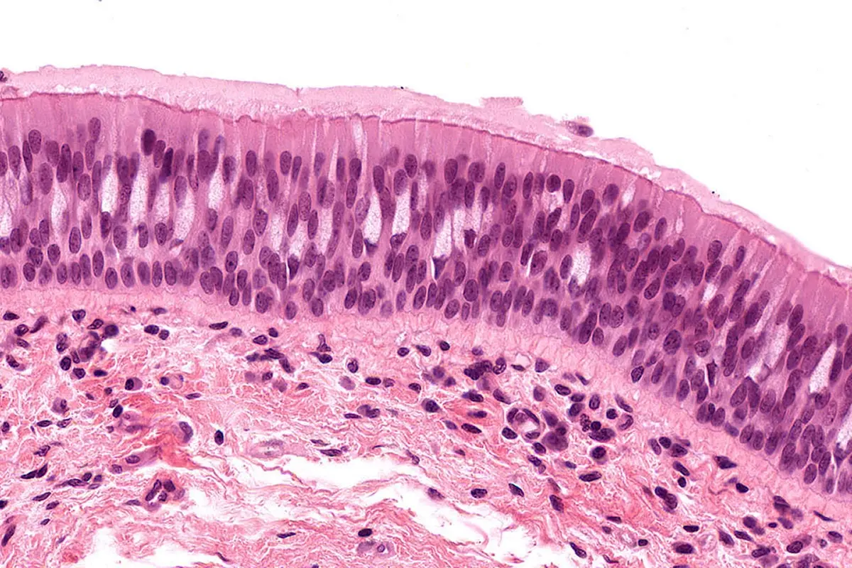
Overview
The Histology Core Facility provides standard and customized research-specific services, including tissue processing, fixed sectioning, and frozen cryosectioning. We offer routine and special histological stains, as well as advanced molecular labeling via immunohistochemistry (IHC), immunofluorescence (IF), and RNA-ISH. Finished slides are returned directly to researchers or digitized using high-throughput brightfield and fluorescent Aperio ScanScopes, which offer remote web access and powerful quantitative image analysis tools.
Additionally, we provide expert training and open access to common-use equipment (paraffin microtomes, cryostats, cytospins). Our staff offers specialized hands-on training for basic histological techniques, including user training for IHC assay optimization.
Services
Conventional Histology
- Aperio high-resolution slide scanning and optional image analysis for bright field and fluorescence samples
- Routine and custom sectioning
- Paraffin and frozen samples
- Standard and special stain (H&E, PAS, Oil Red O , trichome, PSR and more.
Immunohistochemistry
- Double labeling of antibodies and RNA probes
- Immunofluorescence
- Development of custom protocols (overlay assays, competition assays)
- Titration and staining of commercial and custom antibodies
Equipment & Resources
- Leica BOND-RX: Fully automated IHC, IF, and ISH staining.
- Leica Autostainer XL: Automated H&E and special stains.
- Tissue-Tek VIP 5: Automated paraffin tissue processing.
- Aperio AT2 ScanScope: High-throughput brightfield slide scanning.
- Aperio FL ScanScope: Fluorescent slide scanning with remote access.
- CellCut Laser Capture Microscope: Laser-guided single-cell isolation.
- Leica CM 3050S Cryostat: High-quality frozen tissue sectioning down to -40°C.
- Leica Paraffin Microtomes: High-precision sectioning of wax blocks.
- Tissue-Tek Embedding Station: Paraffin embedding and block preparation.
- Leica HistoCore PERMA: Automated high-resolution slide labeling.
- Shandon Cytospin 3: Centrifugal preparation of liquid cytology samples.
- ImageScope Software: Digital slide viewing and quantitative analysis.
Price List
For a complete list of services, please call (858) 646-3100 ext. 3552 or email us.
| Histology Services | Internal Subsidized | Internal | External Non-Profit | External For-Profit | |
|---|---|---|---|---|---|
| Small Combo/Embed/H&E/5unstained | each | $30 | $37.50 | $40.50 | $78.90 |
| Large Combo/Embed/H&E/10unstained | each | $55 | $68.75 | $74.25 | $144.65 |
| Additional Sections/Paraffin | per slide | $5.50 | $6.88 | $7.43 | $14.47 |
| Embedding Only/Paraffin | each | $6.50 | $8.13 | $8.78 | $14.47 |
| Immuno Staining (IHC or IF) | per slide | $35 | $43.75 | $47.25 | $92.05 |
| Aperio 20X scan | per slide | $12 | $15 | $16.20 | $31.56 |
| Aperio 40X scan | per slide | $23 | $28.75 | $31.05 | $60.49 |
| Aperio Fluorescence Scan | per slide | $30 | $37.50 | $40.50 | $78.90 |
| Common Cryostat | per hour | $22.20 | $27.60 | $30 | $58.20 |
Leadership
Maximiliano D’Angelo, PhD
Scientific Director
Guillermina Garcia
Core Manager
ggarcia@sbpdiscovery.org
Contact
Guillermina (Gia) Garcia is an American Society for Clinical Pathology (ASCP) Certified Histotechnician with 20 years of laboratory experience. She manages the day-to-day handling and processing of research samples at the facility. Gia is highly skilled in a wide range of specialized histological staining methods and immunohistochemistry (IHC) protocols. She serves as a primary contact for investigators and is available to answer any questions regarding your active or upcoming research projects.
Please call (858) 646-3100 ext. 3556 or use the button below to send us an email.