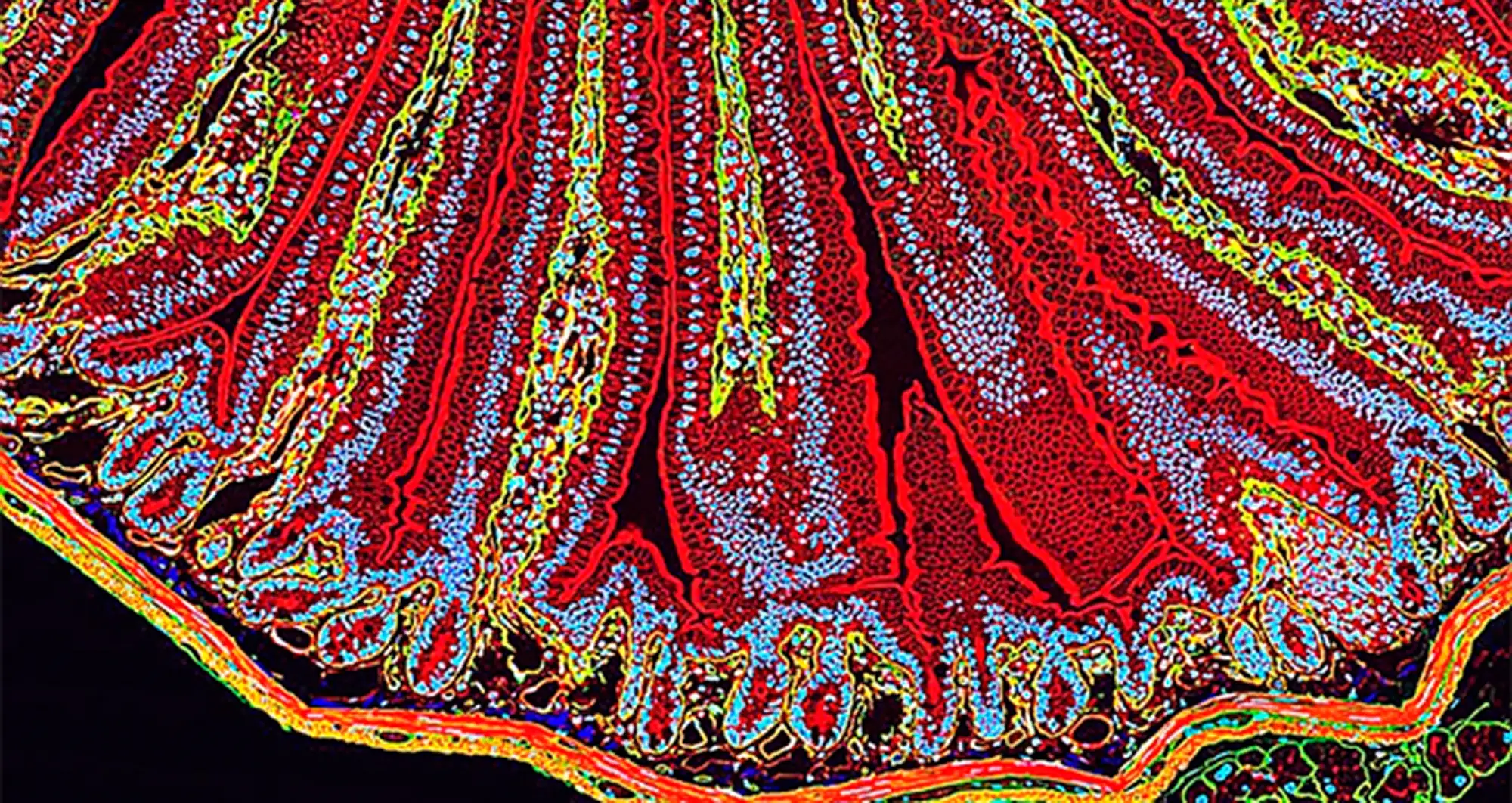
A quantum dot fluorescence image depicts a section of mouse small intestine. Different fluorescent markers indicate different cellular constituents. Actin, a protein that forms the contractile filaments of muscle cells, is shown in red. Lamin (green) are fibrous proteins that provide structural function. Cell nuclei are depicted in blue.
Image courtesy of Thomas J. Deerinck, UC San Diego National Center for Microscopy and Imaging Research.