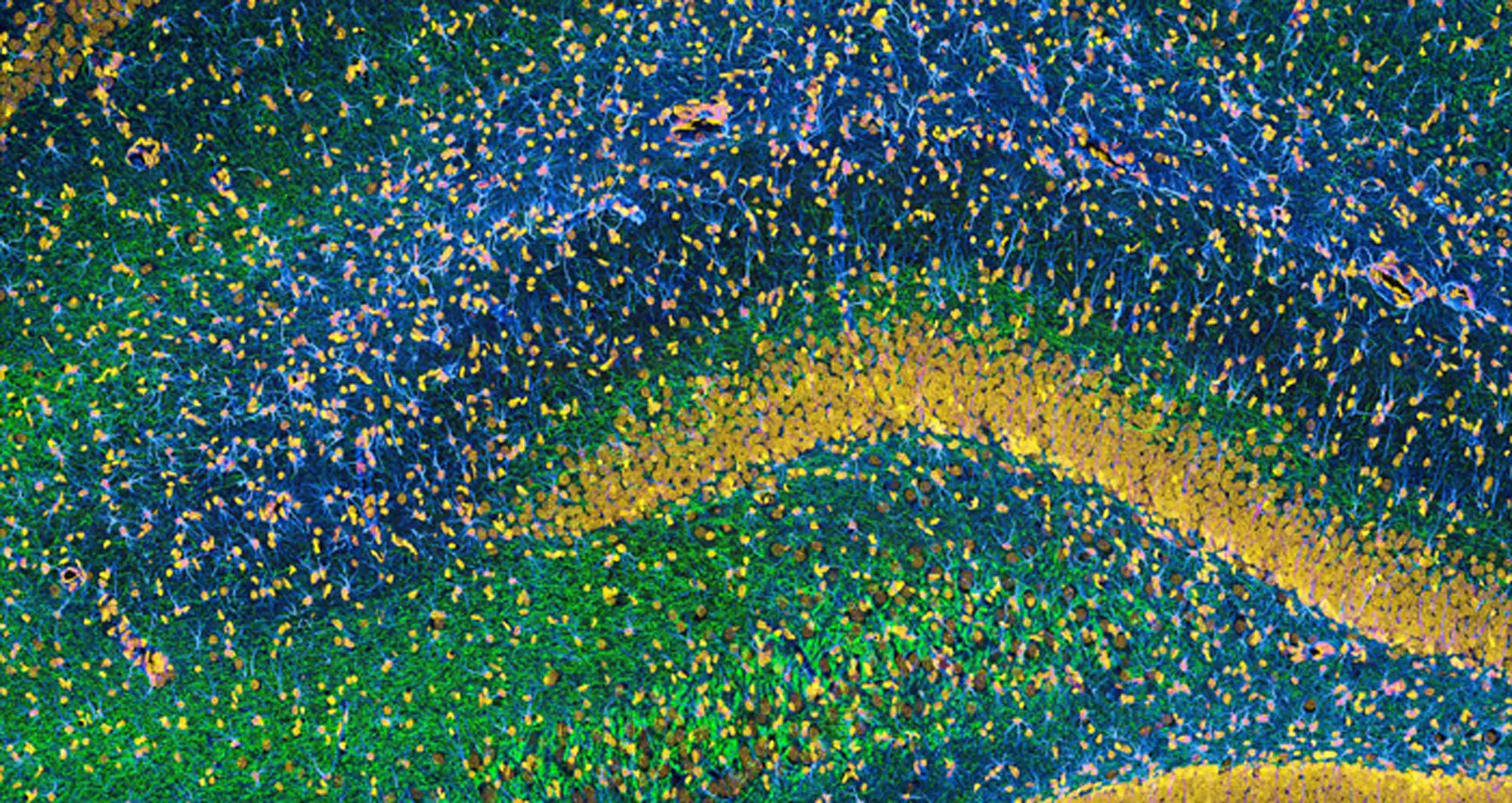
This image of the hippocampus in a rat brain was taken using an ultra-widefield high-speed multiphoton laser microscope. Tissue was stained to reveal the organization of glial cells (cyan), neurofilaments (green) and DNA (yellow).
Image courtesy of Thomas Deerinck, NCMIR and NIH.
