
Structural aging atlas highlights hormonal signaling as a potential conductor
We tend to think of age as a linear progression marked by birthdays and milestones dictating when we can vote, drink, rent a car or have made it beyond the crest of an arbitrary metaphorical hill. When scientists look at how organs age, however, it doesn’t match this idea that we gradually grow older with each tick of the clock.
A new study of aging in our organs instead found that each organ ages on its own schedule. Arteries remodel fastest from age 30-39 while reproductive organs do so at menopause.
Most of our body ages in two waves, and those who experience aging more rapidly in one organ also have accelerated aging in partner organs. The researchers built a map from analyzing 25,000 organ images that points to hormonal signaling as a possible conductor of this symphony of aging throughout the body.
Scientists at Sanford Burnham Prebys Medical Discovery Institute with collaborators at the National Institutes of Health published findings August 31, 2026, in Nature Aging from using a computational model to study rates of aging in 40 types of human organs. They found three major aging patterns and mapped underlying molecular changes that may guide the development of organ-specific treatments to promote healthier aging.
“Our goal is to understand how aging occurs at a structural level,” said Sanju Sinha, PhD, assistant professor in the Center for Data Science and Artificial Intelligence at Sanford Burnham Prebys. “To do this, we needed thousands of normal organ tissues over a wide range of ages.”
The research team found the requisite data in archives hosted by the Genotype-Tissue Expression Project, a major National Institutes of Health Common Fund initiative to accelerate the study of tissue- and cell-specific gene expression and regulation.
“This is a public dataset used by hundreds of groups, but nearly everyone uses the molecular data,” said Sinha. “There are tens of terabytes of imaging data that have gone nearly untouched.”
In this digital warehouse, the investigators found more than 25,000 scanned images of biopsies of 40 types of normal tissue from nearly 1,000 deceased donors. To analyze this visual data, they developed a deep-learning computer vision model called Pathology-based Structural Aging Rate (PathStAR). The model works by segmenting the scanned histopathology slides into more than 30 million smaller images known as patches. PathStAR extracts features from these patches that reflect underlying tissue structure and then uses these features to chart how structural aging occurs over time.
“We hypothesize that clinical imaging provides enough visual information to understand and track how tissues are aging,” said Sinha. “Once we had the model, we started with a proof of concept by applying it to the ovaries.”
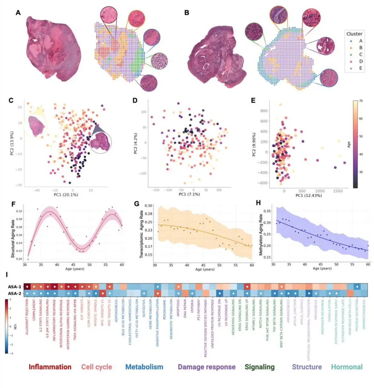
The dataset included 250 scanned histopathology slides from ovarian tissue biopsies. These biopsies came from donors between the ages of 21 and 70. After analyzing the tissue structure in these images, PathStAR revealed that ovary has two accelerated aging periods: ages 35-40, coinciding with fertility decline, and again from ages 55-60, corresponding to postmenopause.
“This showed that the structural information embedded in the clinical images could be measured and deliver an answer that lines up with what we know about the functional aging of a tissue,” said Sinha.
The new study studies how tissue structure ages using a deep-learning computer vision model called PathStAR. The model segments scanned histopathology slides into many smaller images known as patches. This example shows patches from an ovarian tissue biopsy donated by subjects aged 60-69 (top left) and 20-29 (top right).
The patches are analyzed to extract features that reflect underlying tissue structure. PathStAR uses these features to chart how structural aging occurs over time. Image credit: Anamika Yadav, Sanju Sinha, Sanford Burnham Prebys.
“And we learned that the structural changes in tissue with aging are so apparent that the model does not even need any training on chronological age to track them.”
Next, the scientists turned PathStAR’s sights on the remaining organ images after narrowing in on the 15 tissue types that had at least 200 samples representing a wide array of ages across the lifespan. They found that tissues could be subdivided into three groups.
The vascular system was among the tissues considered to experience early structural aging. These tissues experienced expedited aging from ages 30-39 and then the rate of aging declined. Late-aging tissues included the uterus and vagina which were relatively stable in early adulthood before aging most rapidly from ages 50-55.
The most common pattern, however, was similar to that observed during the ovarian tissue pilot test. Nine tissue types featured two distinct periods of aging, which the research team dubbed biphasic structural aging.
“Once we understood these patterns, we could use the gene expression and regulation data from the same Genotype-Tissue Expression Project cohort to see what major molecular changes occur during accelerated aging,” said Sinha.
The research team found that accelerated structural aging periods contained a common molecular signature marked by an increased expression of genes related to inflammation while genes governing energy production, cell growth and cellular quality control were simultaneously suppressed.
“Our findings can be the foundation for building a structural aging atlas to inform the design and assessment of anti-aging interventions,” said Sinha. “Further, more than half the tissues we studied followed the structural aging of the ovaries, so we see the ovaries as a kind of pacemaker for whole-body aging.
“By developing therapies to protect reproductive aging, we see the potential to protect multiple other organs and increase the overall healthspan.”
Anamika Yadav, a research assistant at Sanford Burnham Prebys, led this study.
Additional authors include:
- Kyle Alvarez, Kevin Y. Yip and Caroline Kumsta at Sanford Burnham Prebys
- Eytan Ruppin at the National Cancer Institute
- Jacqueline C. Yano Maher and Veronica Lobo-Gomez at the Eunice Kennedy Shriver National Institute of Child Health and Human Development
The study was supported by the National Cancer Institute-designated Cancer Center at Sanford Burnham Prebys.
The study’s DOI is 10.1038/s43587-026-01200-4.