Cory Dobson, Author at Sanford Burnham Prebys - Page 10 of 41
Institute News

SBP explains: fluorescence microscopy

AuthorMonica May
Date

August 9, 2018

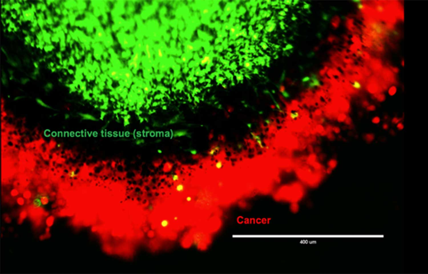
The image above is not only stunning, it’s also helping scientists uncover the secrets to cancer and additional diseases. 

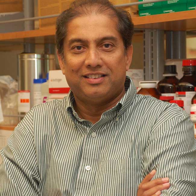
Petrus R. de Jong, MD, PhD
Petrus R. De Jong, MD, PhD 

The technique that created this image—fluorescent microscopy—is so essential to biomedical research we asked Petrus R. de Jong, MD, PhD, research assistant professor at Sanford Burnham Prebys Medical Discovery Institute (SBP), to tell us a bit more about it. [Note: The image above shows a cancer cell, visualized as green, surrounded by structure-supporting stroma cells, visualized as red. DNA is shown in blue.] 

What is fluorescent microscopy? 
First, we must understand that light is a wave that has energy. If you’ve ever been hit by a wave at the beach, you’ve felt wave energy. 

In addition to having energy, light has different speeds. Blue light has more energy than green light, and green light has more energy than red light. [See spectrum visual.] 

We know all energy goes downhill—an overarching scientific principle. So if we shine high-energy blue light on a fluorescent molecule, lower-energy green light will appear.

Fluorescent microscopy harnesses this principle. The microscope beams light on the dyed sample, causing the fluorescent molecules to shine. Scientists then study the images. 

Blue light has more energy than green light.
     Blue light has more energy than green light.

Why would scientists use fluorescent microscopy for their work? 
Using this technique, we can visualize life at all levels—from the single molecule dangling from a cell to our entire stomach. We can see if a drug is helping or hurting our body. We can watch how cancer cells interact with their environment. We can even track cancer cells that spread to other organs, a deadly process called metastasis. All this information helps scientists learn about who we are and how disease affects the body. 

[Read more on how de Jong is using fluorescent microscopy in his research.] 

Is fluorescent microscopy hard? How long would it take a scientist to make this kind of image? 
The technique has gotten easier to use over time, but it still takes a while to optimize. Scientists have to be careful about the natural fluorescence of cells, called background fluorescence, which can affect results. The dyes can also interact with each other. 

It usually takes us a couple of tries over a few weeks to optimize our process. After that, it’s about a day or so to obtain the images—not counting the time to prep our samples. Analysis of the images takes another few days. 

Could you tell us more about the history of this technique? When was fluorescent microscopy invented? 
The first documented observation of fluorescence involved a tree used by the Aztecs for medicinal purposes. When soaked, the bark made water glow bright blue. The tree is called “coatli” by Aztecs and is also known as “Mexican kidneywood.”

Many discoveries later, the first fluorescent microscopes were developed by German scientists Otto Heimstaedt and Heinrich Lehmann between 1911 and 1913. But there were two additional advances that really allowed the technology to take off. 

In 1941, the fluorescent dye was attached to a targeted antibody. Instead of just measuring natural fluorescence levels of a sample, the dye could be directed to specific parts of a cell or organism. However, the dyes were still difficult to create and manipulate, limiting the technique’s use. It was a bit like assembling furniture—it can be done, but it’s not easy. 

That changed twenty years later when a team of San Diego scientists discovered green fluorescent protein in jellyfish in 1962. This protein packages itself—like self-assembling furniture—which really enabled the technique to take off. Today, the technique is essential to biomedical research; nearly every scientist uses it to inform his or her research. 

To learn more about fluorescence, check out the resources below.

Protein Data Bank Molecule of the Month: Green Fluorescent Protein (GFP) 

The Fluorescence Foundation: A Short History of Fluorescence

Interested in keeping up with SBP’s latest discoveries, upcoming events and more? Subscribe to our monthly newsletter, Discoveries.

Institute News

Cancer cells lure in nutrient-rich neighbors, then mug them

AuthorMonica May
Date

August 9, 2018

If your grocery store was out of food, would you still invite your friends to a dinner party? 

Unless you happen to have a flourishing garden, probably not. 

Our cells would have the same answer. Food is hard to come by, so cells in our body rarely share nutrients. The only product a cell releases is waste. 

But cancer cells don’t follow these rules. Despite living in a low-nutrient environment, cancer cells draw neighboring stroma cells—the glue-like cells holding our body together—toward them. In this setting you’d expect cells to keep to themselves, not bring more people to the party.

Petrus R. de Jong, MD, PhD

This odd behavior fascinates Petrus R. de Jong, MD, PhD, research assistant professor at Sanford Burnham Prebys Medical Discovery Institute (SBP), who is studying cancer cells to try to better understand this strange act.

“Scientists are learning more and more about the importance of the tumor’s surrounding environment, called the tumor microenvironment,” says de Jong. “We are finding there are many interactions between stroma and cancer cells. If we could block this cross talk, we might be able to find a new way to treat cancer.”

Many people could benefit from this research, though it is still in its earliest stages. Breast, prostate, ovarian and colorectal cancers are all known to interact with stroma cells. De Jong’s lab is studying pancreatic cancer, which remains one of the deadliest cancers. Less than 10 percent of patients live more than five years after diagnosis. If surgery is not possible, chemotherapy and radiation are the only remaining treatment options. 

Using pancreatic cancer cells, including cells isolated from patients after surgery, de Jong and his team designed an experiment to better understand why the cancer cells draw stroma cells near. They placed the cancer and stroma cells in a special container that separated the two but still allowed them to interact. After two days, they removed the cells and used special dyes to visualize different parts of the cells. DNA shows up bright blue. Cancer cells, a vibrant red. And a nutrient cells crave—fats, called lipids—shows up an intense green. [To learn more about how fluorescence microscopy works, read our primer.] 

Peering under the microscope, de Jong and his team saw green lipids emerge from the stroma and taken up by the red cancer cells. 
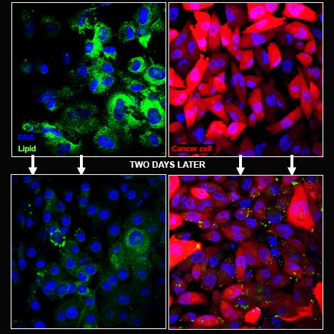
Flourescent cancer cells
    Green lipids emerged from the stroma
    and were taken up by the red cancer     
    cells. 

“Lipids are a valuable source of energy, so it is unusual for the stroma cells to release this nutrient to the cancer cells,” says de Jong. “It appears the cancer cell is sending out a signal that tells the stroma cells to give them this food.”

To try to determine how the cancer cells were taking up these lipids, de Jong used chemicals to halt autophagy—a process that allows cells to destroy and recycle their own guts. Cancer cells are known to use autophagy to break down elements that aren’t useful any longer and reuse the material as building blocks for growth. 

“When the autophagy process was halted, the exchange of lipids stopped,” explains de Jong. “This finding indicates the pancreatic cancer cells forced the stroma cells to start eating themselves, then took up the resulting nutrients.”

De Jong’s team is now focused on the next mystery: how the pancreatic cancer cells are taking up these nutrients. If scientists can identify this process, they might be able to find a medicine that halts cancer cells while leaving healthy cells unharmed, the goal for any cancer treatment. 

In other words, they could stop cancer cells from mugging their unsuspecting neighbors. 

Read the AACR 2017 abstract detailing this research. For the full poster, contact de Jong at pdejong@sbpdiscovery.org. 

Interested in keeping up with SBP’s latest discoveries, upcoming events and more? Subscribe to our monthly newsletter, Discoveries.
Institute News

Taking out a microRNA to thwart melanoma

AuthorSusan Gammon
Date

August 6, 2018

Melanoma is a deadly disease with limited treatment options. However, even when those therapies are initially successful, the cancer often comes back. Researchers continue to hunt for new approaches to make the disease more vulnerable.

Ranjan Perera, PhD, an adjunct professor at SBP’s Lake Nona campus, has been studying melanoma for many years, looking for mechanisms that can help control the disease. These efforts helped his lab discover miR-211, a molecule found in melanocytes, the cells that sometimes go awry and become cancerous. Not surprisingly, miR-211 is sometimes overexpressed in melanoma. 

Ranjan Perara, PhD
     Ranjan Perera, PhD 

Because miR-211 is a microRNA—a small molecule that interferes with the cellular machinery that produces proteins—it can have a big impact on gene expression, and the gene it impacts is pretty interesting.

“MicroRNA-211 is targeted to a gene called PDK4, which is important for mitochondrial energy metabolism,” says Perera. 

Scientists have known for more than 100 years that tumors restructure their metabolisms to compensate for their out-of-control growth. If miR-211 is part of that process, taking it out could make cancer cells more treatable.

To better understand what miR-211 is doing, researchers in Perera’s lab used CRISPR/Cas9 gene editing tools to eliminate it from cancer cell lines. They found that removing the molecule impacted mitochondria, the cells’ energy plants, and made them metabolically vulnerable. In addition, miR-211 loss dampened pathways that drive melanoma growth—so there was a double benefit. In animal models, cells without miR-211 had trouble forming tumors. These results were recently published in the Journal of Investigative Dermatology.

Perera and colleagues were also curious whether removing the microRNA might affect how cancer cells respond to the drug Vemurafenib—a therapy used for the treatment of late-stage melanoma. While the drug is effective in certain patients, tumors often develop resistance after several months. Further study showed that eliminating miR-211 made the melanoma cells much more sensitive to Vemurafenib.

These findings add to the body of evidence that helped Perera and SBP get a patent covering approaches using miR-211 to detect and treat melanoma. Perera’s team will continue to study this molecule, as well as the genes it impacts, to gain more insights and potentially transform these findings into new melanoma diagnostics and treatments.

Though it’s still early, these findings make miR-211 an interesting potential drug target, and Perera believes further investigation is definitely warranted.

“Given that miR-211 loss has a dual anti-cancer effect, by inhibiting both critical growth-promoting cell signaling pathways and rendering cells metabolically vulnerable, it is an extremely attractive candidate for combinatorial therapeutics,” says Perera. “This is especially true if, like here, miR-211 is upregulated in Vemurafenib-resistant melanomas in the clinic, since it provides both a highly specific target.”

Interested in keeping up with SBP’s latest discoveries, upcoming events and more? Subscribe to our monthly newsletter, Discoveries.
Institute News

Protecting motor neurons

AuthorSusan Gammon
Date

July 30, 2018

Neurodegenerative diseases are a major worldwide health problem, and researchers are hunting new strategies to combat them. One approach is to find the mutations that cause these diseases and design therapies to counteract them. But scientists can also pursue another strategy—identify molecules that protect neurons from harm.

SBP’s Laszlo Nagy, MD, PhD, may have found one of these. Nagy is working on gene regulation and epigenomics—so neurons are not his primary beat. But in a recent study published in The Journal of Neuroscience, he and colleagues have shown that the enzyme PRMT8 can protect aging motor neurons from stress, which could have therapeutic implications.

“We stumbled on this molecule PRMT8, which is a protein methyltransferase (an enzyme that adds methyl molecules to control protein expression and activity) while studying stem cell differentiation,” says Nagy. “When the molecule is not there, the motor neurons are less stress resistant. They are vulnerable to changes in their inner circuitry, including DNA damage, and they eventually die.”

Stress management is particularly important in motor neurons, which connect the central nervous system to muscles. Mature motor neurons cannot divide anymore and must live a long time. Once they die, they are lost forever.

In the study, the lab found that motor neurons in animals without PRMT8 developed a number of issues. The cells had DNA breaks and other problems that slowly killed them off. As motor neurons died, the animals gradually lost muscle strength.

“In older animals, they were having difficulty coordinating their movements,” says Nagy.

Because PRMT8 has the potential to protect motor neurons, it could be a compelling drug target. Boosting the enzyme could help slow a number of neurodegenerative conditions, including  ALS. In addition, because the enzyme is only found in the central nervous system, it offers a selective target. This is particularly important, since adding stress protection to dividing cells could cause cancer.

Now that they have identified PRMT8’s neuroprotective benefits, Nagy and colleagues are taking the next step—looking for agents that could make the enzyme more active and potentially help motor neurons resist disease and live longer.

“We’re trying to identify a small molecule that would enhance PRMT8, so we could specifically modulate its activity,” he says.

Institute News

Students conduct real-world research at SBP

AuthorMonica May
Date

July 23, 2018

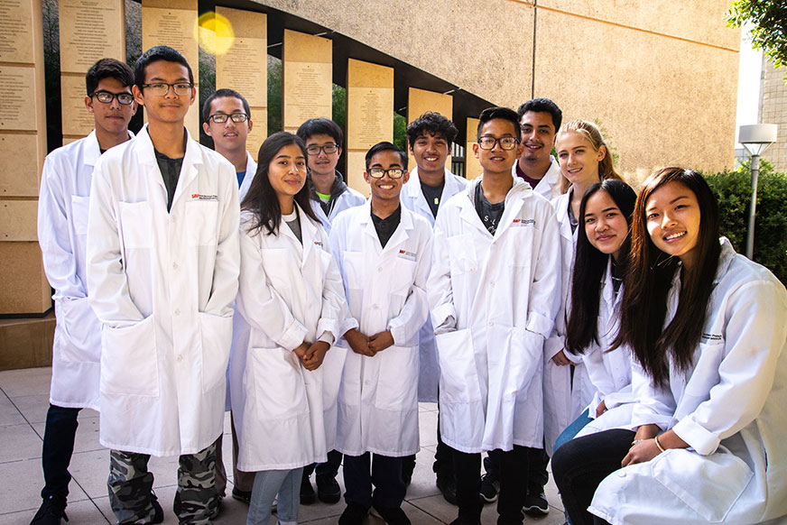
Daydreaming about prom, finally getting that driver’s license and stressing out about the SAT are common rites of passage for teens. Now, twelve high school students from The Preuss School UC San Diego—a distinguished charter school for students who would be the first of their families to graduate from college—can add undertaking real-world biomedical research to their list of high school memories.

These soon-to-be juniors spent two weeks in July working directly with SBP scientists as part of the SBP Preuss program. Thanks to the generous contributions of Peggy and Peter Preuss and Debby and Wain Fishburn, for more than a decade this summer program has introduced high school students to laboratory research with the hope of inspiring future scientists.

Rotating between four labs during a period of two weeks, these students used laboratory techniques such as micropipetting, microscopy and gel electrophoresis to help reveal the underlying causes of diabetes, aging and heart disease.

At the end of the program, the students presented their findings and experiences to SBP scientists and staff, including SBP’s president, Kristiina Vuori, MD, PhD; the Preuss School’s director of development, Tamika Franklin; program donors; and the students’ family members. For students interested in taking an even deeper dive into the science, they have the opportunity to return for a separate six-week, full-time program at SBP.

We spoke with three students to hear more about their experiences during the two-week program, if a science career is in their future and any surprises they encountered along the way.

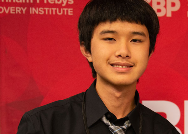
Kevin Vo, 16

Kevin Vo
   Kevin Vo, 16

Tell me a bit more about what you have experienced at SBP.
“What I remember most is taking DNA from three mice and figuring out which mouse had been genetically changed. We used enzymes to amplify the DNA, which is a technique called PCR (polymerase chain reaction). Then we waited, and a day later we used a paper [gel electrophoresis] to determine which mouse was wild type (meaning normal, or found in the wild), which had one gene missing (heterozygous) and which of our samples was just water (a negative control).”

Were you surprised by any of your experiences at SBP?
“I learned that there is a lot of waiting in science! But it’s all for a good reason.”

Do you think you want to become a scientist?
“I’ve always been interested in science, but I’m still conflicted about whether I want to be an engineer or a scientist. That’s why I enrolled in this program, so I could “try before I buy.” I learned I really liked how hands-on science is. We’ll see!”

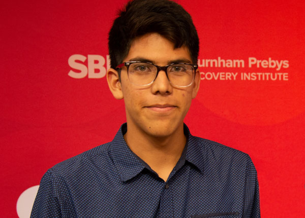
Leo Gonzalez Cazares, 16 

Leo Gonzalez Cazares, preuss
   Leo Gonzalez Cazares, 16

Tell me a bit more about what you have experienced at SBP.
“We worked with zebrafish to visualize different parts of the animal. We added a colored dye and used a microscope to see different structures, like DNA and red blood cells. I hadn’t ever used a microscope before, so that was a really interesting experience.”

Were you surprised by any of your experiences at SBP?
“People usually think science is someone working by themselves at their desk. But there is so much communication in science. You have to talk to people about your project, why it works and what you are doing. I really liked that—there is more interaction than I had thought.”

Do you think you want to become a scientist?
“I definitely want to be a scientist, but I’m not sure what kind yet, I also like chemistry and biochemistry. I really liked being part of this program because I could learn what being a scientist is like and see the actual environment. It’s so great that I get to experience this as an incoming junior and learn now if this is what I want to do in the future.”

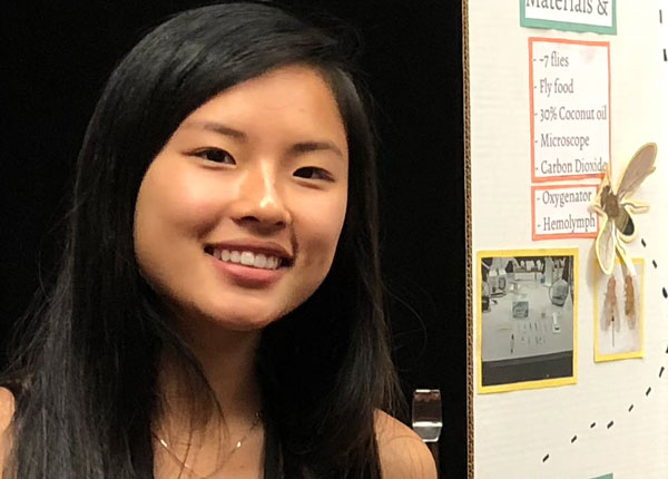
Ngoc Vo, 16

Ngoc Vo
   Ngoc Vo, 16

Tell me a bit more about what you have experienced at SBP.
“We visited a lab studying fruit flies to see how they can help people with diabetes, which affects a lot of Americans. They fed the flies a high-fat diet of coconut oil, and their heartbeats are abnormal. We used a microscope to look at the flies’ heartbeats, recorded the heart rate on video and analyzed how fast it was, which was pretty cool.”

Were you surprised by any of your experiences at SBP?
“There were so many fruit flies! There were hundreds of vials of flies. That was pretty incredible to see.”

Do you think you want to become a scientist?
“It was really cool to see how science is practiced. But right now I think I want to be a doctor who delivers babies.” 

Interested in keeping up with SBP’s latest discoveries, upcoming events and more? Subscribe to our monthly newsletter, Discoveries.

Institute News

Duc Dong honored at Alagille Syndrome Alliance Gala

AuthorSusan Gammon
Date

July 10, 2018

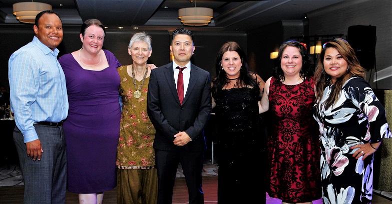
Associate Professor Duc Dong, PhD, was the guest of honor at the Gala of Dreams, the inaugural fundraiser for the Alagille Syndrome Alliance held June 30 at the San Diego Marriott Del Mar. Dong is a trailblazer in the field of Alagille syndrome research—he is working toward a cure for the extremely rare genetic condition that affects approximately one in 30,000 births.

Babies born with Alagille syndrome have too few bile ducts—which are essential for the transport of waste out of the liver. This causes toxins build up in the liver and throughout the body, leading to constant severe itching, and more critically, liver damage and failure. Alagille syndrome patients also have many other life-threatening developmental defects in other parts of their bodies, including the heart, kidneys, vertebrae, and blood vessels. There is no cure for this debilitating disease, and up to 50 percent of patients eventually need a liver transplant, often during childhood.

Dong and his team have been studying JAGGED1, the gene implicated in Alagille syndrome. Taking advantage of an unusual animal model, the zebrafish, he has been able to uncover a novel genetic mechanism for the disease—opening new potential therapeutic avenues. Further, his team has surprisingly discovered that the bile ducts lost can be regenerated after turning the affected gene back “on.”

“The implication is that these developmental defects in Alagille syndrome patients could potentially be reversible and therefore curable,” says Dong. “We will now start screening for drugs that may be used to restore the function of this genetic pathway and hopefully allow for these lost bile ducts to regenerate. We will continue to challenge the science of Alagille syndrome to move closer to a cure.”

The theme of the event, “The Dawn of a Dream,” signified new advances in Alagille syndrome research and the anniversary of the organization’s 25th year in existence. The evening gave advocates, families, doctors and pharma representatives an opportunity to interact in a fun, casual setting and participate in a silent auction to raise money for research. Dong’s lab was presented with the Champion of Alagille Syndrome Award and funds raised by the Alliance through crowd sourcing.

Institute News

Seeing is believing: cancer imaging

AuthorSusan Gammon
Date

June 28, 2018

SBP’s recent Cancer Center Open House event gave guests a unique opportunity to see cancer in a different light—through the eyes of scientists. More than 120 guests took guided tours of faculty labs, giving attendees a behind-the-scenes look into our scientists’ approach to finding new pathways to combat cancer—the second-leading cause of death in the U.S.

SBP Cancer Center Open House Guests

SBP Cancer Center Open House Guests

Nicholas Cosford, PhD, deputy director of SBP’s NCI-designated Cancer Center, welcomed visitors with an introduction, including an overview of how Dr. William Fishman and his wife, Lillian, moved from Tufts University in Boston to found the Institute with a pioneering spirit that helped make SBP into the renowned center of discovery it is today.

After light refreshments and mingling with cancer scientists, survivors and research advocates, guests signed up for tours:

Picture This: MRI Imaging
Magnetic resonance imaging (MRI) helps scientists analyze the structures and functions of proteins and their interactions with drugs. This information is essential for developing new, powerful therapies to treat cancer.
Francesca Marassi, PhD

Eye on Crystals: Crystallography
Using atomic models of proteins, scientists can visualize how molecules interact to create signals that promote cancer, and design drugs to block those interactions.
Robert Liddington, PhD

Getting Big to See Small: Cryo-Electron Microscopy
By assembling 3D maps of cells and their components, scientists can derive models to understand mutations that cause irregularities in cell functions leading to cancer.
Dorit Hanein, Ph.D.

In Focus: Optical Imaging of Cancer Cells
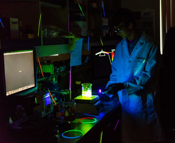
Fluorescent staining of cell proteins helps researchers visualize the cell signals and pathways that drive cancer progression.
Petrus de Jong, MD, PhD

Fluorescent science at SBP's cancer center open house

“It’s an honor to host the supporters of SBP’s Cancer Center,” said Cosford. “On evenings like this, we learn so much about what the public wants—and needs to know about cancer research. The questions we get from them are always refreshing and out-of-the-box, which is a very valuable experience for us as cancer researchers.”

Special thanks to SBP’s Cancer Advisory Board for hosting the event and their support of our postdocs and graduate students who present their research. And thank you as well to Bobbie Larraga and Heather Buthmann who helped coordinate the very special evening.

Institute News

Preeminent scientists present at SBP’s Cancer Metabolism Symposium

AuthorSusan Gammon
Date

June 27, 2018

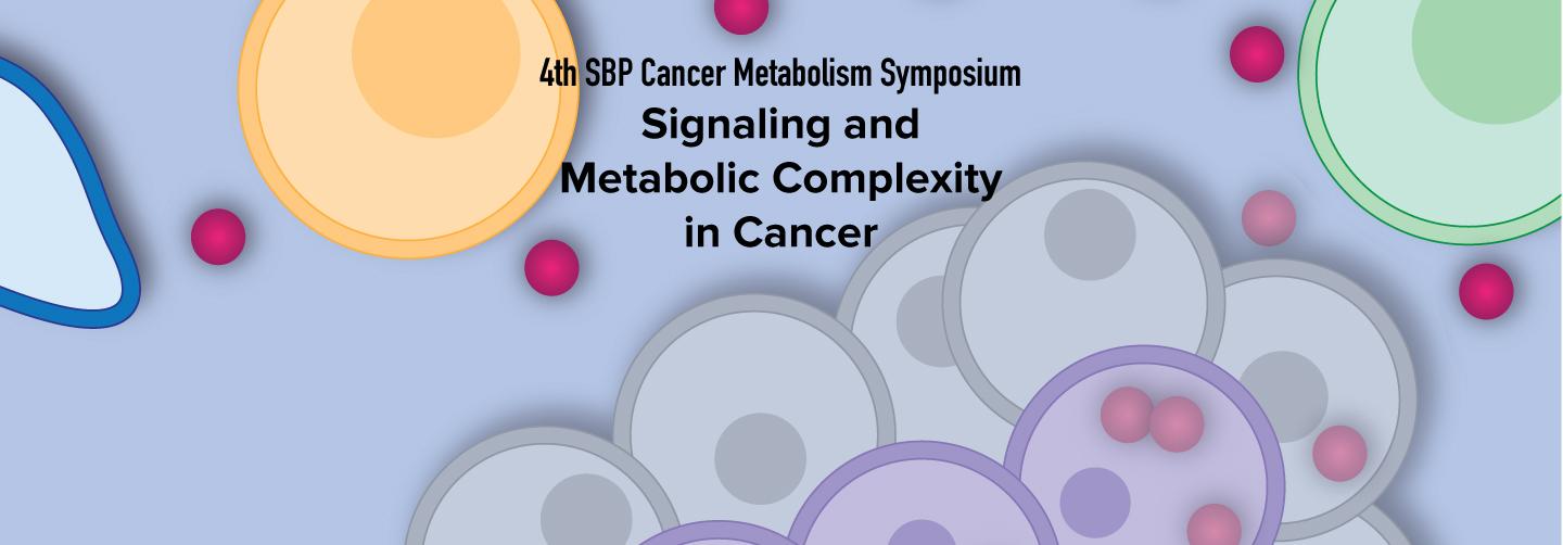
SBP’s 4th Cancer Metabolism Symposium attracted nearly 150 attendees—all eager to learn more about how the nation’s top-tier cancer scientists are looking to target tumor metabolism.

Research on cancer metabolism is critical to identify new therapeutic targets to starve tumors of the fuels and building blocks they need to grow. Recognition and understanding of the impact of cancer metabolism will increasingly and positively affect the development of novel anti-cancer therapeutics.

The event featured 21 speakers who presented the latest concepts and models in the field of tumor metabolism, which expands to other areas of cancer biology, including microenvironment, immunometabolism and cell bioenergetics. All of the presentations addressed fundamental mechanisms of cancer as well as how this emerging field of science is impacting translational research and personalized medicine. 

“It’s an honor to host the top experts in the field and discuss what we know about metabolic wiring in tumors and their environment,” said Jorge Moscat, PhD, conference co-organizer and director and professor of the Cancer Metabolism and Signaling Networks Program at SBP. “These events will lead us to strategies to exploit tumor vulnerabilities and better ways to treat cancer.”

Keynote speaker M. Celeste Simon, PhD, who studies cancer metabolism and the influence of oxygen availability on tumor growth, presented her recent data on “Metabolic Symbiosis in the Hypoxic Tumor Microenvironment.” Simon, a recipient of a National Cancer Institute Outstanding Investigator Award, is a leader in biomedical research on cancer metabolism, specifically renal cancer, which is one of the 10 most common cancers in both men and women. She is scientific director of the Abramson Family Cancer Research Institute of the Perelman School of Medicine at the University of Pennsylvania.

“These conferences are important because they give scientists an opportunity to share new ideas and promote collaborations that can enhance accelerated discovery and development of new therapeutic approaches to target cancer metabolism,” says conference co-organizer Maria Diaz-Meco, PhD, professor in the Cancer Metabolism and Signaling Networks Program at SBP.

Robert Abraham, PhD, Pfizer Worldwide Research & Development, represented the industry side of research with a presentation titled “Probing Cancer Metabolism with Cancer Drugs.”

Academic biomedical research institute speakers were represented by the Symposium’s organizers, Moscat and Diaz-Meco; and a broad range of acknowledged leaders in cancer research, including Ronald DePinho, MD (MD Anderson Cancer Center); Ramon Parsons, MD, PhD (Mount Sinai); John Blenis, PhD (Weill Cornell Medicine); Reuben Shaw, PhD (Salk Institute); Eileen White, PhD (Rutgers Cancer Institute of New Jersey); Tak Wah Mak, PhD (University of Toronto and The Campbell Family); Alec Kimmelman, MD, PhD (NYU Langone Health); Karen Vousden, PhD (The Crick Institute); Ralph J. DeBerardinis, MD, PhD (University of Texas SW Medical Center); Roberto Zoncu, PhD (UC Berkeley); Christian Metallo, PhD (UC San Diego); Douglas Green, PhD (St. Jude Children’s Research Hospital; Matthew Vander Heiden, MD, PhD (David H. Koch Institute at MIT); Dafna Bar-Sagi, PhD (NYU Langone Health); Michael Karin, PhD (UC San Diego); Jeffrey Rathmell, PhD (Vanderbilt University); and Davide Ruggero, PhD (UC San Francisco).

“We wish to thank all the speakers, attendees and support staff that helped pull this amazing conference together,” said Moscat. “We look forward to planning SBP’s next cancer metabolism conference to continue sharing breakthrough research advances that will ultimately improve the lives of patients.”

Institute News

SBP women awarded American Heart Association Fellowships

AuthorSusan Gammon
Date

June 15, 2018

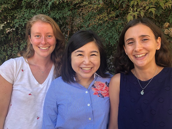
There has never been a more exciting time to embark on a career in biomedical research. Fortunately, the American Heart Association (AHA) is supporting early-career scientists with passion, commitment and focus by providing fellowships that fund their pursuit of cardiovascular research. Recently, three SBP scientists were awarded AHA grants to finance projects that align with the AHA mission of building healthier lives, free of cardiovascular disease and stroke.

Katja Birker (left)
Birker, a graduate student in the lab of Rolf Bodmer, PhD, will be studying genes that could possibly contribute to hypoplastic left heart syndrome (HLHS)—a condition that affects roughly 2–4 out of every 10,000 babies. Today, the cure for HLHS is a three-step invasive surgery that begins two weeks after the baby is born.

Birker will be collaborating with the Mayo Clinic to identify and test whether candidate HLHS genes found in patients have similar consequences in the hearts of fruit flies, which are an established model organism for cardiovascular research. She will use the flies to work toward her goal of validating novel genes that could be used in the future for diagnostic and therapeutic purposes related to cardiovascular diseases. 

EePhie Tan, PhD (middle)
Tan’s research is taking a deeper dive into previous research showing that the cell recycling process called autophagy provides health benefits—including life extension—in response to reduced food intake. This project will examine the cell networks that govern autophagy, and a specialized form of autophagy called lipophagy (fat recycling). Lipophagy is a relatively new field of biomedical research, but scientists have already learned that malfunctions in lipophagy can lead to the accumulation of toxic fat deposits and contribute to heart disease.

Tan, a postdoc in the lab of Malene Hansen, PhD, will use a small worm called C. elegans as a model system to study proteins involved in the lipophagy process. Since the core machinery of lipophagy is conserved in all organisms (from humans to C. elegans), Tan’s findings may be used to find future treatments that target toxic fat deposits in heart disease.

Clara Guida, PhD (right)
Guida will study why children from obese parents have an increased risk of developing cardiovascular disease. The research may lead to the development of biomarkers that can predict heart conditions caused by parents that eat a high-fat diet (HFD), and may lead to new drugs that can prevent the negative effects of a parental HFD on the heart function of offspring.

Guida, a postdoc in Bodmer’s lab, will study the inheritance of DNA modifications called “epigenetic marks” in fruit flies fed a HFD. These epigenetic marks are thought to cause heart problems in the next generation. She will be testing potential drugs to see if they can erase the inherited abnormal gene changes and prevent the negative effects of a parental HFD. The research is especially relevant to lipotoxic cardiomyopathy—a condition associated with fat accumulation in the heart.

Institute News

Usue Etxaniz Irigoien awarded Fishman Fund Fellowship

AuthorSusan Gammon
Date

June 13, 2018

Congratulations to SBP postdoc Usue Etxaniz Irigoien, PhD—the recipient of the 2018 Fishman Fund Fellowship. This prestigious award is a “super stipend” given to exceptional young researchers in recognition of their research accomplishments and in support of their future research plans. Etxaniz Irigoien will use the financial support to continue her research on muscle biology—explorations that may lead to treatments for disorders such as muscular dystrophy and amyotrophic lateral sclerosis (ALS).

“I’m honored to receive this special fellowship,” says Etxaniz Irigoien. “I came to SBP to pursue my interest in muscle development and regeneration, and have been so fortunate to work with world-renowned, collaborative scientists with similar interests. This award makes the whole experience even better, and secures my ability to continue making discoveries that may one day improve human health.”

Etxaniz Irigoien, a postdoc in the laboratory of Pier Lorenzo Puri, MD, PhD, professor in the Development, Aging and Regeneration Program at SBP, studies a type of muscle cell called fibro-adipogenic progenitors, or FAPs. These are the cells that act as intramuscular sensors and effectors, which means that FAPs can detect “alert” signals and generate different responses by orchestrating other cells’ activity upon different muscle perturbations. In healthy conditions, when muscle suffers an injury, FAPs cue muscle stem cells to repair the damaged muscle fibers. However, in disease (i.e., muscular dystrophies or neuromuscular disorders such as ALS), FAPs’ activity results in fibrosis, fat deposits and other detrimental events that contribute to disease progression.

“If we can begin to uncover how FAPs support muscle regeneration, or contribute to muscle degeneration in different environments, i.e., healthy versus disease tissue, we can look for potential therapeutics that will move the process toward the healthy state,” says Etxaniz Irigoien. “This is so important because today there are no effective therapeutics for dystrophies or ALS, and it’s time we make progress to help these patients.”

Etxaniz Irigoien has come a long way from her hometown of Getaria, a small fishing village located in the Basque Country of Northern Spain. She says, “I had a biology teacher who inspired my interest in science, and I have always known I wanted a career in research. My family, most of whom still live in Getaria, are very supportive and excited about my career and this award. In fact, some of them will be traveling to San Diego for the Fishman Fund ceremony in September. I’m very excited for them to visit SBP and meet some of my colleagues.”

Getaria, located in the Basque Country of Northern Spain
 
Usue as young girl in Getaria

The Fishman Fund Fellowship
The Fishman Fund Fellowship was established in honor of Dr. William and Mrs. Lillian Fishman, co-founders of SBP. Applicants must have a doctoral degree, no more than five years of training at any institution, and at least one full year of postdoctoral study at SBP. Fellowship support is for two years in length and covers salary, benefits and a professional-development allowance.

SBP is thankful for the generous Fishman Fund donors who make career development awards possible. If you would like to donate to the Fishman Fund to support young scientists click here.