Researchers from Sanford Burnham Prebys have collaborated the University of Pittsburgh Cancer Institute to reveal a new approach to enhance the effects of immunotherapy in glioblastoma, one of the most aggressive and treatment-resistant forms of brain cancer.
The study, published recently in Cancer Discovery, describes a novel method to ‘turn off’ cancer stem cells—the malignant cells that self-renew and sustain tumors—enabling the body’s own defense system to take charge and destroy tumors.
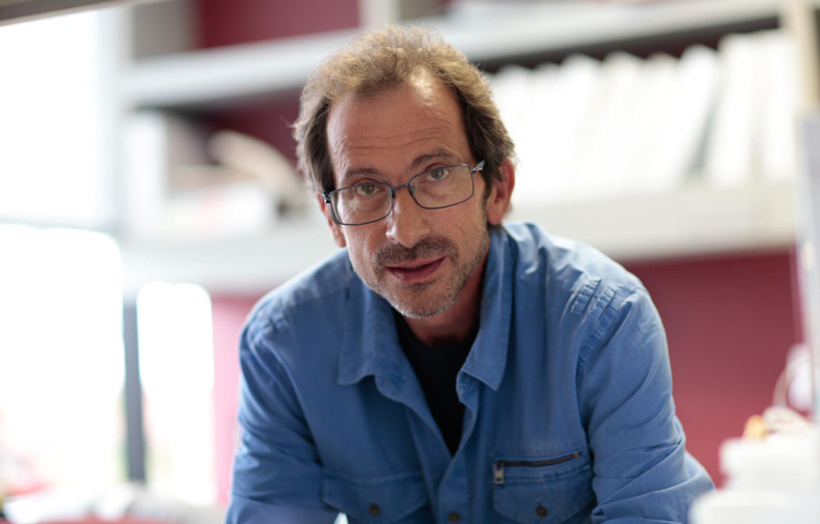
“Tumors are more than just masses of cells—each one is a complex system that relies on a vast network of chemical signals, proteins and different cell types to grow,” says senior author Charles Spruck, PhD, an assistant professor at Sanford Burnham Prebys. “This is part of why cancer is so difficult to treat, but it also presents us with opportunities to develop treatment strategies that target the machinery powering tumor cells rather than trying to destroy them outright.”
Glioblastoma is an extremely aggressive form of cancer that affects the brain and the spinal cord. Occurring more often in older adults and forming about half of all malignant brain tumors, glioblastoma causes worsening headaches, seizures and nausea. And unfortunately for the thousands of people who receive this diagnosis each year, glioblastoma is most often fatal.
“We haven’t been able to cure glioblastoma with existing treatment methods because it’s just too aggressive,” says Spruck. “Most therapies are palliative, more about reducing suffering than destroying the cancer. This is something we hope our work will change.”
Immune checkpoint inhibitors—which help prevent cancer cells from hiding from the immune system—can be effective for certain forms of cancer in the brain, but their results in glioblastoma have been disappointing. The researchers sought a way to improve the effects of these medications.
“Modern cancer treatment rarely relies on just one strategy at a time,” says Spruck. “Sometimes you have to mix and match, using treatments to complement one another.”
The researchers used genomic sequencing to investigate glioblastoma stem cells. These cells are the source of the rapid and consistent regeneration of glioblastoma tumors that make them so difficult to treat.
The team successfully identified a protein complex called YY1-CDK9 as essential to the cells’ ability to express genes and produce proteins. By modifying the activity of this protein complex in the lab, the team was able to improve the effectiveness of immune checkpoint inhibitors in these cells.
“Knocking out this transcription machinery makes it much more difficult for the cells to multiply” says Spruck. “They start to respond to chemical signals from the immune system that they would otherwise evade, giving immunotherapy a chance to take effect.”
While the approach will need to be tested in clinical settings, the researchers are optimistic that it may provide a way to improve treatment outcomes for people with glioblastoma.
“What our results tell us is that these cells are targetable by drugs we already have, so for patients, improving their treatment may just be a matter of adding another medication,” adds Spruck. “For a cancer as treatment-resistant as glioblastoma, this is a great step forward.”
Though DNA is essential for life, it can also wreak havoc on our bodies as we age
DNA is one of the essential building blocks of life, giving our cells instructions for virtually everything they do, but researchers at Sanford Burnham Prebys are investigating what happens to our cells when DNA ends up in places where it shouldn’t normally be, particularly as we age.
The answer – as described in their recent review in the journal Cell—is disease-causing inflammation. And the researchers hope that targeting this rogue DNA will lead to new therapeutic strategies for a range of age-related illnesses, including cancer, diabetes, rheumatoid arthritis, cardiovascular disease and neurodegenerative disorders.
“Age is the primary risk factor for all of these diseases, but they share another risk factor – chronic inflammation,” says first author Karl Miller, PhD, a postdoctoral researcher in the lab of Peter Adams, PhD, Sanford Burnham Prebys. “We’re trying to understand the underlying processes behind this inflammation so we can potentially treat all these age-related diseases together”
Typically, cells have DNA safely sequestered in their nucleus and in the mitochondria, where the DNA can do its job without interfering with the rest of the cells’ activities. When cells detect DNA in other areas, they unleash a series of biochemical responses designed to protect the cell from invaders. This response is a component of the innate immune system, our body’s first line of defense against infection.
Scientists have known about this system for decades, but until recently it was mostly thought to respond to foreign DNA, such as during a bacterial or viral infection. However, over the last decade, researchers have discovered that pieces of our own DNA, called endogenous cytoplasmic DNA, can escape from the nucleus or mitochondria and trigger this inflammatory response in our own cells, even in the absence of infection. The resulting ‘sterile’ inflammation can accumulate over time, contributing to a range of age-related diseases in all systems of the body.
But this inflammation is not without its upsides. Cytoplasmic DNA is actually an important short-term protective strategy against cancer formation. The inflammation can alert the immune system at the first sign of cancer, preventing its formation. But over the long term, the sterile inflammation caused by cytoplasmic DNA is also thought to contribute to cancer risk. In fact, we’ve only been able to observe the damage associated with sterile inflammation because people are now living long enough to experience it.
“Systems like this exist because they’re beneficial in youth, but as we age, they break down,” says Miller. “100 years ago, a lot more people died from infectious diseases early in life. Over time, we’ve become better and better at treating these acute infections, and we’re living much longer. It’s in this later period in life that we see chronic diseases emerging that used to be much less common.”
Miller’s review describes four different types of cytoplasmic DNA fragments, classified according to when and how they appear. Some arise from the nucleus during mistakes in cell division. Others emerge because of errors in DNA repair or replication. Some even escape from mitochondria—energy-producing parts of the cell that have their own separate DNA. Others still are of unknown origin.
“They all look similar under a microscope, and they all can cause similar effects. That’s one of the major problems in this field. The benefit of studying how the different types emerge is that it gives us more points to target for therapeutics,” says Miller.
In the Adams Lab, Miller and his colleagues look specifically at cytoplasmic chromatin fragments, one of the four types of cytoplasmic DNA. These fragments appear in the cell when the membrane surrounding the nucleus is weakened by senescence, a cellular stress response. Senescence is also associated with aging.
“We’ve shown how this pathway works in mice, and now we’re actually moving forward with therapeutic applications for humans by doing drug screening to find compounds that can target it,” adds Miller.
And while there is still a lot of work left for the researchers, their progress is encouraging. Adams, senior author on the Cell review, was recently awarded a $13 million grant by the NIH to study the effects of aging, including the role of cytoplasmic DNA, on the progression of liver cancer.
“We like to call what we’re doing here ‘increasing the healthspan’, as opposed to the lifespan,” says Miller. “We’re hoping to maximize the healthy period of people’s lives.”
Institute News
How a breast cancer advocate shapes research at Sanford Burnham Prebys
An end goal of all biomedical research is improving outcomes for patients living with illness, but far too often, patient’s voices are not heard in the process. Advocacy programs, such as those offered by the Susan G. Komen Breast Cancer Foundation, help give individuals battling illness a voice in the lab. They also provide critical insights for the researchers doing the science.
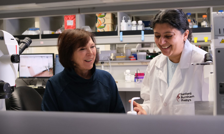
To learn more about the role of patient advocates in cancer research, we spoke with Svasti Haricharan, PhD, an assistant professor at Sanford Burnham Prebys, and Karen McDonald, a patient advocate working with Haricharan’s team. Karen, a retired computer applications professor, has been working with Svasti’s group since 2020 and has battled three forms of cancer in her life, including her current fight with metastatic breast cancer, which began in 2020.
Today, she is helping in the broader fight against cancer by bringing her unique perspective to the lab, both as a retired scholar and as a woman living with cancer.
What do patient advocates bring to the lab and why are they so important?
Karen: I was a neurology technician many years ago, so I have some science background, but I’m not an expert in the type of science that Svasti and her team do. I look at things from a patient’s point of view – not as a scientist or a doctor. I ask questions most patients would ask—but not necessarily questions scientists think about.
One of the things I’ve learned is that I need to have research presentations ahead of time—before a lab meeting—so I can figure out the technical terms. Once I’ve done that, I can bring a real-life perspective to the research—because the scientists aren’t treating patients. Svasti and her team thankfully welcome my input.
Svasti: I agree completely. Having an advocate really helps me and everybody else on the team see things in a completely different way. I think the experience also helps humanize scientists, because it’s easy, especially in biomedical research, to become so focused on the next paper or the next grant that we forget we came into research not to publish papers, but to do amazing science and help people. And having Karen’s perspective does influence what we do.
For example, there was a project on nutraceutical research, or using food as medicine, which a lot of funders don’t want to support because some people think it’s just made up. I was ready to give up on the project because it wasn’t getting support, but Karen brought up the point that patients would love to see this transitioned into the clinic because it’s less toxic with fewer side effects. I went to the top at Sanford Burnham Prebys and actually got funding to develop a drug to mimic the nutraceutical’s effects. I’m not sure this would have happened without Karen’s input—and now we are hopeful for the results.
How far have we come toward giving patients their due voice, and what are some hurdles we need to overcome?
Karen: As times have changed, physicians have stopped being thought of as gods and have started to be more human. They make mistakes. And women in particular have become more active in their healthcare because we were ignored for so long. When women started to speak up, doctors started to listen. I think is why patient advocacy started with breast cancer. Women are communicators and take an integral role in their family’s healthcare. But we still have a long way to go in terms of giving advocates’ voices full consideration.
Svasti: Karen brings up a great point here that women are more used to having to fight to have their voices heard, and that’s why breast cancer helped start the patient advocacy movement through organizations like Susan Komen. It’s beginning to spread beyond breast cancer as more funding agencies are including advocates as grant reviewers, because these are the people who are going to benefit directly from the research.
I think one thing that’s still a problem is not taking an advocate’s input seriously enough. I often see grant applications where the advocate says that a project is very significant, but other reviewers find some nitpicky aspect of the research strategy they don’t like, and the grant doesn’t get funded. There must be a better way for patient advocates’ voices to be included, as opposed to just having them on a review panel to check a box.
What’s something you’d want to tell people who may not know much about cancer research or patient advocacy?
Karen: People need to take it upon themselves to learn more about how research is done. There is such a big divide between scientists and patients, and that’s part of why patients go unheard. Even when you ask your physician about the latest research, they don’t always know.
It’s great that we have the internet now to help. I have a friend with lymphoma and that’s what we spend our time doing – researching the latest science, because we want to make sure we’re in charge of driving our own bus, not letting others have full control.
We need an environment of open-mindedness and willingness to learn. And that goes for physicians as well. We need bridges to connect cancer researchers with the oncologists who are actually going to implement their work and help humanity.
Svasti: That’s such an important idea because just like patient advocates, working with clinicians is sometimes a check box for researchers as well. It’s essential that we have meaningful collaborations—between science and medicine—that can advance research breakthroughs and improve patient outcomes.
I once spoke at a conference that had both patient advocates and researchers, and an advocate came up to me after my talk and asked, “Well what are you doing about this? If your research is real and important, why aren’t you bringing this to clinicians to get this into a clinic to help me?” That really blew my mind, because even though my role is to study cancer in the lab, she was right. Just like we need patient advocates in the lab, scientists need to advocate for research that will help patients the most.
Saying Goodbye to Dawn Dunsmore: A reflection from Josh Baxt
In September 2021, we lost Dawn Dunsmore to breast cancer, a disease she fought for a decade. Dawn was one of Sanford Burnham Prebys’ many committed administrators, most recently in Carl Ware’s lab, before stepping down to pursue treatment. She was a mom, an adventurer, an animal lover, a stubborn fighter and a friend to many, myself included.
Dawn had the bad luck of developing triple-negative breast cancer—one of the deadliest—and the good luck of being surrounded by people who loved her. She had been working for Carl for about two years when she told him about the lump in her chest. She was quickly diagnosed and treated, but the tumor soon returned.
“Dawn was approaching the end very quickly,” says Bobbie Larraga, Community Relations Manager and one of Dawn’s closest friends. “She was having seizures and difficulty breathing, and we were starting to make end-of-life plans.”
In the background, Carl, a world-renowned immunologist, helped Dawn get into a Moores Cancer Center clinical trial for an immunotherapy (PD-1 inhibitor) that takes the brakes off T cells, allowing them to attack tumors. PD-1 inhibitors work for about a third of patients and, fortunately, Dawn was one of them.
“It’s even crazier because breast cancer is not one that typically responds well to checkpoint inhibitors,” says Carl, who directs the Infectious and Inflammatory Disease Center at Sanford Burnham Prebys. “She was lucky to have that response.”
When they work, immunotherapies are like a little miracle, and Dawn did not take that lightly. She’d been given a reprieve and had things to do.
“She was just crazy for travel,” says Bobbie. “She went with her daughter to Spain. Italy, Indonesia, India, Central America. She went skydiving and was able to really check off things on her bucket list.”
But the cancer never quite went away. There were more treatments and surgeries and at 51, she finally ran out of options. Even in September, when the hospital would not release her, she was planning a trip to Yosemite.
Thinking Forward
There are so many stories. Bobbie shared how Dawn interviewed her when she first applied at Sanford Burnham Prebys (then the Burnham Institute); how they had an instant connection. Carl described the incredible work they accomplished, and how her support helped him keep it together when his wife was dying of Alzheimer’s.
I don’t usually insert myself into the articles I write – it’s just not appropriate – but Carl asked if I would, and that got me thinking. I joined Sanford Burnham Prebys in 2008, and the first piece I wrote here was a news release for the faculty member Dawn was working for. I was pretty green and Dawn helped me through, the first time of many. She was a generous soul.
Dawn was one of our own and it hurts that she’s gone. Like many at our Institute, she worked long hours to shepherd papers and grants through and helped manage the labs where she worked. She didn’t complain, even when she had the right.
Her experience underscores Sanford Burnham Prebys’ important work. Immunotherapies were first tested in academic labs, much like ours. It also brings home that the statistics we read so often, five-year survival rates or whatever, are representations of real people. The long hours, the stress of so many deadlines, the weekends in the lab, there’s a reason for those. And it’s a good one.
Institute News
This enzyme is one of the hardest working proteins in the body
Researchers from Sanford Burnham Prebys have shown that a protein they identified plays a major role in the breakdown of hyaluronic acid, a compound found in the scaffolding between our cells. The findings, published recently in the Journal of Biological Chemistry, could have implications for epilepsy, cancer and other human diseases associated with hyaluronic acid and similar compounds.
They also shed light on one of the most active biochemical processes in the body.
“Our body turns over hyaluronic acid at an extremely rapid rate, far faster than the other compounds surrounding our cells,” says senior author Yu Yamaguchi, MD, PhD, a professor in the Human Genetics Program at Sanford Burnham Prebys.
Hyaluronic acid, a common ingredient in cosmetic anti-aging products, is a one of several large sugar molecules known as glycosaminoglycans (GAGs). These are found naturally in the extracellular matrix, the complex network of organic compounds surrounding our cells that gives structure to our tissues. In addition to its structural role, the extracellular matrix is involved in regulating the immune system and is critical in the early development of connective tissues like cartilage, bone and skin.
“The extracellular matrix is found in every organ and tissue of the body, and malfunctions in its biochemistry can trigger or contribute to a variety of diseases, some of which we don’t even know about yet,” says Yamaguchi. His team studies how GAGs affect childhood diseases including congenital deafness, epilepsy and multiple hereditary exostoses, a rare genetic disorder that causes debilitating cartilage growths on the skeleton.
Hyaluronic acid is also known to be correlated with several health conditions, depending on its concentration in certain tissues. Reduced levels of hyaluronic acid in the skin caused by aging contribute to loss of skin elasticity and reduced capacity to heal without scarring. Levels of hyaluronic acid in the blood dramatically increase in alcoholic liver disease, fatty liver and liver fibrosis. In addition, hyaluronic acid levels have been correlated with increased tumor growth in certain cancers.
“These compounds are literally everywhere in the body, and we continue to learn about how GAG’s influence disease, but there’s also a lot we still don’t know about how these molecules are processed,” says Yamaguchi, “Research like this is about understanding what’s happening at the molecular level so we can later translate that into treatments for disease.”
For this study, the team focused on a protein called TMEM2, which they had previously found to break down hyaluronic acid by cutting the longer molecule into manageable pieces for other enzymes to process further. Using mice as a research model, they selectively shut off the gene that codes for TMEM2 and were able to successfully measure precisely how much the absence of TMEM2 affects the overall levels of hyaluronic acid.
The answer: a lot.
“We saw up to a 40-fold increase in the amount of hyaluronic acid in the study mice compared to our controls,” says Yamaguchi. “This tells us that TMEM2 is one of the key players in the process of degrading this compound, and its dysfunction may be a key player in driving human diseases.”
The team further confirmed this role of the TMEM2 protein by using fluorescent compounds that detect hyaluronic acid to determine where the TMEM2 protein is most active. They found the most activity on the surface of cells lining blood vessels in the liver and lymph nodes, which are known to be the main sites of hyaluronic acid degradation.
“These findings refine our understanding of this critical biochemical process and set us up to explore it further in the interest of developing treatments for human diseases,” says Yamaguchi. “Hyaluronic acid is so much a part of our tissues that there could be any number of diseases out there waiting to benefit from discoveries like these.”
Institute News
How Sanford Burnham Prebys is helping map the brain
By joining forces with hundreds of researchers across the country, a team from the Chun Lab at Sanford Burnham Prebys are working to create a comprehensive map of the human brain, in the hopes of leveraging that knowledge to better treat brain disorders.
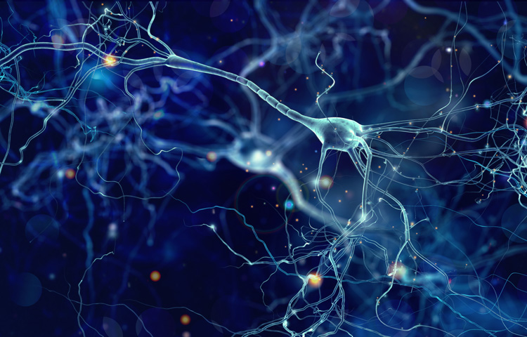
Researchers in the lab of Sanford Burnham Prebys professor Jerold Chun, MD, PhD, have helped the NIH create a cellular atlas of the motor cortex – the area of the brain responsible for movement. Their work, published recently in the journal Nature, is the flagship paper for the NIH’s BRAIN initiative, a massive multi-institution project to unravel the mysteries of the human brain.
“There are hundreds of billions of cells in the brain, and identifying and classifying all the different types of brain cells is just too big a job for any single lab,” says Chun, who is a coauthor on the study. “Similar to efforts in particle physics, hundreds of neuroscientists have now come together and it’s really exciting for us to be part of this major effort.”
The NIH Brain Research through Advancing Innovative Neurotechnologies® (BRAIN) Initiative aims to revolutionize our understanding of the human brain to more effectively diagnose, treat and prevent neurological diseases and disorders. Since its launch in 2013, the BRAIN Initiative has awarded more than 900 grants to research institutions across the country, totaling $1.8 billion.
Chun is one of the principal investigators of the BRAIN Initiative Cell Census Network (BICCN), a subset of the Initiative that aims to develop a database of all the brain cell types in humans, mice, and non-human primates.
“While the project is about exploring the brain, what we’re really interested in over the long term is the clinical applications,” says study coauthor Carter Palmer. “Understanding the nuances of the brain and how the trillions of neural connections really work is going to lead us to new targets for therapies and diagnostics so we can help people heal.” Palmer is a graduate student in Chun’s lab, alongside fellow co-authors Christine Liu and William Romanow.
There are over 150 billion cells in the average human brain and well over a thousand different cell types, depending on how you characterize them. With such a vast landscape to track, many different types of data are needed to develop a comprehensive atlas of the brain.
For their part, the Chun Lab provided single-cell transcriptomes for human brain cells, focusing on the motor cortex. Single cell transcriptomes provide a measure of how hundreds to thousands of genes are expressed in individual cells and can provide hints as to what functions those cells are serving. This process also provides a molecular definition of cell types, making it easier for researchers to identify and classify them.
“Looking at how genes are expressed gives us a wealth of information on what cells are doing, how they develop and how they’re interacting with other cells,” says Palmer. “And when our data feed into the data from other teams, we start to get a much clearer picture of what’s happening in the brain than has ever been possible.”
Their flagship Nature paper is one of seventeen in a special edition of the journal, chronicling recent advances by hundreds of BICCN researchers. The team also contributed to a second paper in the issue, which expands on the first by comparing the motor cortex cells of humans, mice, and marmosets. These publications speak not only to the expertise of Chun and his colleagues, but to the power of collaborative, interdisciplinary work to achieve previously unheard-of research goals.
“Fifty years ago, a project like this would have been impossible, because we just didn’t have the technology or even basic knowledge to collaborate on such a large scale,” says Chun. “Huge initiatives like BRAIN are an important part of the future of scientific research, and we’re thrilled we were able to contribute to this milestone in neuroscience.”
Institute News
How microbes shape human health: an interview with Andrei Osterman
In his work on the human microbiome, Sanford Burnham Prebys professor Andrei Osterman, PhD, has shown how the organisms living within us can be leveraged to boost human health for a humanitarian cause – the plight of malnourished children.
Describe your research aimed to improve the gut microbiome in malnourished children. In infants, it’s been well-established that conditions of severe poverty and food insecurity cause a delayed development of gut microbes, and that this results in stunted growth, numerous syndromes, and even death. My team has been collaborating with researchers at Washington University in St. Louis to develop foods that are designed to enhance the microbiome, and we’ve found that these can actually work to correct some of these pathologies.
One study involved introducing microbes from undernourished Bangladeshi children into the guts of mice. When these mice were then fed a typical Bangladeshi diet, they exhibited a weaker immune response to the oral cholera vaccine. More importantly, this poor response could be repaired by establishing a more normal gut microbiome in the mice and providing them supplements to boost these microbes’ propagation.
What else can we learn from this research that could be applied more broadly? From a humanitarian perspective, the progress we’ve made is so valuable that there is no question we will continue the work. But studying the microbiome in infants, regardless of their food security, can also provide us with new insights into the importance of the microbiome in human health.
In a more recent study with collaborators from University of California San Diego and University of Southern California, we found that adding corn syrup to infant formula can enhance the populations of beneficial microbes they might otherwise have gotten from breast milk.
We hope this is the first of many studies we work on with this team, because the transition of infants from breast milk or formula to conventional foods is thought to be the most drastic example of how the microbiome changes with diet. Studying infants and their diets at this early point in life could help reveal fundamental truths that we’ll be able to translate to other syndromes related to the microbiome in children and adults worldwide, regardless of food security.
And this isn’t just speculation. Another study with the team in St. Louis used the same methods as the malnutrition study to develop supplementary foods, called “fiber snacks,” to correct microbiome imbalances in people with obesity. One might think that obesity would be the total opposite of malnutrition, but the microbiome is a key player in both.
More broadly, gut microbes are already the most well-studied part of the human microbiome, and the list of health associations with these microbes extend well beyond the digestive tract, even into the immune system, affecting the risk for diseases like cancer or diabetes. There’s also a growing body of evidence that suggests that gut microbes can have a direct effect on the brain. For example, the microbiome is being studied closely in connection to autism spectrum disorder, since many people on the spectrum experience concurrent gastrointestinal syndromes.
What would you say is important to know for people not familiar with the subject? We need to acknowledge that our body and many of its problems have a huge microbiome component. The human body is a complex organism, and we are still learning how the microbiome influences and is influenced by different health conditions. The next step is to incorporate the role of the microbiome into the design of new diagnostics and therapeutics—because this undoubtedly influences their effectiveness. We can’t ignore this aspect of our biology, and the time is ripe to improve our understanding of it and leverage it to our advantage. Moving forward, this is going to help us solve so many problems—from issues we’ve already started looking at like obesity and malnutrition, all the way through to problems we aren’t even aware of yet.
What are the next steps for you and your team? What we’re really interested in now is exploring new genomic technologies that are starting to revolutionize the field. The latest development is something called MAG genomics, short for metagenomically assembled genomes. This involves looking at the big picture, sequencing DNA from the whole microbiome at once in a way that is much faster and of much better quality than we’ve ever been capable of before. It’s like the difference between watching a movie on a clunky pixelated monitor from the 80’s and seeing that same movie on an HD monitor. Methods like this are moving us into a new paradigm in biomedical research, one that may be more complex, but also one that has the potential to substantially improve health outcomes for people around the world.
Institute News
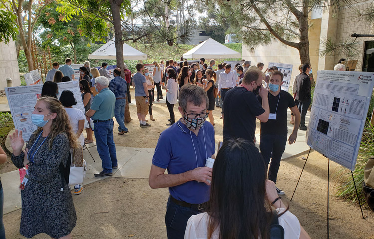
Early-career scientists showcase their work at the 20th Annual Trainee Research Symposium
The scientific leaders of tomorrow gathered to showcase their work at the 20th Annual Trainee Research Symposium on September 23rd. The talented group of presenters included postdoctoral researchers, graduate students, and staff scientists from labs across Sanford Burnham Prebys.
After introductory remarks by Sanford Burnham Prebys Student Network co-chairs Stephen Sakuma and Marie Berenguer, PhD, as well as president Kristiina Vuori, MD, PhD, the young scientists gave 20-minute podium presentations about their research, which were judged by a panel of Sanford Burnham Prebys faculty and staff. Speakers included:
Cynthia Lebeaupin, PhD and Valeria Guglielmi, PhD were respectively awarded first and second place by the judges for their presentations, for which they’ll each receive a cash prize to go towards career development activities. Lebeaupin’s research focuses on the progression of fatty liver disease to liver cancer, and Guglielmi studies the role of nuclear pores the development of bone marrow cells.
After the first two sessions of presentations, the keynote speech for the Symposium was given by Katherine Thompson-Peer, PhD, an assistant professor of developmental & cell biology at the University of California at Irvine.
Following the last podium presentation was a poster session where 30 early-career scientists were given the chance to present their work. A panel of judges selected the top three posters—presented by Shaun Lim from the Kumsta and Hansen labs, Aleksandr Arzamasov from the Osterman lab, and Michaela Lynott from the Colas lab—and they will also receive a cash prize. The day was capped off with closing remarks from Sanford Burnham Prebys CEO C. Randal Mills, Ph.D.
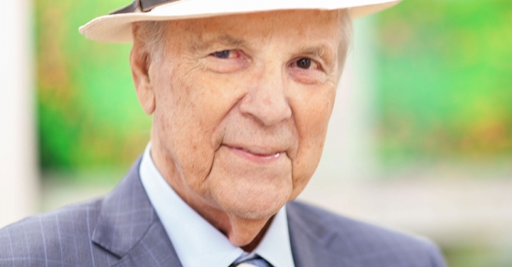
Conrad Prebys was an extraordinary man and a passionate philanthropist. Today, his generosity extends beyond his life through the Conrad Prebys Foundation.
This year, the Foundation provided $3 million to Robert Wechsler-Reya, PhD, and his team of researchers to advance a potential drug to treat medulloblastoma—the most common malignant brain tumor in children.
Children with medulloblastoma often receive aggressive treatment (surgery, radiation and chemotherapy), but many still die of their disease, and survivors suffer long-term effects from therapy. Safer and more effective therapies are desperately needed.
Wechsler-Reya recently combined forces with Michael Jackson, PhD, senior vice president of Drug Discovery and Development, to find a drug(s) that would inhibit the growth of Group 3 medulloblastoma, the most aggressive form of the disease. Using high-throughput screening technology, they identified a compound that reduces levels of a protein called MYC, which is found at exceptionally high levels in Group 3 medulloblastoma, as well as in cancers of the blood, breast, lung and prostate.
“An effective MYC inhibitor could have a major impact on the survival and quality of life of patients with medulloblastoma,” says Wechsler-Reya. “We identified a compound that reduces levels of MYC in medulloblastoma cells, but now we need to learn how it works to optimize it as an anti-cancer drug and advance studies toward the clinic.
“Historically, pharmaceutical companies and funding agencies have under-invested in childhood cancers, and the majority of drugs currently used to treat these cancers were originally developed for adult cancer,” adds Wechsler-Reya. “We believe that effective drugs for pediatric brain tumors must be developed—and this award from the Foundation will help us achieve this goal.”
“We are profoundly grateful to Conrad for his generosity over the years,” says President Kristiina Vuori, MD, PhD “He has a special legacy at our Institute, which was renamed Sanford Burnham Prebys in 2015 to honor him. We are now thankful to his Foundation for including us in their inaugural grant cycle, and for supporting the critical work we do to benefit children and others suffering from cancer.”
The Conrad Prebys Foundation allocated $78 million in its inaugural grant cycle to fund 121 projects. The awards reflect areas of personal interest to Conrad Prebys—including visual and performing arts, higher education, health care, youth development and animal conservation.
Sanford Burnham Prebys joins a long list of recipients, which included other prominent San Diego institutions such as Rady Children’s Hospital, KPBS, San Diego State University, Scripps Research, Museum of Contemporary Art San Diego and the La Jolla Music Society.
Institute News
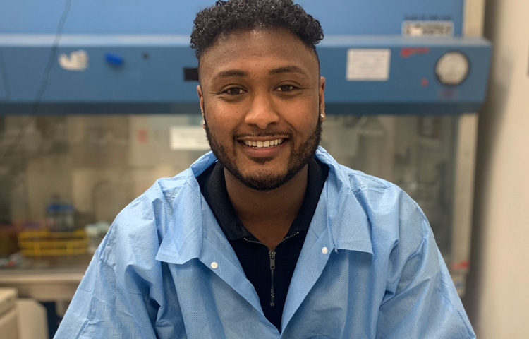
Alumni adventures: Catching up with student intern Daniel Million
Million reflects on how a summer internship at Sanford Burnham Prebys accelerated his scientific career.
Daniel Million was always fascinated by biology. But, like most high school students, he wasn’t sure what life as a scientist was really like. That all changed when he had the opportunity to complete a summer internship at Sanford Burnham Prebys.
“We thought it would be a strict environment where we were all very serious, with nobody talking,” says Million. “But my mentors both made the work in the research lab exciting. They taught me early on that you can have fun while doing great science.”
For six weeks during the summer of 2013, he and nine other classmates from the Preuss School at UC San Diego—a charter school for students who would be the first in their families to graduate from college—gained valuable laboratory skills while working directly with cancer researchers.
“Now that I’ve had the opportunity to do biological research in college and grad school, I look back and am amazed at what we were able to accomplish while in high school,” says Million. “We were doing PCRs, gel electrophoresis—techniques you usually don’t get to experience until college. They gave us a great preview of what it’s like to work in science.”
Million believes that this experience gave him a leg up that led to his acceptance to the University of Southern California, and to his receipt of a prestigious GATES Millennium scholarship, which covered all of his college costs through graduation. The benefits also extended to when he arrived on campus to start his degree.
“When you go into a research lab, that can be an intimidating place,” says Million. “If I didn’t get the chance to build my confidence in the research setting, I don’t feel that I would have performed as well when I got to college.”
Today, Million is wrapping up his master’s degree in infectious disease at Keck Graduate Institute. Whatever his future holds—perhaps medical school, or a master’s degree in public health—he remains a supporter of the internship program.
“This experience not only changed my life but changed a lot of students’ lives at Preuss,” says Million. “For a student who is going to be a first-generation college student, and who is already going to have a lot of barriers entering higher education, this is the extra push and extra knowledge they need to be successful.”
This internship was funded by the National Cancer Institute’s Continuing Umbrella of Research Experiences (CURE) program, which supports training and career-development opportunities from middle school through junior investigator levels with the goal of increasing diversity in the cancer research workforce.
Institute News
Fighting rare diseases: Finding treatments and bringing hope to families
The majority of rare diseases affect children, most of whom have an underlying genetic cause for their condition that is incurable.
The majority of rare diseases affect children, most of whom have an underlying genetic cause for their condition that is incurable.
Often, their own doctors have never heard of their disease, let alone know how to treat it.
But there is someplace they can turn to for help. The Human Genetics Program at Sanford Burnham Prebys provides insights into the genes and environmental factors that play a role in the development of childhood diseases. Their work often leads to better ways to diagnose, treat, and sometimes, even cure children.
On March 18, 2021, two patients whose lives were saved by discoveries made by Hudson Freeze, PhD, and José Luis Millán, PhD, joined the scientists for a conversation about what this work means to them and how their lives have been impacted. Watch the full discussion below.