Cardiovascular Pathobiology Archives - Page 2 of 2 - Sanford Burnham Prebys
Institute News

Sanford-Burnham wins GlaxoSmithKline drug discovery challenge

Authorpbartosch
Date

December 1, 2014

We’re excited to announce that a joint team of scientists from Sanford-Burnham at Lake Nona and Mayo Clinic has been selected as a winner of GlaxoSmithKline (GSK)’s 2014 Discovery Fast Track Challenge. The Challenge is designed to accelerate the translation of academic research into novel therapies. Researchers from the two organizations will work with partners at GSK to screen the pharmaceutical company’s library of compounds for potential new drugs to treat resistant hypertension, blood pressure that remains high despite treatment with current medications. The Challenge provides resources for small-molecule discovery and offers the opportunity for long-term collaboration. Continue reading “Sanford-Burnham wins GlaxoSmithKline drug discovery challenge”

Institute News

Sanford-Burnham welcomes two new scientists to Lake Nona

Authorpbartosch
Date

October 29, 2014

We’re excited to announce that we have recruited two cardiometabolic experts to our Medical City campus in Lake Nona (Orlando), Fla. Peter A. Crawford, MD, PhD, and Andre d’Avignon, PhD, join our Cardiovascular Pathobiology Program from Washington University in St. Louis, Mo. The increasing density of local scientists and clinicians in Orlando is accelerating the growth of the region’s bio-medical industry, promoting both economic development and the quality of health care. Continue reading “Sanford-Burnham welcomes two new scientists to Lake Nona”

Institute News

A signature for early-stage heart failure could improve diagnosis and prevent disease progression

AuthorGuest Blogger
Date

September 30, 2014

This is a post by our guest writer Janelle Weaver, PhD

Heart failure affects about five million people in the United States, and about half of these individuals die within five years of diagnosis. This condition occurs when the heart can’t pump enough blood to meet the body’s needs, and evidence suggests that abnormalities in energy metabolism play an important role. However, many past studies addressing the underlying molecular mechanisms have focused on severe, late-stage heart failure, potentially missing out on early events that could guide the development of treatment strategies for early disease stages. Continue reading “A signature for early-stage heart failure could improve diagnosis and prevent disease progression”

Institute News

New insights into how the heart forms may help identify heart defects

AuthorGuest Blogger
Date

September 29, 2014

This is a post by our guest writer Janelle Weaver, PhD

The formation of the heart during development is a highly complex process that requires precise coordination between cells and molecular signaling pathways. The fruit fly has been widely used for studying the underlying cellular and molecular mechanisms, and a great deal is known about how the fate of heart cells is controlled by signaling pathways and transcription factors—proteins that control gene activity. But beyond that, events that regulate heart formation have not been clear. Continue reading “New insights into how the heart forms may help identify heart defects”

Institute News

The bright side of free radicals

Authorsgammon
Date

September 17, 2014

In a new study by Rolf Bodmer, Ph.D., director of the Development, Aging, and Regeneration Program at Sanford-Burnham, and Hui-Ying Lim, Ph.D., assistant member of the Free Radical Biology and Aging Program at the Oklahoma Medical Research Foundation as lead author, researchers report a previously unrecognized role for reactive oxygen species (ROS) in mediating normal heart function. The findings show how under normal physiological conditions, ROS produced in non-muscle heart cells act on nearby muscle cells to maintain normal cardiac function. The results provide vital insight on how ROS direct cell communications, and in addition to the heart, may be important for the function of other organs.

“Until now, scientists knew that ROS in non-muscle heart cells affected nearby muscle cells in conditions of cellular damage and stress,” said Bodmer. “We have shown that ROS have an essential role in normal cardiac health. Understanding the fundamental communication systems in healthy and damaged hearts has important implications for developing protective and therapeutic interventions for cardiac diseases.”

ROS—a reputation of destruction ROS are free radicals that are usually associated with diseases such as cancer, cardiovascular, and neurodegenerative disorders. ROS have atoms with an unpaired electron in their orbit which can send them on a rampage to pair with other molecules, including DNA—causing mutations that contribute to disease. Antioxidants are molecules that soak up the extra electron and remove free radicals, raising the possibility that antioxidant vitamins and supplements might have a protective role in human health.

Opinions on antioxidant supplements are highly polarized. Several large-scale randomized trials of supplements have had inconsistent results and the antioxidant pendulum appears to be swinging from healthy to insignificant, and in some cases even toxic. More reliable data is needed to better define the role of antioxidants in the prevention of cardiovascular and other diseases.

ROS regulate cardiac function by cell-to-cell signaling The new study, published in Cell Reports, illustrates a previously unappreciated role for ROS signaling in the heart and supports the critical concept that optimal levels of ROS are needed in the body to provide protection to the heart and other organs.

“Interestingly, we found that ROS do not diffuse from non-muscle cells into cardiac muscle cells to exert their function. Instead, ROS in the non-muscle (pericardial) cells exert their function by starting a specific signaling cascade within the cell that in turn acts on nearby cardiac muscle cells to regulate their proper function,” said Lim. “Although the precise mechanism by which ROS maintain cardiac functions has yet to be established, our research provides a more complete understanding of the functional interactions between cardiac muscle cells and non-muscle cells—and possibly cell-to-cell (paracrine) communications in other tissues.”

The research team used Drosophila melanogaster—the common fruit fly—to decipher the ROS signals that impact the cell function. The Drosophila heart shares many of the same genes, proteins, and structural characteristics with humans, and has been used for decades as a model to understand the human genes that govern healthy development as well as those involved with disease.

A link to the paper can be found at: http://www.cell.com/cell-reports/abstract/S2211-1247(14)00143-0

Institute News

The hungry heart

Authorsgammon
Date

September 4, 2014

A normal healthy heart has the ability to choose its fuel from the menu of bioavailable substrates in the body. Glucose and fatty acids are the most common substrates, and the healthy heart switches seamlessly between the two depending on which is most available. For example, if you eat a candy bar, there is ample glucose in the blood, and the heart primarily uses that as its fuel source. In contrast, after going without food for some time, blood-sugar levels drop and the heart switches to fatty acids to provide its energy. The heart needs energy to contract, relax, repair, and rejuvenate itself. Continue reading “The hungry heart”

Institute News

Genes promote hardening of arteries in type 2 diabetes

Authorsgammon
Date

July 15, 2014

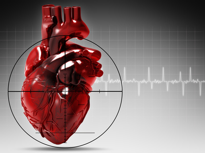
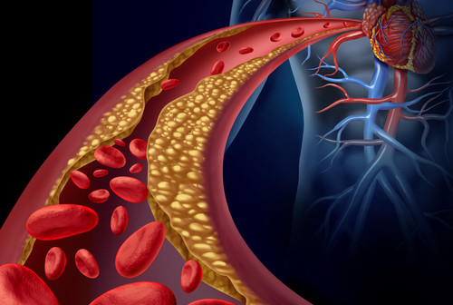
Type 2 diabetes has become a national epidemic, affecting nearly 26 million children and adults in the U.S. and approximately 170 million worldwide. According to the American Diabetes Association, $245 billion in costs are associated with diabetes, and 1 in 5 health-care dollars is spent caring for diabetics. A significant portion of the health costs associated with diabetes are those attributed to complications of the disease—including heart attacks, heart failure, stroke, dementia, chronic kidney disease, and amputations of the lower limbs. These complications emerge partly from hardening of the arteries caused by calcium deposits—a process known as arterial calcification—and are much more common in type 2 diabetics than in non-diabetics.

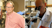
Dwight Towler, MD, PhD, professor and director of the Cardiovascular Pathobiology Program at Sanford-Burnham, has been actively researching the molecular causes of arterial calcification for more than a decade. Finding a way to prevent cardiovascular calcification could improve the vascular health of type 2 diabetes and prevent many of the associated medical complications.

In Towler’s previous work, he found that the assumption that arterial calcification was a natural, passive process that happens when cells die was incorrect. Instead, he showed that when the metabolism is disturbed—as in diabetes—calcium deposits are made by an active process that happens when key regulatory proteins erroneously trigger bone-formation genes in the arteries. Today, he is focused on those regulatory proteins, coded in the DNA by the Msx genes. Under normal conditions, Msx genes are essential for the formation of bones and teeth in the skull. But, in inflammatory conditions such as those associated with type 2 diabetes, the genes trigger the formation of calcium deposits in the arteries.

In his most recent study published on July 16 in the journal Diabetes, in collaboration with Dr. Robert Maxson of the University of Southern California, Towler’s research team examined the impact of Msx genes in mice genetically engineered to develop diabetes when fed high-fat diets. Previously, Towler showed how high-fat diets up-regulated the Msx genes in the aorta and coronary vessels of these mice, and caused calcium deposits via the Wnt paracrine signaling cascade. Now the question was: What would happen if Msx genes were absent in these mice?

“We were pleased to find that down-regulation of the Msx genes did indeed reduce the arterial calcification and vascular stiffness associated with diabetes,” said Towler. Our results are important because currently, there are no drugs to treat cardiovascular calcification. We have now identified four signaling pathways that represent targets for new drugs to intervene and inhibit the process.”

As a board-certified internist, Towler is committed to advancing these research findings to improve patient health and health care. “Our next step is to biochemically and genetically validate these pathways in human vascular disease—and identify drugs that improve vascular structure and function in mice. We are starting with lead compounds already tested in humans for other indications to see if we can repurpose those drugs to minimize the time it takes to get a treatment to the patients that suffer from this devastating complication of diabetes,” added Towler.

The study was performed in collaboration with the Norris Cancer Center, University of Southern California (CA),  Washington University in St. Louis (MO), the Translational Research Institute for Metabolism and Diabetes (FL), and MD Anderson Cancer Center (TX).

Funding for the study was provided by NIH grants HL69229 and HL81138, the Barnes-Jewish Hospital Foundation, and Sanford-Burnham Medical Research Institute.