Research News Archives - Page 2 of 8 - Sanford Burnham Prebys
Institute News

Closing in on the causes of Alzheimer’s disease

AuthorGuest Blogger
Date

April 5, 2016

This post was written by Nicole Le, a guest blogger.

Imagine if we could clear the brain of plaque that accumulates and causes Alzheimer’s disease (AD) as simply as having the plaque removed from our teeth? The body has a natural clearing mechanism in place to rid the brain of these deposits, but if this mechanism gets overwhelmed or disrupted, the plaques can accumulate and lead to neurodegeneration. Continue reading “Closing in on the causes of Alzheimer’s disease”

Institute News

Generating good fat by pushing the right buttons

Authorjmoore
Date

March 30, 2016

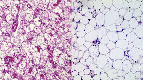
Researchers at SBP have identified a protein complex that is required for conversion of “bad” white fat to “good” brown fat. The findings, published in the Journal of Clinical Investigation, could help treat metabolic disorders such as obesity. Continue reading “Generating good fat by pushing the right buttons”

Institute News

Research points to new ways to treat inflammatory bowel diseases

Authorjmoore
Date

March 16, 2016

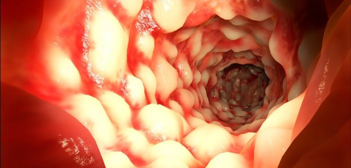
A new paper in Nature co-authored by SBP’s Randal Kaufman, PhD, reveals how briefly reducing dietary amino acids could help patients with inflammatory bowel diseases (IBD) such as Crohn’s disease and ulcerative colitis. The research shows for the first time that a specific amino acid sensor controls gut inflammation.

These results, generated in the laboratory of Bali Pulendran, PhD, of Emory University, could also lead to new drugs for IBD, which are sorely needed—currently available drugs don’t work for many patients.

What was known before this study

The amino acid sensor studied, GCN2, is one of four sensors that activate the integrated stress response (ISR) (the other sensors respond to different types of cellular stress). Activation of the ISR alters gene expression in ways that help cells survive: facilitating efficient utilization of nutrients, combating oxidative stress, and repairing DNA damage. While previous research had shown that the ISR helps limit gut inflammation, this is the first to implicate GCN2.

Significance

The findings “highlight the capacity of the immune system to sense and adapt to environmental changes, such as nutritional starvation, that cause cellular stress,” said Kaufman.

“This response may have evolved as a negative feedback mechanism to limit inflammation. This mechanism ensures that sufficient building blocks are available for the tissue regeneration required to repair the damage caused by inflammation and prevents the immune response from getting out of control.”

Implications for IBD

While GCN2’s anti-inflammatory activity can be triggered by a low-amino acid diet, reducing protein intake is not a feasible long-term treatment for IBD because it would generally impair the immune system. If human studies confirm that GCN2 is also protective in human disease, drugs that target GCN2 or later steps in the ISR could be developed to treat IBD.

Next steps

“We plan to investigate whether this pathway is involved in regulating other types of inflammation. If it is, this discovery could be important for treating other diseases like rheumatoid arthritis or multiple sclerosis,” Kaufman added.

The paper is available online here.

Institute News

Super advanced microscopy reveals how cellular anchors are activated

Authorjmoore
Date

March 15, 2016

Integrins are indispensable cell-surface proteins that form bridges with the surrounding protein matrix and other cells. They also transmit the biochemical and mechanical signals that regulate essential cellular processes, such as proliferation, differentiation, migration and cell death. Understanding how integrins become activated is important for developing ways to modulate their function, which is relevant to many disease processes, including cancer, autoimmunity, and fibrosis (hardening of tissue).

The laboratory of Dorit Hanein, PhD, in collaboration with Niels Volkmann, PhD, both professors in SBP’s Bioinformatics and Structural Biology Program, have made an important contribution in this area. Their recent study fundamentally changes our understanding of how integrins are activated—instead of a binary one-step process, it turns out to be a much more nuanced process involving multiple states.

The details

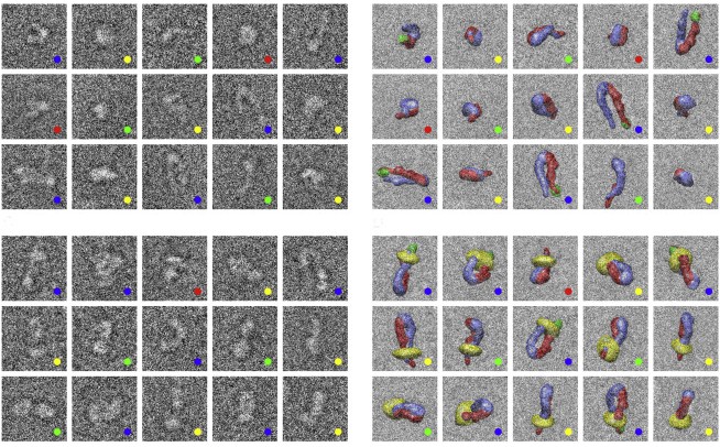
Publishing in the Biophysical Journal, the team used cryo electron microscopy (cryoEM) and advanced computational methods to determine the three-dimensional structures of full-length human integrin, αIIbβ3, which mediates blood clotting. CryoEM allows scientists to look at how a protein works in a near-physiological environment—surrounded by water and interacting with other proteins.

“Because cryoEM captures multiple states, we were able to show that the activation of integrins is much more complex than we thought—they exist in an equilibrium of conformations between bent and upright, and binding to their partners makes them more likely to straighten. Also, the matrix-binding domain is accessible even in the bent form, making it clear how integrins can be initially activated by extracellular signals,” explained Hanein.

Until now, the model for integrin activation was based on structures generated using other techniques that have value, but significant drawbacks as well. For example, X-ray crystallography constrained the protein to a highly compact and bent conformation, which implied an inactive configuration. Another type of electron microscopy revealed an upright conformation, assumed to be the active form that’s triggered by a one-step switch.

What the results mean

The new model aligns better with integrins’ ability to integrate multiple signals to control key cell processes. Instead of one binding event turning the integrin all the way on, each additional signal shifts it towards the upright form, which transmits mechanical signals more easily.

This refinement of the model is critical for designing and interpreting experiments involving integrins. According to Volkmann, “To do translational research, you have to understand the language, and in this case the language is structure.”

The paper is available online here.

Institute News

New drug combination may lead to treatment for childhood brain cancer

AuthorJessica Moore
Date

March 14, 2016

Researchers at SBP have identified a new combination therapy for the most aggressive form of medulloblastoma, a fast growing type of pediatric brain cancer. The study, published  in Cancer Cell, is expected to lead to a clinical trial to confirm the benefits of the novel drug combination. Continue reading “New drug combination may lead to treatment for childhood brain cancer”

Institute News

Study triples the number of known cases of a rare disease

Authorjmoore
Date

March 10, 2016

A recent paper from the laboratory of Hudson Freeze, PhD, characterizes 39 previously unreported cases of a specific type of congenital disorder of glycosylation (CDG). CDGs, the focus of research in Freeze’s lab in SBP’s Sanford Children’s Health Research Center, are rare inherited disorders. CDG symptoms, which can include developmental delay, movement problems, and impaired function of multiple organs, differ depending on the underlying mutation. Continue reading “Study triples the number of known cases of a rare disease”

Institute News

New insight on the development of T cells that promote autoimmune disease

Authorjmoore
Date

February 8, 2016

Generally, T cells, the policemen of the body, activate immune responses upon finding infected or diseased cells, but some T cells respond the same way to normal cells in the body. If unchecked, such T cells can cause autoimmune diseases such as psoriasis, rheumatoid arthritis, inflammatory bowel disease, and multiple sclerosis. These self-reactive T cells are a recently discovered type of T cell, the TH17 cell. Preventing young T cells from becoming TH17 cells is of major interest as a means to treat autoimmune diseases. Continue reading “New insight on the development of T cells that promote autoimmune disease”

Institute News

A sugar found in seaweed may help treat skin cancer

Authorsgammon
Date

December 8, 2015

New research from scientists at Sanford Burnham Prebys Medical Discovery Institute (SBP) suggests that a rare sugar found in seaweed, mushrooms, seeds and other foods may be able to help treat skin cancer. The sugar, called L-fucose, has previously been linked to a number of pathological conditions including inflammation and certain cancers. The current study, published in Science Signaling, is the first to associate L-fucose with melanoma, the most dangerous form of skin cancer.

“Our findings offer new, unprecedented detail into the sugar’s role in cancer,” said Ze’ev Ronai, PhD, senior author and scientific director of SBP’s La Jolla campus. “We found that by tampering with L-fucose metabolism, we could inhibit melanoma tumor metastasis. Not only were the tumors affected but also their microenvironment—the cells surrounding the tumor that play a critical role in sustaining the cancer—making the discovery even more impactful.”

Sugars, such as glucose and sucrose, come from many different sources and are used by the body in unique ways. Some sugars, including L-fucose, provide crucial tags on cell-surface proteins that signal inflammation and help direct cell migration. Previous research has shown that changes in the amount of L-fucose on cells are associated with breast and stomach cancers.

The study started with a broader investigation of activating transcription factor 2 (ATF2), a protein that controls the expression of many other proteins and that has been implicated in the development of melanoma and other cancers. Ronai’s group has been studying ATF2 for more than 20 years.

“To our surprise, one of the genes found to be regulated by ATF2 was fucokinase (FUK), which controls the ability of cells to process the dietary sugar, L-fucose, into a form that is useable for the modification (fucosylation) of proteins, many of which are on the cell surface, said Ronai.”

“In human samples, we found reduced fucosylation in metastatic melanomas and a better prognosis for primary melanomas with increased fucosylation. We suspect that the absence of L-fucose on melanoma cells makes them less sticky and more mobile in the body, making them more likely to metastasize,” Ronai explained.

Importantly, in mice with melanoma, the researchers were able to increase fucosylation either by adding the sugar to their drinking water or by genetic manipulation. Both methods inhibited the growth and metastasis of the tumors.

“Many patients develop resistance to current melanoma drugs. If we can add something like L-fucose to enhance these therapies, that’s very exciting, and it’s something we’re actively looking into,” said lead author Eric Lau, PhD, who is extending studies on the role of L-fucose in melanoma at the H. Lee Moffitt Cancer Center in Tampa, Florida,

“The dietary result was especially gratifying, because it suggests that modifying fucosylation could be achieved by the simple addition of L-fucose to drinking water.

“Our results further suggest that the addition of dietary sugar may help fight melanoma by boosting numbers of helpful immune cells. We are continuing our exploration of how fucosylation and other sugar coatings affect the immune system and impact cancer,” added Ronai.

To read the paper click here

Institute News

Enhancing drug delivery to tumors: CendR’s game

AuthorGuest Blogger
Date

November 25, 2015

This post was writing by Kelly Chi, a freelance writer.

New research shows the uniqueness of a promising technology that SBP scientists are employing to deliver large payloads of drugs to tumors. The findings, published recently in the journal Science Advances, not only lend insight into how large molecules get into cells, but also show how a compound the team previously discovered may work to target cancer cells.

One main challenge in treating cancer is delivering sufficient amounts of drug to the intended target. Often, cancer drugs do not penetrate the tumor but instead hit only a few cells. Surviving cells proliferate, and those given only a small dose can evolve resistance to treatment.

Erkki Ruoslahti, PhD, distinguished professor in SBP’s Tumor Microenvironment and Metastasis Program, and his team at SBP have been hunting for small-protein drugs that make drugs do a better job at penetrating tumors. The way the scientists do this is to inject an entire ‘library’ of different small proteins (or peptides) into mice with cancer, and see which ones, if any, reach the tumor.

More than a decade ago, they found a unique peptide that took only minutes to reach the tumor after being injected into the bloodstream.

Only later, in 2009, having discovered another peptide, iRGD, which had similar properties, did they find out what made these peptides special. They included a short string of amino acids in a particular pattern that gave it the ability to penetrate tumors. The pattern, called CendR, has been a major focus of Ruoslahti’s research.

“The iRGD peptide was special because by using it we could get a drug to go deeper into a tumor and hit all the tumor cells, not just the ones that are close to the blood vessels, which is usually what happens,” said Ruoslahti.

Last year, the group learned even more about iRGD. In Nature Communications, they showed that the peptide triggers a unique mode of transport into the cell, unlike what has been seen before. It does so by binding to a receptor on the cell surface called neuropilin1, which is itself involved in crucial functions, including the movement of fluid and other molecules across walls of blood vessel cells.

There are many ways that molecules can get into cells. iRGD appears to trigger cells to swallow large vesicles. This process is similar to a mechanism known as ‘macropinocytosis’ (macro meaning ‘large’; pino ‘drink’; and cyte ‘cell’) yet distinct in some ways, Ruoslahti said.

In the new study, the research team did additional work to characterize CendR’s actions, comparing them with another known peptide technology (TAT) that is often used to get drugs into cells.

“That pathway [TAT] is not specific for tumors and can’t be made specific for tumors as ours can. That’s one difference, and we show other differences between the two pathways in this paper,” Ruoslahti said.

Another key feature of the CendR pathway is that it’s affected by the nutrient status of the tissue.

“If the tissue, in our case a tumor, is deficient in nutrients and needs more, it activates the pathway, which fits perfectly with the idea of why this pathway exists: it brings in nutrients. In contrast, TAT is not affected by nutrient status, so it probably has another function.

“We turn on the pathway with iRGD only in the tumor, not in normal tissues. At the same time, we slip a drug into the pathway as a stowaway, and the result is that more of the drug accumulates in the tumor than would get there otherwise, and the drug also penetrates deeper into the tumor. These features make iRGD promising as an adjunct to various kinds of chemotherapy,” adds Ruoslahti.

The iRGD peptide is undergoing preclinical development, namely mouse toxicology studies that are needed before the team can apply to investigate it in humans. The team has another year’s worth of work ahead. But they already have reason to believe that iRGD might work: it binds to the human version of its receptor. In addition, they have seen promising results after testing the peptide in tumor tissue from people with cancer.

Other authors on the paper are staff scientists Hongbo Pang, PhD, and Gary Braun, PhD, both of SBP.

The full paper can be found at: http://advances.sciencemag.org/content/1/10/e1500821

Institute News

Can your heart prevent diabetes?

AuthorGuest Blogger
Date

November 19, 2015

This article was written by guest blogger Crystal Woodard, PhD

Can your heart prevent diabetes? Being overweight or obese is currently deemed the single best predictor of type 2 diabetes. With the prevalence of obesity on the rise, estimates suggest that one in three American adults could have type 2 diabetes by 2050. Weight loss is key to preventing this epidemic. At SBP, scientists are investigating how hormones released by the heart may help the body burn more calories to prevent obesity and type 2 diabetes.

What color is your fat? All fat is not created equal. Excess weight is held in energy-storing fat cells called white adipose tissue as well as energy-burning fat cells called brown adipose tissue. Increasing a person’s brown fat could improve the risks associated with obesity.

Two compounds released by the heart in response to high blood pressure—human atrial natriuretic peptide (ANP) and B-type natriuretic peptide (BNP)—have been found to play a direct role in “browning” white adipose tissue. By browning, white fat starts to burn more calories, mimicking what occurs in brown fat. Sheila Collins, PhD, professor in the Integrative Metabolism Program and her research team, are investigating how these natriuretic peptides activate fat browning with the goal of tapping into the process to help promote weight loss and prevent diabetes.

In collaboration with Dr. Richard Pratley at the Florida Hospital – SBP Translational Research Institute for Metabolism and Diabetes, the teams are conducting clinical trials with obese and lean volunteers to test whether BNP can increase energy expenditure and improve glucose tolerance. Since recombinant human BNP is an FDA-approved drug prescribed for acute heart failure patients, the costs, and development and approval times for using BNP for these conditions may be reduced.

How does BNP work? Investigators in Italy almost 20 years ago discovered that binding sites for BNP, called natriuretic peptide receptors (NPRs), were expressed in human adipose tissue. The natriuretic peptide ‘signaling’ receptor, NPRA, binds the natriuretic peptides, while the natriuretic peptide ‘clearance’ receptor, NPRC, removes them from circulation. Since then, several studies have reported that BNP levels are lower in the blood of obese patients compared to their lean counterparts. Additional research suggests BNP can lead to increased release of adiponectin, an insulin-sensitizing hormone produced by fat cells and that low levels of BNP in the bloodstream might contribute to insulin resistance.

According to Collins, “Early studies proposed that increased clearance is responsible for the lower peptide levels observed in obese individuals in comparison to lean individuals; however, there are no definitive studies to actually prove this or not. Important efforts are currently underway to understand how NPRs are regulated and how the peptides can be best used for their fat-burning capacity.”

Dr. Sheila Collins is a professor at Sanford Burnham Prebys Medical Discovery Institute (SBP) in Lake Nona, Fla. and a recipient of an American Diabetes Association research award. Dr. Richard Pratley is a senior investigator at the Florida Hospital – SBP Translational Research Institute, Medical Director of the Florida Hospital Diabetes Institute, and adjunct professor at SBP in Lake Nona. This post was written by Crystal Woodard, PhD, a post-doctoral fellow in Dr. Collins’s lab.