Research News Archives - Page 3 of 8 - Sanford Burnham Prebys
Institute News

Antioxidant-rich diet could help stave off type 2 diabetes

AuthorGuest Blogger
Date

November 12, 2015

Type 2 diabetes affects about 8% of all adults and is a leading cause of death worldwide. Despite its prevalence, relatively little is known about underlying molecular causes of the disease. SBP researchers now show that defects in a major cell stress pathway play a key role in the failure of pancreatic beta cells, leading to signs of diabetes in mice. The findings, published recently in PLOS Biology, also suggest that a diet rich in antioxidants could help to prevent or treat type 2 diabetes.

“The findings open new therapeutic options to preserve beta cell function and treat diabetes,” said senior study author Randal Kaufman, PhD, director of the Degenerative Diseases Program at SBP. “Because the same cell stress response is implicated in a broad range of diseases, our findings suggest that antioxidant treatment may be a promising therapeutic approach not only for metabolic disease, but also neurodegenerative diseases, inflammatory diseases, and cancer.”

Excess cell stress

Type 2 diabetes is caused by the failure of pancreatic beta cells to produce enough insulin—a hormone that helps to move a blood sugar called glucose into cells to be stored for energy. A major cause of type 2 diabetes is obesity, which can lead to abnormalities in insulin signaling and high blood glucose levels. Beta cells try to compensate by producing up to 10 times the usual amount of insulin, but this puts extra stress on a cell structure called the endoplasmic reticulum to properly fold, process, and secrete the hormone.

An increase in protein synthesis in beta cells also causes oxidative stress—a process that can lead to cell damage and death through the build-up of toxic molecules called reactive oxygen species. If the stress is too great, the beta cells will eventually fail. Approximately one-third of individuals with abnormal insulin signaling eventually develop beta cell failure and diabetes.

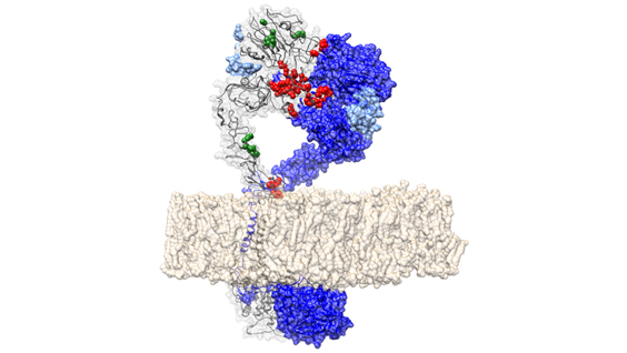
In the new study, Kaufman and his collaborators discovered that beta cell failure is caused by deficiency in a protein called IRE1α, which would otherwise help to protect cells against the stress of increased insulin production. Mice that lacked IRE1α in pancreatic beta cells did not produce enough insulin and developed high blood glucose levels, similar to patients with type 2 diabetes. IRE1α deficiency also caused inflammation and oxidative stress, which was the primary cause of beta cell failure. But treatment with antioxidants, which prevented the production of reactive oxygen species, significantly reduced metabolic abnormalities, inflammation and oxidative stress in these mice.

Taken together, the findings suggest that IRE1α evolved to expand the capacity of beta cells to produce insulin in response to increases in blood glucose levels. The study also implicates this major cell stress pathway in the development of type 2 diabetes and suggests that a diet rich in antioxidants could help to prevent or reduce the severity of the disease.

“Currently, we are testing the effects of antioxidants on glucose levels and beta cell function in mice,” Kaufman said. “If these studies prove successful, they could pave the way for clinical trials in humans and eventually lead to a new therapeutic approach for dealing with a major pandemic of the 21st century.”

This post was written by guest blogger Janelle Weaver, PhD

Institute News

Is there a type 3 diabetes?

AuthorGuest Blogger
Date

November 10, 2015

This article was written by guest blogger Jessica Frisch-Daiello, PhD

People with type 2 diabetes are twice as likely to develop Alzheimer’s disease—a type of dementia affecting behavior, memory, and cognitive functions. According to the Centers for Disease Control and Prevention, in 2013 Alzheimer’s ranked sixth and diabetes was seventh as the leading causes of death in the United States. Recent studies are suggesting a link between insulin resistance in the brain and Alzheimer’s disease, prompting some researchers to consider a new classification for the disease: type 3 diabetes.

People with diabetes can’t effectively break down blood sugar. Either their bodies don’t produce enough insulin (type 1 diabetes) or their bodies become desensitized to insulin (type 2 diabetes).

The exact mechanisms between insulin resistance and Alzheimer’s disease are not well understood and research is on-going. However, studies suggest that insulin resistance in the brain leads to the formation of two pathological hallmarks of Alzheimer’s disease—the formation of tau tangles and the build-up of clusters of beta amyloid peptides called plaques in the brain. The degree of insulin resistance is correlated with the amount of plaques deposited between nerve cells. Plaques create a blockade that inhibits cell-to-cell signaling in the brain. Additionally, insulin dysfunction has also been shown to affect the formation of tau tangles by mediating the activity of an important enzyme in the body, GSK-3β (glycogen synthase kinase 3).

Juan Pablo Palavicini, PhD, an SBP postdoctoral fellow in the lab of Xianlin Han, PhD, is studying the role of a particular class of molecules found in the body that might give more clues to the mechanisms connecting these two seemingly disparate diseases. According to Palavicini, “We have found that a specific lipid class called sulfatide is severely deficient in the brains of both Alzheimer’s disease patients and type 2 diabetics. Moreover, our research shows that when sulfatide is removed, there is a dramatic change in insulin levels, beta amyloid peptides, and tau tangles. We are currently exploring therapeutic techniques to restore sulfatide content as a treatment for both diseases.”

Sulfatide serves many functions in the body, including aiding neural plasticity and memory. It also plays a role in insulin secretion. A change in the expression of sulfatide has been associated with a number of conditions, including Alzheimer’s disease, Parkinson’s disease, and diabetes.

Given the association between Alzheimer’s disease and diabetes, it is important for people to incorporate healthy habits in everyday life. Both the American Diabetes Association and the Alzheimer’s Association say that daily exercise, social interaction, and a diet emphasizing fruits, vegetables, and whole grains may reduce the risk of developing, or slowing the progression of, these diseases.

Dr. Palavicini and Dr. Han are pursuing this research as part of a mentor-based postdoctoral fellowship awarded by the American Diabetes Association. This article was written by Dr. Jessica Frisch-Daiello, a postdoctoral associate in Dr. Han’s laboratory at SBP.

Institute News

‘Big Data’ used to identify new cancer driver genes

Authorsgammon
Date

October 20, 2015

In a collaborative study led by Sanford Burnham Prebys Medical Discovery Institute (SBP), researchers have combined two publicly available ‘omics’ databases to create a new catalogue of ‘cancer drivers’. Cancer drivers are genes that when altered, are responsible for cancer progression. The researchers used cancer mutation and protein structure databases to identify mutations in patient tumors that alter normal protein-protein interaction (PPI) interfaces. The study, published today in PLoS Computational Biology, identified more than 100 novel cancer driver genes and helps explain how tumors driven by the same gene may lead to different patient outcomes.

“This is the first time that three-dimensional protein features, such as PPIs, have been used to identify driver genes across large cancer datasets,” said lead author Eduard Porta-Pardo, PhD, a postdoctoral fellow at SBP. “We found 71 interfaces in proteins previously unrecognized as cancer drivers, representing potential new cancer predictive markers and/or drug targets. Our analysis also identified several driver interfaces in known cancer genes, such as TP53, HRAS, PI3KCA and EGFR, proving that our method can find relevant cancer driver genes and that alterations in protein interfaces are a common pathogenic mechanism of cancer.”

Cancer is caused by the accumulation of mutations to DNA. Until now, scientists have focused on finding alterations in individual genes and cell pathways that can lead to cancer. But the recent push by the National Institutes of Health (NIH) to encourage data sharing has led to an era of unprecedented ability to systematically analyze large scale genomic, clinical, and molecular data to better explain and predict patient outcomes, as well as finding new drug targets to prevent, treat, and potentially cure cancer.

“For this study we used an extended version of e-Driver, our proprietary computational method of identifying protein regions that drive cancer. We integrated tumor data from almost 6,000 patients in The Cancer Genome Atlas (TCGA) with more than 18,000 three-dimensional protein structures from the Protein Data Bank (PDB),” said Adam Godzik, PhD, director of the Bioinformatics and Structural Biology Program at SBP. “The algorithm analyzes whether structural alterations of PPI interfaces are enriched in cancer mutations, and can therefore identify candidate driver genes.”

“Genes are not monolithic black boxes. They have different regions that code for distinct protein domains that are usually responsible for different functions. It’s possible that a given protein only acts as a cancer driver when a specific region of the protein is mutated,” Godzik explained. “Our method helps identify novel cancer driver genes and propose molecular hypotheses to explain how tumors apparently driven by the same gene have different behaviors, including patient outcomes.”

“Interestingly, we identified some potential cancer drivers that are involved in the immune system. With the growing appreciation of the importance of the immune system in cancer progression, the immunity genes we identified in this study provide new insight regarding which interactions may be most affected,” Godzik added.

The study was performed in collaboration with the European Bioinformatics Institute (UK), Centro de Investigación Principe Felipe (Spain), and CIBER de Enfermedades Raras (Spain).

Institute News

How proteins age

Authorsgammon
Date

October 19, 2015

SBP researchers and colleagues discover a mechanism that regulates the aging and abundance of secreted proteins.

Physiological processes in the body are in large part determined by the composition of secreted proteins found in the circulatory systems, including the blood. Each of the hundreds of proteins in the blood has a specific life span that determines its unique range of abundance. In fact, measurements of their quantities and activities contribute to many clinical diagnoses. However, the way in which normal protein concentrations in the blood are determined and maintained has been a mystery for decades.

Biomedical scientists at Sanford Burnham Prebys Medical Discovery Institute (SBP) and UC Santa Barbara (UCSB) have now discovered a mechanism by which secreted proteins age and turnover at the end of their life spans. Their findings, which shed light on a crucial aspect of health and disease, appear today in the Proceedings of the National Academy of Sciences (PNAS).

“This is a fundamental advance that is broadly applicable and provides an understanding of how secreted proteins, which are involved in many important physiological processes, normally undergo molecular aging and turnover,” said senior author Jamey Marth, PhD, professor in SBP’s NCI-designated Cancer Center.

“When a secreted protein is made, it has a useful life span and then it must be degraded — the components are then basically recycled,” added Marth, also director of UCSB’s Center for Nanomedicine and a professor in the campus’s Department of Molecular, Cellular, and Developmental Biology. “We can now see how the regulation and alteration of secreted protein aging and turnover is able to change the composition of the circulatory system and thereby maintain health as well as contribute to various diseases.”

This newly discovered mechanism encompasses multiple factors, including circulating enzymes called glycosidases. These enzymes progressively remodel N-glycans, which are complex structures of monosaccharide sugars linked together and attached to virtually all secreted proteins.

It is the N-glycan structure itself that identifies the protein as nearing the end of its life span. Subsequently, multiple receptors known as lectins — carbohydrate-binding proteins — recognize these aged proteins and eliminate them from circulation.

Marth and colleagues identified more than 600 proteins in the bloodstream that exhibit molecular signs of undergoing this aging and turnover process. Many of these proteins are regulators of proteolysis (the breakdown of proteins), blood coagulation and immunity.

Honing in on individual examples, the researchers were able to track each of them through time and watch the process unfold. “In these studies we further saw that the different life spans of distinct proteins are accounted for by the different rates of aging due to N-glycan remodeling,” said lead author Won Ho Yang, PhD, a postdoctoral associate at SBP and at UCSB’s Center for Nanomedicine.

“Altering this aging and turnover mechanism is the fastest way to change the abundance of a secreted protein, which we increasingly note is occurring at the interface of health and disease,” Marth explained. “In retrospect from published literature and from studies in progress, we can now see how sepsis, diabetes and inflammatory bowel disorders can arise by the targeted acceleration or deceleration of secreted protein aging and turnover.”

“The discovery of this mechanism provides a unique window into disease origins and progression,” Marth added. “It has been known that circulating glycosidase enzyme levels are altered in diseases such as sepsis, diabetes, cancer and various inflammatory conditions. The resulting changes in the composition and function of the circulatory systems, including the blood and lymphatic systems, can now be identified and studied. We are beginning to see previously unknown molecular pathways and connections in the onset and progression of disease.”

Institute News

Unraveling the mystery of muscle regeneration could lead to effective treatment for muscular dystrophies

Authorsgammon
Date

September 22, 2015

By shedding light on the distinct functions of a protein complex that controls the formation of skeletal muscle tissue, SBP researchers could pave the way for the development of novel therapies for neuromuscular diseases.

Continue reading “Unraveling the mystery of muscle regeneration could lead to effective treatment for muscular dystrophies”

Institute News

Researchers reawaken sleeping HIV in patient cells to eliminate the virus

Authorsgammon
Date

September 9, 2015

Researchers at Sanford Burnham Prebys Medical Discovery Institute (SBP) have identified a new class of drugs that may be used to purge pockets of dormant HIV from a patient’s body, eliminating the virus once and for all. Fortuitously, these agents are already being explored in clinical trials for treating cancer, which could speed up the route to approval for treating HIV.

Antiretroviral therapies have made it possible for people to live with AIDS for decades. However, small reservoirs of a patient’s cells hide the virus. That is, HIV’s genes live in the cells, but its genetic code is never read to make protein, and so the virus goes undetected by the immune system.

“If you take people off the antiretroviral therapies, some of these dormant cells reawaken to make more virus,” said lead author Lars Pache, PhD, a postdoctoral fellow in the lab of Sumit Chanda, PhD, director of the Immunity and Pathogenesis Program at SBP. “The key for a cure for HIV is to purge these cells that have dormant HIV.”

Reactivating latent HIV-infected cells so that they can be killed off once and for all is called ‘shock and kill.’ The approach has remained elusive so far, because drugs that reawaken the virus could also trigger massive immune system activation, which itself could be deadly, Chanda said.

The new study, published September 9 in the journal Cell Host & Microbe, “uses a class of drug called Smac mimetics to tap into a cell pathway that can be used to wake up the virus but, based on clinical studies and our data, doesn’t appear to activate the immune system,” Chanda added.

The study started with a broad search of genes within the host cells that help keep the virus silent. Chanda’s group identified 651 genes. They then created batches of cells in which each one of those genes was silenced, and they measured how much HIV the cells produced after they were exposed to the virus.

The scientists whittled the list of candidate genes down to 139, to 24, and then 12 using increasingly stringent criteria. The absence of one gene in particular, BIRC2, boosted the activity of HIV. Even better, Smac mimetics—already proven safe in early-stage clinical trials for cancer—works by inhibiting BIRC2 and related molecules.

“These experiments led us to develop a strategy of using Smac mimetics to reawaken dormant HIV so that we could then kill it with anti-viral therapy,” said Chanda.

Chanda’s colleague at SBP, Nicholas Cosford, PhD, professor in the Cell Death and Survival Networks Program, had recently described a potent BIRC2 inhibitor, SBI-0637142. “This drug is about 10-100 times more potent than the small molecules currently in clinic development, making it a promising candidate to tackle HIV latency,” says Chanda.

Part of the reason that HIV’s genes stay hidden in its host is that they cover themselves with tightly wound DNA. A class of drugs called histone deacetylase inhibitors, which unfurls the DNA, is used to treat a variety of conditions. Although most of these inhibitors haven’t worked well on their own to reactivate latent HIV, they might work well with Smac mimetics including SBI-0637142, Chanda’s group reasoned.

The key question was whether they could reactivate the virus in cells from HIV-infected patients undergoing antiretroviral therapy. They combined SBI-0637142 with a histone deacetylase inhibitor (panobinostat) and saw signs that the virus had reawakened without triggering immune cell death.

“We anticipated that we would see a synergy because the drugs work along parallel pathways. What we didn’t expect was the level of activation—the potency and efficacy with which we were able to reverse latency in patient samples,” Chanda said.

They saw similar results in patient cells treated with a combination of LCL161—a Smac mimetic that is already in phase 1 and 2 trials for treating cancer—and panobinostat. “This is a one-two punch for HIV,” said Chanda, adding that ultimately, a cocktail of drugs will be necessary to cure HIV.

The scientists hope to partner with a pharmaceutical company to develop these molecules for testing in animal models of HIV and then move them into the clinic if they meet the safety and efficacy criteria.

In addition to SBP, the study consortium included the University of Utah School of Medicine, The Salk Institute for Biological Studies, the Perelman School of Medicine at the University of Pennsylvania, the Icahn School of Medicine at Mount Sinai, the Paul-Ehrlich-Insitut, and the German Center for Infection Research.

This post was written by Kelly Chi, a freelance science writer. 

Institute News

How protein tangles accumulate in the brain and cause neurological disorders

Authorsgammon
Date

September 2, 2015

A new Sanford Burnham Prebys Medical Discovery Institute (SBP) study takes a step forward in understanding how similar, yet genetically unrelated neurodegenerative diseases, such as Alzheimer’s disease, frontal temporal dementia, and progressive supranuclear palsy (PSP) are caused by the protein tau. The findings, published today in Neuron, create new opportunities to target this key protein that leads to the brain lesions found in patients with impaired motor functions and dementia. Continue reading “How protein tangles accumulate in the brain and cause neurological disorders”

Institute News

10 years of studying metabolism, nutrition, and human energy—what have we REALLY learned?

Authorsgammon
Date

August 17, 2015

Every day we read or hear something about a food that is bad for us, a fruit that will help us lose weight, or a supplement that will extend our lives beyond their natural endpoint. Unquestionably, every year a significant amount of money, research, and time is spent exploring the cause and prevention of obesity, diabetes, heart disease, and the myriad of other metabolic conditions that affect our health and well-being. But what do scientists think are the truly important things we have learned about our metabolism, diet, and exercise over the last decade?  And how is this leading to the next-generation of medicines to treat metabolic disorders? Continue reading “10 years of studying metabolism, nutrition, and human energy—what have we REALLY learned?”

Institute News

Pathway that controls cancer cell proliferation discovered

Authorsgammon
Date

August 13, 2015

In a new study by SBP, researchers have identified a novel kinase cascade that regulates mTORC1, a protein complex implicated in the control of cancer cell growth in response to nutrients. The study, published in Cell Reports, provides further insight into the control of mTORC1 activation, and highlights several new potential drug targets to treat human pathologies linked to mTORC1 deregulation. Continue reading “Pathway that controls cancer cell proliferation discovered”

Institute News

Scientists solve structure of important protein for tumor growth

Authorsgammon
Date

August 5, 2015

In a collaborative study between SBP and the Argonne National Laboratory, scientists have used a highly specialized X-ray crystallography technique to solve the protein structure of hypoxia-inducible factors (HIFs), important regulators of a tumor’s response to low oxygen (hyopoxia). The findings, published today in the journal Nature, open the door to search for new drugs to treat tumors by cutting off their supply of oxygen and nutrients. Continue reading “Scientists solve structure of important protein for tumor growth”