Research News Archives - Page 6 of 8 - Sanford Burnham Prebys
Institute News

New study sheds light on cancer stem cell regulation

Authorsgammon
Date

February 5, 2015

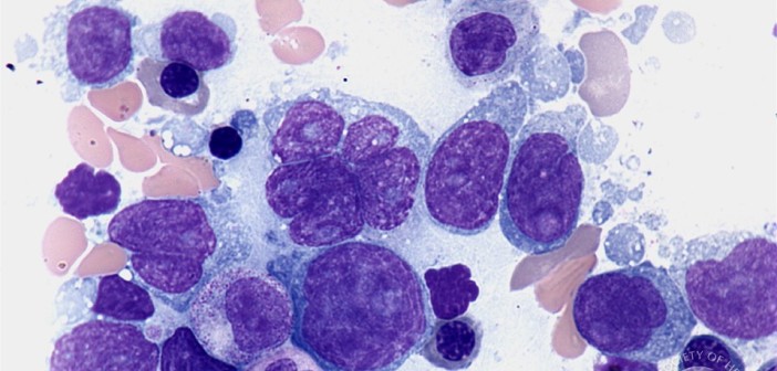
Researchers at Sanford-Burnham have discovered a precise stem cell signaling process that can lead to intestinal tumors if disrupted. The findings add to our understanding of how stem cells give rise to tumors and identify specific stem cell molecules that may be targeted to prevent the onset, progression, and recurrence of intestinal cancers. The results of the study appear online in Cell Reports today. Continue reading “New study sheds light on cancer stem cell regulation”

Institute News

Stem cells grow new hair – Arabic translation

Authorsgammon
Date

January 27, 2015

“دراسة جديدة لنمو الشعر”

في دراسة جديدة، إستخدم الباحثون في معهد سان- برونهام الخلايا الجذعه المحفزة1 في جسم الإنسان لتعمل على توليد ونمو الشعر الجديد. وتمثل هذه الدراسة خطوة أولى نحو تطوير علاج عن طريق الخلايا الجذعية للأشخاص الذين يعانون من مشكلة فغداً الشعر “الصلع” .في الولايات المتحدة وحدها، يعاني أكثر من 40 مليون رجل و 21 مليون إمرأة من مشكلة فقدان الشعر، ولقد تم نشر هذا البحث على الإنترنت في بلوس وان. 

تبرعكم سوف يصنع الفرق الرجاء التبرع للدكتور ألكسي للمضي قدما في هذا الإنجاز.

“لقد قمنا بتطوير طريقة إستخدام الخلايا الجذعية المحفزه لخلق خلية جديدة قادرة على بدء نمو الشعر عند الإنسان”. و قال ألكسي ترسكي و هو دكتور أستاذ مشارك في معهد التنميه، والشيخوخة و برامج التجديد ” طريقة إستعمال الخلايا الجذعية توفر مصدرا غير محدود من الخلايا من المريض لعملية زراعة الشعر وهي ليست محدودة كالطرق المستعملة حالياً عن طريق إستخدام ونزع بصيلات الشعر المتواجدة من الأصل. طور فريق البحث نظام يعمل على تمايز الخلايا الجذعية المحفزة عند الإنسان لتصبح وتتحول إلى خلايا جلدية حليمة. هذه الخلايا الجلدية تعد خلايا فريده من نوعها وتعمل على تنظيم تشكيل بصيلات الشعر و دورة نموه. هذه الخلايا الجلدية لا تعد مناسبة لنمو الشعر من تلقاء نفسها لأنها لا تتوفر بالكميات المطلوبة وتفقد قدرتها بسرعة على حث تشكيل بصيلات الشعر بدون عمله التمايز مع الخلايا الجذعية. 

عند البالغين، هذه الخلايا الجلدية يمكن تضخيمها خارج الجسم، وأنها سرعان ما تفقد خصائصها في عملية تحفيز الشعر”.يقول الدكتور ألكسي “لقد قمنا بإبتكار نظام يعمل لدفع الخليا الجذعية المحفزه عند الإنسان لتتمايز أو تتحول إلى خلايا جلدية حليمة، وقد أكد نجاح التجربة حيث أكد على قدرة هذه الخلايا على حث نمو الشعر عند زرعها عند الفئران”. خطوتنا التاليه هي زراعة الخلايا الجذعية المحفزة عند الإنسان مرة أخرى إلى جسم الإنسان، و نحن حالياً نسعى إلى شركات لتنفيذ الخطوة الأخيرة”. 

شاهد المزيد على الرابط 
 

تبرع


Institute News

Stem cells grow new hair – Chinese translation

Authorsgammon
Date

January 27, 2015

用干细胞促生毛发

在一项新的研究中,桑福德 – 伯纳姆的研究人员利用人类多能干细胞促生出新的头发。该研究代表了以细胞为基础的治疗人类脱发的第一步。仅在美国,就有超过4000万男性和21万女性有着脱发困扰。这项研究在PLOS ONE在线发表。 

您的资助很重要。请帮助捷列克博士继续这项研究!

“我们已开发出一种使用人类多能干细胞的方法,来创造能够引发人体毛发生长的新的细胞。该方法相对于目前那种依赖于将毛囊从头的一个部位移植到另一个部位的方法有显着的改善,”阿列克谢-捷列克博士说。身为桑福德 – 伯纳姆的研究院发展、老化、再生项目副教授的捷列克博士还指出:“我们的干细胞的方法为移植提供了无限多的来自病人的细胞源,而不受现有的毛囊的限制。” 

该研究小组开发了促使多能干细胞成为真皮毛乳头细胞的程序。真皮毛乳头细胞是一种调节毛囊形成和生长周期的独特细胞。如果只靠本身的话,毛乳头细胞并不适合于头发移植,原因是:它们的采集量不够,而且在采集后便很快失去其诱导毛囊形成的能力。 

“对成人来说,真皮毛乳头细胞不易在体外扩增,而且会很快失去它们的生发功能,”捷列克博士说。他还说:“我们开发了一个驱动人类多能干细胞分化为毛乳头细胞的程序,并通过移植到小鼠身上的实验证实了它们的生发能力。” 

“我们的下一个步骤是将那些由人类多能干细胞衍生出的人类真皮毛乳头细胞移植回人体。目前,我们正在寻求合作伙伴来实现这最后一步。” 

更多信息见 

捐款


Institute News

Using stem cells to grow new hair

Authorsgammon
Date

January 27, 2015

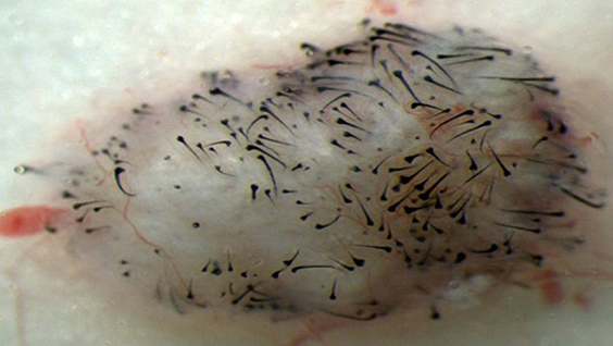
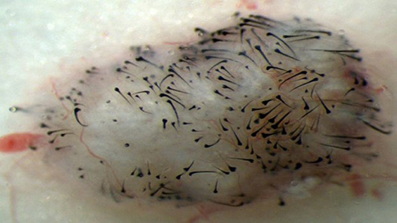
In a new study, Sanford-Burnham researchers have used human pluripotent stem cells to generate new hair. The study represents the first step toward the development of a cell-based treatment for people with hair loss. In the United States alone, more than 40 million men and 21 million women are affected by hair loss. The research was published online in PLOS ONE.

“We have developed a method using human pluripotent stem cells to create new cells capable of initiating human hair growth. The method is a marked improvement over current methods that rely on transplanting existing hair follicles from one part of the head to another,” said Alexey Terskikh, PhD, associate professor in the Development, Aging, and Regeneration Program. “Our stem cell method provides an unlimited source of cells from the patient for transplantation and isn’t limited by the availability of existing hair follicles.”

The research team developed a protocol that coaxed human pluripotent stem cells to become dermal papilla cells. They are a unique population of cells that regulate hair-follicle formation and growth cycle. Human dermal papilla cells on their own are not suitable for hair transplants because they cannot be obtained in necessary amounts and rapidly lose their ability to induce hair-follicle formation in culture.

“In adults, dermal papilla cells cannot be readily amplified outside of the body and they quickly lose their hair-inducing properties,” said Terskikh. “We developed a protocol to drive human pluripotent stem cells to differentiate into dermal papilla cells and confirmed their ability to induce hair growth when transplanted into mice.”

“Our next step is to transplant human dermal papilla cells derived from human pluripotent stem cells back into human subjects,” said Terskikh. “We are currently seeking partnerships to implement this final step.”

View translations of this story


Donate Now


Institute News

Targeting protein could improve diagnosis and treatment of lymphoma

AuthorGuest Blogger
Date

January 26, 2015

This post was written by Janelle Weaver, PhD, a freelance writer

Lymphoma is the most common blood cancer in the United States and is responsible for about 20,000 deaths each year. This type of cancer begins in white blood cells called lymphocytes, which normally play an important role in the immune system by recognizing and responding to pathogens such as bacteria and viruses. To control infections, these cells must multiply in a process that depends on a protein called MALT1. But when inappropriately activated, MALT1 drives the survival of lymphoma cells, underscoring the need to monitor the activity of this protein to develop novel diagnostic tests and facilitate drug discovery. Continue reading “Targeting protein could improve diagnosis and treatment of lymphoma”

Institute News

A new approach to treating osteoarthritis

AuthorSusan Gammon
Date

January 20, 2015

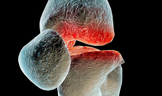
In a recent collaborative research study between two brothers—one a rheumatologist and the other a medical engineer—novel shaped nanoparticles were able to deliver anti-osteoarthritis drugs directly to the cells that drive the onset and progression of osteoarthritis (OA). The findings show promise to improve the treatment options for the nearly 21 million Americans, 25 years of age and older, that suffer from this chronic, often debilitating disease.

“We are excited to have developed nanoparticles which can efficiently and safely bring anti-OA drugs into the cells called chondrocytes that cause OA,” said Massimo Bottini, Ph.D., adjunct assistant professor in the Bioinformatics and Structural Biology Program at Sanford-Burnham. “Our method not only delivered the drug effectively, but stayed in the joint for a prolonged time without causing side effects. This is a significant improvement over previous attempts to deliver anti-OA drugs to affected joints.”

About osteoarthritis Under normal conditions, the extracellular matrix of the joint is maintained through a continual remodeling process in which low levels of different enzymes that produce and degrade cartilage are maintained. However, with increasing age and general wear and tear on joints, OA can occur when the enzymes that degrade cartilage are overproduced, creating an imbalance that leans toward the loss of collagen and joint impairment.

“For advanced OA, joint-replacement surgery is the only option for patients to regain comfortable and normal joint functions. For less severe cases, there is currently no medical therapy that can slow down or halt progression of the disease. This makes OA one of the largest unmet clinical needs in the field of rheumatology,” said Nunzio Bottini, MD, Ph.D., an associate professor in the Division of Cellular Biology at the La Jolla Institute for Allergy and Immunology (LIAI), who is also a practicing rheumatologist—and Massimo’s brother.

“The goal of treating OA is to restore the balance of the enzymes that control the matrix environment. Since there is no blood supply to the joint, drugs to treat the disease must be injected directly into the joint,” said Nunzio.

“Until now, scientists have tried using spherical nanoparticles to deliver anti-OA drugs. But the physical shape and size of the spheres predisposes them to diffuse into the synovial fluid and be flushed out of the joint before they can be effective,” said Massimo. “We have designed a one-dimensional linear nanoparticle made of graphite that is 100,000 times thinner than a human hair. This unique nanoparticle is engineered to travel through the negatively charged extracellular matrix and carry molecules to the nucleus of chondrocytes to turn off the genes that cause the disease.”

The study Using a mouse model for OA, the brothers injected the novel nanoparticle loaded with a gene inhibitor into the knees of affected mice. The nanoparticle delivered large amounts of the gene inhibitor to the cytoplasm and the nucleus of chondrocytes. Importantly, particles remained in the joint for two weeks compared to only few days for spherical nanoparticles.

“This is a significant improvement over previous attempts to deliver drugs to OA joints,” said Massimo. “Our next step is to further optimize the nanoparticle, see how long it remains in the body, and move to clinical studies in humans,” said Massimo.

Arthritis is a complex disease and integrated work between technologists—such as my brother Massimo—and biologists like me significantly increases the chance to make major treatment advances. Our next objective is to secure NIH funding to continue applying our complementary expertise to the quest to improve the lives of those suffering from arthritis,” added Nunzio.

The collaborative study was published in ACS Nano and performed at both Sanford-Burnham and LIAI. Cristiano Sacchetti, PhD, a shared postdoctoral fellow in Massimo and Nunzio Bottini’s laboratories was lead author on the paper.

A link to the paper can be found at: http://pubs.acs.org/doi/abs/10.1021/nn504537b.

Institute News

Ketogenesis prevents fatty liver disease

Authorsgammon
Date

January 12, 2015

A new study, published in the Journal of Clinical Investigation, suggests that ketogenesis may prevent non-alcoholic fatty liver disease (NAFLD). NAFLD is term used to describe the accumulation of fat in the liver of people who drink little or no alcohol. It affects approximately one billion individuals worldwide, has become a leading cause of cirrhosis, and increases the risk of cardiovascular disease, including heart attacks and stroke. Continue reading “Ketogenesis prevents fatty liver disease”

Institute News

Newly discovered cell stress pathway could hold therapeutic promise for diverse diseases

AuthorGuest Blogger
Date

January 5, 2015

This post was written by Janelle Weaver, PhD, a freelance writer.

When cells are faced with unfavorable environmental conditions, such as limited nutrient availability, the activation of adaptive stress responses can help protect them against damage or death. For example, stressed cells can maintain sufficient energy levels for survival by degrading and recycling unnecessary or dysfunctional cellular components. This survival mechanism, known as autophagy (literally, ‘self-digestion’), also plays key roles in a variety of biological processes such as development and aging, and is often perturbed in various diseases. Even though tight control of autophagy is key to survival, relatively little is known about the signaling molecules that regulate this essential process.

Sanford-Burnham researchers have made important progress in addressing this gap in knowledge by discovering that proteins called STK3 and STK4 regulate autophagy across diverse species. As reported recently in Molecular Cell, the newly identified mode of autophagy regulation could potentially have important clinical implications for the treatment of a broad range of diseases, including cancer, diabetes, Alzheimer’s disease, cardiac dysfunction, and immune-related diseases.

“Our discovery is fundamental to our molecular understanding of how autophagy is regulated,” said senior study author Malene Hansen, PhD, associate professor of the Development, Aging, and Regeneration Program at Sanford-Burnham. “Because impairment in the autophagy process has been linked to many disorders in humans, we believe that pharmacological agents targeting this novel regulatory circuit may hold great therapeutic potential.”

Critical kinases

Autophagy is a cellular recycling process involving a highly intricate and complex series of events. Cellular components such as abnormal molecules or damaged organelles are first sequestered within vesicles known as autophagosomes. These vesicles then fuse with organelles called lysosomes, which contain enzymes that break down various molecules. This fusion process results in the formation of hybrid organelles called autolysosomes, where the defective cellular components are enzymatically degraded and recycled. A protein called LC3 plays crucial roles in the formation of autophagosomes and the recruitment of dysfunctional cellular components to these vesicles. The signaling events that coordinate LC3’s various functions in autophagy have not been clear, but new research from the Hansen lab now proposes a novel and essential role for the mammalian Hippo kinases STK3 and STK4 in regulating autophagy by targeting LC3 for phosphorylation.

In their study, Hansen and her team describe that deficiency in both STK3 and STK4 impairs autophagy not just in mammalian cells, but also in nematodes and yeast. When exploring how the kinases regulate autophagy in mammalian cells, the researchers discovered that phosphorylation of LC3 by STK3 and STK4, specifically on the amino acid threonine 50, is critical for fusion between autophagosomes and lysosomes—an essential step in the autophagy process. “Collectively, the results of this study strongly support a critical and evolutionarily conserved role for STK3 and STK4 in regulating autophagy, by phosphorylating the key autophagy protein LC3, at least in mammalian cells,” Hansen said.

Killing bacteria

Previous studies have shown that STK4 also plays a role in regulating antibacterial and antiviral immunity in mammals, including humans. Moreover, autophagy is known to play a role in the clearance of intracellular pathogens. “These findings, taken together with our discovery that deficiency in STK3 and STK4 severely compromises autophagy, led us to test whether STK4 also plays a role in antimicrobial immunity through its function in autophagy,” said lead study author Deepti Wilkinson, Ph.D., a postdoctoral fellow in Hansen’s lab.

To test this notion, the researchers collaborated with Victor Nizet MD, professor of Pediatrics and Pharmacy  at UC San Diego and found that indeed mouse embryonic cells deficient in both STK3 and STK4 were unable to efficiently kill intracellular group A streptococci—bacteria known to be cleared by autophagy. However, an LC3 mutation that resulted in constant phosphorylation at threonine 50 restored the ability of the STK3/STK4-deficient cells to kill the bacteria. “This finding suggests that the same STK4-LC3 signaling pathway involved in autophagy also contributes to the response of mammalian cells to infection with intracellular pathogens and could play a role in human immune-related disease,” Wilkinson said.

Correcting defects

Moving forward, the researchers plan to further probe the molecular mechanisms by which STK3 and STK4 regulate autophagy. They will also investigate the therapeutic implications of the STK3/STK4 signaling pathway for tumor suppression as well as immune-related disorders such as bacterial and viral infections. “Understanding how autophagy works and why it sometimes stops to function optimally is essential for fighting diseases such as cancer, diabetes and neurodegeneration,” Hansen said.

“We have made a major contribution towards this endeavor by showing that STK3 and STK4 play an essential role in keeping the process of autophagy running smoothly by directly phosphorylating the key autophagy protein LC3. We hope our discoveries will lead to the development of effective drugs that can help correct autophagy defects that commonly occur in these diseases,” added Hansen.

A copy of the paper can be found at: http://www.ncbi.nlm.nih.gov/pubmed/25544559
Institute News

The switch that promotes kidney cancer progression and metastasis

Authorsgammon
Date

December 12, 2014

Kidney cancer is the sixth most common cancer in adults in the United States. Clear-cell kidney cancer, or clear-cell renal cell carcinoma (CRCC)—the cells appear pale and clear under a microscope—accounts for seven out of 10 people with kidney cancer, and is the most aggressive form of the disease. Because kidney cancer does not cause symptoms until the tumor has already grown, and is very difficult to detect in a routine physical examination, approximately 25 to 30 percent of patients aren’t diagnosed until the disease is metastatic. Unfortunately, metastatic clear-cell kidney cancer is currently incurable. Continue reading “The switch that promotes kidney cancer progression and metastasis”

Institute News

Enzymes could be key for brain cancer and Alzheimer’s disease

AuthorGuest Blogger
Date

December 9, 2014

This post was written by Janelle Weaver, PhD, a freelance writer.

Animals ranging from fish to humans produce a vitamin-A metabolite called retinoic acid, which plays an important role in growth and embryonic development and protects against diseases such as cancer. By regulating the activity of key genes, retinoic acid causes immature cells called embryonic stem cells to turn into mature, specialized cells such as neurons. “Neurons—the building blocks of the nervous system—are particularly important cell types in therapy, due to the fact that they normally don’t reproduce or replace themselves after they become damaged,” said Laszlo Nagy, MD, PhD, director of the Genomic Control of Metabolism Program and professor in the Diabetes and Obesity Research Center at Sanford-Burnham’s Lake Nona campus. “Despite their crucial role, we still have a limited understanding regarding the molecular programs that coordinate their functionality.” Continue reading “Enzymes could be key for brain cancer and Alzheimer’s disease”