Study shows neurons from grafted stem cells contain intrinsic codes for navigating and forming connections, may improve cell therapy for a brain short-circuited by stroke.
Some parts of our bodies bounce back from injury in fairly short order. The outer protective layer of the eye—called the cornea—can heal from minor scratches within a single day.
The brain is not one of these fast-healing tissues or organs. Adult brain cells are stable and last for a lifetime barring trauma or disease, whereas some cells lining our guts last only five days and must be continually replaced.
Scientists and physicians would like to use stem cell therapy to boost the brain’s ability to regenerate damage due to concussion or stroke. So far, these treatments have been stymied by changes in the brain due to injury, as well as difficulties with integrating regenerated cells into existing brain circuits to restore functions such as memory retention or motor skills.
Scientists at Sanford Burnham Prebys Medical Discovery Institute and Duke-National University of Singapore (NUS) Medical School published findings January 8, 2026, in Cell Stem Cell from testing a therapy derived from human stem cells. When transplanted into mice, the cells matured, integrated into existing circuits and restored function. By tracing the cells and sequencing their gene expression patterns, the researchers also revealed how transplanted cells find where they need to go and form connections with the nervous system.
The first challenge faced by hopeful regenerative medicine therapies for stroke and other forms of brain damage is the lack of a nurturing environment. Whereas the developing brain is a welcoming and instructive place for stem cells forming neurons and wiring the nervous system’s circuits, therapeutic cells arriving after a stroke find more hostility than hospitality.
“In the adult brain after a stroke, you see the formation of a cyst, a cavity that is filled with all sorts of inflammatory molecules, so it is a bit like the therapeutic cells are swimming in a dangerous swamp full of threats,” said Su-Chun Zhang, MD, PhD, the Jeanne and Gary Herberger Leadership Chair in Neuroscience and the director of and professor in the Center for Neurologic Diseases at Sanford Burnham Prebys.
“If that wasn’t enough, scar tissue surrounds the cavity to protect the brain from further damage, but it also forms a barrier against any potential regeneration.”
Some cell therapy strategists try grafting new cells next to the damaged region of the brain where it is easier for the cells to survive and grow. The goal is to eventually reestablish circuits by bypassing the damaged region. Zhang feels that this trauma needs to be healed rather than side-stepped to reach the potential benefits of regenerative medicine.
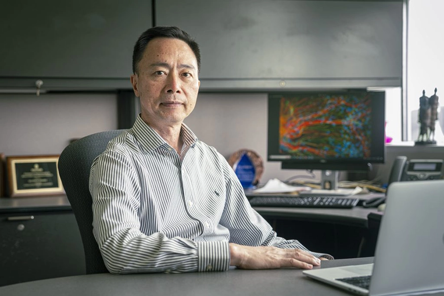
Su-Chun Zhang, MD, PhD, is the Jeanne and Gary Herberger Leadership Chair in Neuroscience and the director of and professor in the Center for Neurologic Diseases at Sanford Burnham Prebys Medical Discovery Institute. Image credit: Sanford Burnham Prebys.
“Following a stroke, the damaged lesion is often very large and presents an immense challenge to efforts to functionally reconnect the brain to the brain stem and spinal cord.”
Zhang and the research team sought to span this gap by developing a method to support the survival of therapeutic cells grafted directly into the harsh environment of the stroke cavity. Using a mixture of small molecule drugs and structural proteins, the scientists found that transplanted cells succeeded in surviving and growing to fill the damaged region.
“Once transplanted cells can survive and become neurons, then we started asking whether those neurons can break through the scar tissue and grow functioning nerves by making new connections and reconstructing the disruptedcircuits,” said Zhang.
While the researchers had proven it was possible to transplant cells and grow new neurons, they knew it would be of little benefit if they didn’t form the correct kinds of connections. Were they rebuilding bridges that had been demolished, orcreating new bridges to nowhere?
“We found that different types of transplanted neurons found their own partners even in the complicated context of the mature brain environment,” said Zhang. “They still can find their targets in a very specific manner.”
After conducting three-dimensional reconstruction of the transplanted neurons, the scientists observed that the patterns of long, spiny projections neurons use to form connections in the nervous system resembled the patterns seen innormal neurons populating the pathway between the cerebral cortex and spinal cord.
Next, the scientists sought to better understand the navigational abilities of these regenerated neurons. They used a genetic barcode to label and trace the transplanted cells. This data was combined with the results of sequencing the transplanted cells’ gene expression profiles.
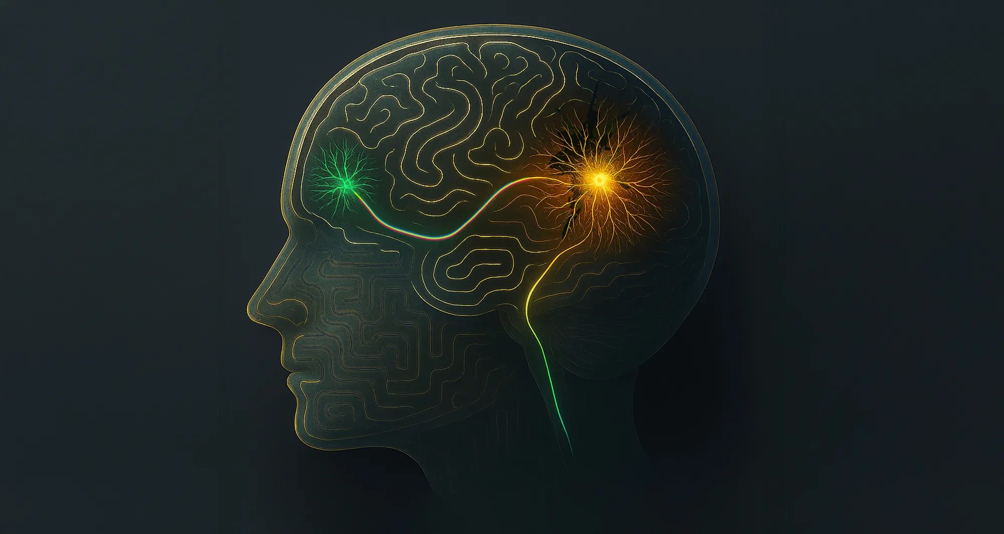
“We revealed that each cell type has its own code and, once the cells become neurons, this code tells each cell to send its projections or axons to different parts of the brain and spinal cord,” said Zhang.
“It’s the first time this striking phenomenon has been reported, and it is significant because it basically tells us that if we have the right types of transplanted cells, they already know where to go and what to do to repair what has been lost.”
The scientists used machine learning to identify four subtypes of neurons that develop from transplanted therapeutic cells. Each subtype has a distinct expression of genes known to guide the growth of axons, which explains why most neurons of a particular subtype send axons to form circuits with the same brain region.
The research team also validated how axonal projection patterns are affected by transcription factor proteins that modify gene expression. They tested stem cells modified without a transcription factor called Ctip2. These transplanted cells’ projection patterns varied significantly from those with the factor, with more axons seeking to form connections with the hippocampus and amygdala.
“By learning more about these subtypes of transplanted neurons, we may be able to predict their projections and connectivity in order to select appropriate neuronal cell types for targeted circuit reconstruction in patients,” said Zhang.
“It opens a promising future for cell therapy to help the millions of people that suffer from stroke and other devastating neurological conditions.”
Zhifu Wang, PhD, a research fellow at Duke-National University of Singapore (NUS) Medical School, shares first authorship of the study with Danyi Zheng, PhD, a 2025 graduate of Duke-NUS Medical School.
Additional authors include:
- Phil Jun Kang, staff scientist at Sanford Burnham Prebys
- Shu-Min Chou and Fei Ye from Duke-NUS Medical School
The study was supported by the National Medical Research Council of Singapore and Duke-NUS Medical School.
The study’s DOI is 10.1016/j.stem.2025.12.008.