Dr. Mao Tian is a Research Assistant Professor at Sanford Burnham Prebys Medical Discovery Institute. She is a computational biologist whose research focuses on developing artificial intelligence (AI) and machine learning approaches to understand cancer genomics, tumor evolution, and the tumor microenvironment. Her work integrates multi-omics data, including whole-genome sequencing, transcriptomics, and single-cell sequencing, to identify molecular signatures and uncover mechanisms underlying cancer development and progression. Her research also explores the application of deep learning and genomic foundation models to improve the interpretation of cancer-associated genetic alterations and advance precision oncology.
Education and Training
2021–2025: Postdoctoral Researcher, University of California, Los Angeles; with Dr. Paul C. Boutros 2017–2021: PhD in Medical Biology, University of Southern California; with Dr. Pinghui Feng 2012–2016: BS in Life Sciences and Technology, Sichuan University, Chengdu, China
Related Disease
Breast Cancer, Cancer, Head And Neck Cancer, Prostate Cancer, Thyroid Cancer
Techniques and Technologies
Computational Biology, Genomics, Machine Learning
Dr. Mao Tian’s research focuses on developing computational and artificial intelligence (AI) approaches to understand cancer development, tumor evolution, and the molecular mechanisms underlying disease progression. Given the extensive genetic and cellular heterogeneity of cancer, her work integrates large-scale genomic, transcriptomic, proteomic, and single-cell datasets to uncover the mechanisms driving tumor progression and treatment response. By combining multi-omics analysis with machine learning and AI foundation models, her research aims to identify novel cancer drivers, characterize tumor heterogeneity, and advance precision oncology. Her research program is organized into three major areas:
Cancer evolution and multi-omics integration
AI foundation models for cancer genomics
AI-driven computational methods for biomedical discovery
Select Publications
Showing 0 of 0
Michael Karin received a PhD in Molecular Biology from UCLA in 1979 and conducted postdoctoral research with Prof. Beatrice Mintz at Fox Chase Cancer Center from 1979 to 1980 and with Prof. John Baxter at UCSF from 1980 to 1983. He served as an Assistant Professor in the Department of Microbiology at USC School of Medicine from 1983 to 1985 and, in 1986, joined the Department of Pharmacology at UCSD School of Medicine, where he rose through the ranks to become a Distinguished Professor until his retirement in 2024. That same year, Karin joined the Sanford Burnham Prebys Medical Research Institute as the Director of the Center for Metabolic and Liver Diseases.
Throughout his academic career, Karin investigated stress and inflammation signaling, employing a full range of experimental approaches from biochemistry and molecular cell biology to animal pathophysiology. By discovering how infection, inflammation, radiation, or environmental stressors activate AP-1, NF-κB, and other transcription factors, he explored how these processes contribute to the pathogenesis of cancer, as well as degenerative and metabolic diseases. His group elucidated fundamental mechanisms by which inflammation and obesity promote tumor development and contribute to type II diabetes and insulin resistance, and was among the first to highlight the role of inflammation in metabolic disease.
Karin’s group established how members of the IL-6 cytokine family control the development of colorectal (CRC) and liver (HCC) cancers through activation of STAT3 and YAP, and identified cell type–specific mechanisms by which NF-κB activation via IκB kinases (IKK)—which his group was the first to discover—affects the pathogenesis of various cancers. His research also demonstrated that not only innate immune cells, such as macrophages, but also adaptive immune cells, including T regulatory cells and B lymphocytes, contribute to tumorigenesis. This body of work helped to establish the field of Inflammation and Cancer.
More recently, Karin’s group demonstrated the existence of immunosuppressive plasma cells and their role in negatively regulating immunosurveillance and immunotherapy. His current work focuses on HCC, CRC, and pancreatic ductal adenocarcinoma (PDAC). The team has developed robust mouse models for studying non-alcoholic steatohepatitis (NASH)-induced HCC and chronic pancreatitis–accelerated PDAC, using them to discover how adaptive immunity controls HCC development and to investigate the role of the autophagy substrate p62 in inflammation and HCC/PDAC pathogenesis.
They discovered that activation of the p62–KEAP1–NRF2 cascade confers resistance to autophagy inhibitors, leading to the development of a new PDAC and HCC therapeutic approach targeting autophagy and macropinocytosis. These mouse models have also been used to study how metabolism and nutrition affect NASH and HCC development, with a focus on the adverse effects of fructose and alcohol intake and exposure to toxicants.
Board Appointments, Conference Organization or Other Memberships
National Academy of Sciences, 2005 National Academy of Medicine, 2011 Associate member of the European Molecular Biology Association, 2007
Honors and Recognition
American Association for Cancer Research (AACR) G.H.A. Clowes Award for Outstanding Basic Cancer Research, 2020 William B. Coley Award for Distinguished Research in Basic and Tumor Immunology, 2013 Charles Rodolphe Brupbacher Prize for Cancer, 2013 Harvey Prize for Human Health, 2010 Morton I. Grossman Lectureship, 2003
Education
PhD, University of California Los Angeles, Molecular Biology, 1979 Postdoctoral fellowship, Fox Chase Cancer Center, 1980 Postdoctoral fellowship, University of California San Francisco, 1983
Related Disease
Cancer, Liver Cancer, Liver Diseases, Metabolic Diseases
Research in the Karin lab is currently focused on three general research areas: 1. Inflammation biology and how it is modulated by the individual metabolic and nutritional status. 2. Inflammation and metabolic disease, such as type 2 diabetes and metabolic dysfunction associated steatotic liver disease (MASLD). 3. The role of inflammation in cancer, including pancreatic, liver and gastrointestinal cancers. We have also started investigating the role of interorgan communication in metabolic disease and cancer.
The Karin lab has established that the main type of primary liver cancer hepatocellular carcinoma (HCC) arises in the context of chronic liver injury and inflammation. To better understand this process we have developed methods for the isolation of HCC Progenitor Cell (HcPC) and have been studying how they are generated from normal, healthy, hepatocytes. This investigation has led us to make the surprising discovery that HcPC are generated from senescent hepatocytes, questioning whether cellular senescence is an effective tumor suppressive mechanism as was assumed till now. Curiously, in the context of MASLD and its more aggressive manifestation, MASH, metabolic liver diseases that affect 30% of Americans, hepatocyte senescence is triggered as a protective response that limits diet-induced DNA damage. Yet, by limiting hepatocyte DNA damage, senescence prevents the death of damaged hepatocytes keeping them as “ticking time bombs” that give rise to cancer once they have found ways to re-enter the cell cycle and start proliferating. Induction of hepatocyte senescence also seems to give rise to insulin resistance, and we are currently studying the mechanistic connection between the two, focusing on the role autocrine type I interferon signaling, which is activated in response to hepatocyte DNA damage.
Another, but less common, liver cancer that arises in chronically injured livers is intrahepatic cholangiocarcinoma (ICC). Although ICC is a cancer of the small intrahepatic bile ducts its cell of origin remains somewhat mysterious as it can either be the cells that form the bile ducts (cholangiocytes), hepatocytes or bipotential hepatobiliary progenitors. We have recently developed a new mouse model for a rare liver, inflammatory liver disease called Primary Sclerotizing Cholangitis (PSC) that shows progression to ICC and tumors with a mixed HCC-ICC phenotype. We are using this model to gain novel insights to the pathogenesis of both PSC and ICC.
MASLD/MASH, the two most common chronic liver diseases, which are caused by over-nutrition and obesity, are accompanied by substantial alterations to liver macrophage populations, leading to disappearance of resident liver macrophages or Kupffer Cells and the appearance of lipid associated macrophages (LAM), which are derived from recruited monocytes. We have recently found that LAMs are composed of two subclusters, pathogenic LAM-1 and protective LAM-2. We are investigating the diversification of LAM into LAM-1 and LAM-2 and investigating how LAM-2 exert their protective activity which leads to a marked reduction in MASH severity. We are also testing the therapeutic potential of LAM-2 transplantation.
Another inflammation promoted cancer is the most common pancreatic cancer -pancreatic ductal adenocarcinoma (PDAC). We have been studying how chronic inflammation and stress allow KRAS induced premalignant lesions progress to full blown invasive PDAC focusing on an EZH2-NRF2 controlled autoregulatory loop and activation of the collagen receptor DDR1, which is specifically engaged by a fragment of collagen I and other fibrillar collagens that is generated by matrix metalloproteases (MMPs). We are currently identifying the MMPs that are most critical for collagen cleavage in the context of PDAC and also investigating how the inhibition of collagen cleavage results in downregulation of DDR1 and interference with PDAC growth and progression. Moreover, we examining whether and how MASLD stimulates PDAC metastasis to liver.
Alexander Strongin earned his PhD from Moscow State University in Russia in 1972 and his D.Sci. degree from the Institute of Microbial Genetics in Moscow in 1983. From 1982 to 1988, Dr. Strongin was head of the Laboratory of Functional Enzymology at the Institute of Genetics of Microorganisms in Moscow. He served as head of the Department of Biotechnology and Laboratory of Protein Engineering, Institute of Molecular Genetics, Russian Academy of Sciences, Moscow, from 1988 to 1990. From 1990 to 1994, he was a visiting professor of biochemistry in the Division of Dermatology at Washington University School of Medicine, St. Louis, Missouri. Dr. Strongin has worked in the La Jolla area since 1994, as senior staff scientist in the Biology Division at General Atomics, 1994-1995, and as senior staff scientist at the La Jolla Institute for Experimental Medicine, 1995-1999. Dr. Strongin joined Sanford Burnham Prebys on September 1, 1999.
Related Disease
Anthrax, Arthritis, Brain Cancer, Breast Cancer, Cancer, Hepatitis C, Multiple Sclerosis, Prostate Cancer, Type 1 Diabetes
Tumors produce unique enzymes, which by degrading the normal tissue provide an opportunity for tumor to grow in size and metastasize. These enzymes are called matrix metalloproteinases or MMPs. MMPs are a primary target for the design of anti-cancer pharmaceuticals. Our work aims to lay the foundation from which efficient therapeutics could ultimately be derived. Dr Strongin’s research is focused on the fundamental mechanisms involving MMPs in the processes of cell migration, cell proliferation and metastasis. The far-reaching goals of this pioneering research are to gain an understanding of how MMPs by cleaving the surrounding tissue and by affecting cell surface receptors govern locomotion of malignant cells. This knowledge is critical for design of efficient pharmaceuticals that may find applications in a variety of disease conditions including cancer, arthritis and stroke.
Alex Strongin’s Research Report
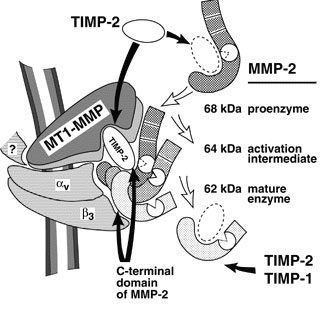
Degradation of the extracellular matrix and tissue remodeling play critical roles in tumor progression, particularly in invasion, metastasis, and neovascularization. Matrix metalloproteinases (MMPs) are essential for matrix proteolysis. Understanding the roles of MMPs in tumor growth and angiogenesis is of paramount importance to novel strategies for treatment of malignant tumors. Our primary goal is to characterize mechanisms of MMP activation involved in spatial and temporal control of focal proteolysis and to increase knowledge of cell-mediated matrix remodeling. Recently, we showed that heteromolecular assemblies involving MMPs and integrins that can simultaneously activate and dock MMPs are present on the surfaces of tumor cells. Apparently, these mechanisms of MMP activation are instrumental in clustering activated proteinases and integrins at the protrusions at the invasive front of migrating cells and at the invasive front of tumors (see Figure).
Interactions between MMPs and integrins, critical to spatial and temporal control of proteolysis, most likely are common for many cell types. To test our hypotheses and to outline novel approaches to control the activity of MMPs, we are examining mechanisms involved in activation, docking, and cross talk of MMPs and integrins in vivo and in vitro. We expect to experimentally show at the cell and protein sequence levels how the temporal and spatial regulation of matrix degradation is linked to dynamic changes of cell shape and cytoskeleton and to identify molecular mechanisms of cell motility. The immediate results of these studies will be improved diagnosis and treatment of malignant neoplasms.
Schematic depiction of the MMP-integrin molecular assembly. Inhibitory effects of tissue inhibitors of metalloproteinases (TIMPs) and the C-terminal domain of MMP-2 are shown by black arrows. The conversions of MMP-2 are shown by open arrows. The question mark stands for a cytoskeletal protein involved in a complex with membrane type-1 matrix metalloproteinase (MT1-MMP). This assembly is critical for docking, activation, and cross talk of MMPs and integrins and is intimately involved in regulating focalized matrix degradation and cell locomotion. MT1-MMP is in immediate proximity to integrin alpha vbeta 3. Both MT1-MMP and the integrin associate with the cytoskeleton. TIMP-2 links MT1-MMP (“a receptor”) and the secretory MMP-2 proenzyme. The second molecule of MT1-MMP (“an activator” that is free of TIMP-2) activates integrin alpha vbeta 3 by limited proteolysis of the beta 3 integrin subunit. The activator initiates the activation of pro-MMP-2 by cleaving the N-terminal part of the 68-kDa MMP-2 latent zymogen. The 64-kDa activation intermediate of MMP-2 efficiently associates with activated integrin alpha vbeta 3 via the C-terminal domain of the enzyme. The 64-kDa to 62-kDa autocatalytic maturation occurs if MMP-2 is complexed with the integrin. The mature MMP-2 enzyme transiently associated with the integrin at the surface of tumor cells accelerates the directional invasion of the cells. Additionally, if transiently complexed with the integrin at the surface of host stromal cells, the enzyme facilitates tumor neovascularization. TIMPs, including TIMP-2 and TIMP-1, efficiently inhibit the activity of soluble MMP-2. By providing links between integrins, MMPs, and TIMPs and, more generally, between cell shape and focal proteolysis, this model represents basic mechanisms of pro-MMP-2 activation and illustrates a coordinated interplay of inhibitors, proteinases, and integrin adhesion receptors at cell surfaces.
Arnold C. Satterthwait earned his PhD In Biochemistry with William Jencks from Brandeis University in 1973. He carried out postdoctoral research in Chemistry at Harvard University with Frank Westheimer, Imperial College with Alan Fersht and MIT with the Nobel laureate Gobind Khorana. In 1984, he joined The Scripps Research Institute in La Jolla, CA as an Assistant Professor. He moved to Sanford Burnham Prebys in 1998.
Related Disease
Anthrax, Breast Cancer, Cancer, HIV/AIDS, Prostate Cancer
The development of diagnostic reagents, drugs and vaccines is the visible outcome of a long process that spans the researcher’s laboratory and doctor’s office. The translation of disease discoveries into early detection, treatment, and prevention both tests and shapes our understanding of disease. Traditionally, drug companies have screened large collections of compounds against diseases to identify drugs. The Satterthwait lab seeks to take advantage of the explosion of new discoveries at the molecular level. We have developed synthetic methods that allow us to independently make and manipulate the critical three-dimensional regions of proteins that are being implicated in many diseases. These mini proteins are being used to assess new theories of disease at the molecular level to identify targets for various uses. We are currently using mini proteins to identify new antibodies (HIV-1), cancer drugs (prostate, breast and lung), and vaccines (anthrax).
Arnold Satterthwait’s Research Report
Peptide engineering relies on synthetic procedures to fold peptides into bioactive structures. It seeks to bridge a gap between chemistry and molecular biology by reducing the active sites of proteins to smaller molecules. Although synthetic peptides show occasional activity they are, unlike proteins, disordered and because of this often inactive. By refolding peptides into three-dimensional structures, they become active, opening up new avenues for studies on protein structure and function as well as providing leads for drugs and vaccines.
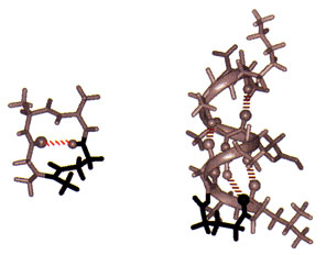
Solid-phase synthesis of a peptide constrained with a hydrazone covalent hydrogen bond mimic. The NMR structure of Sar-Ala-Ala-Gly (left) stabilized as a Type I turn (10% of protein structure) with an amidinium link (in black). The NMR structure of acetyl-Gly-Leu- Ala- Gly-Ala-Glu-Ala-Ala-Lys-Ala-amide (right) stabilized as an a-helix at its N-terminus with a hydrazone link (in black).
To fold peptides, we developed covalent hydrogen bond mimics. On average, greater than 60% of the amino acids in globular proteins engage in main-chain to main-chain hydrogen bonding (NH –> O=CRNH). In addition, protein substructures are defined by distinct hydrogen bonding patterns. Because hydrogen bonds are weak bonds, insufficient for stabilizing peptide structure, we replace them at structure defining positions in peptides with amidinium (N-C(R) = NH-CH2CH2) and hydrazone (N-N=CH-CH2CH2) covalent links. To simplify these transformations, we developed machine-assisted, multiple-peptide-synthesis procedures for inserting the hydrazone link into peptides which we link to automated multiple purifications. While these procedures, like peptide synthesis, remain labor intensive and often problematic, the critical problems have been breached.
Conformational analysis is as much a part of peptide engineering as synthesis because any claim to structure requires rigorous proof and further advances rely on understanding the relation between structure and chemistry. We have made considerable use of 2D NMR spectroscopy for structural analysis and calculations and with the help of collaborators, X-ray crystallography. These studies show unequivocally that covalent hydrogen bond mimics can stabilize peptides as b-turns, the a-helix, and even complex loops, which together make up the majority of protein substructures found on the surfaces of globular proteins.
Because protein substructure mimetics are now accessible, we have been examining the relationship between structure and activity by comparing the activities of peptides with substructure mimetics. From the several examples we have studied in detail, it is clear that remarkable gains in activity can be achieved.
Guy Salvesen earned his PhD in biochemistry from Cambridge University in 1980. He conducted postdoctoral research at Strangeways Laboratory and MRC Laboratory of Molecular Biology in Cambridge, followed by further post-doctoral training at the University of Georgia. In 1991 he was appointed Assistant Professor at Duke University. Dr. Salvesen was recruited to Sanford-Burnham Medical Research Institute in 1996, where he is professor and director of the Apoptosis and Cell Death Research Program and dean of the Graduate School of Biomedical Sciences. He also holds an adjunct position as professor in the Department of Pathology at the University of California, San Diego.
Education
1981: PhD, Cambridge University, England, Biology 1977: B. Sc., London University, London, England, Microbiology
Other Appointments
Adjunct Professor, Department of Pathology, University of California, San Diego
Honors and Recognition
2014: Organizer, Keystone Meeting on Cell Death, February 2013: IUBMB Gold Medal Recipient, October 2010: Keynote Speaker, European Cell Death Organization Conference, 2010: Keynote Speaker, Gordon Research Conference on Cell Death 2009: Lifetime Achievement Award of the International Proteolysis Society 2008: Keynote Speaker, Queenstown Molecular Biology Conference 2008: Chair, Gordon Research Conference on Cell Death 2005: Helmut Holzer Memorial Prize 1999: International Proteolysis Society, Elected Secretary 1999: Keynote Speaker, Gordon Research Conference on Matrix Metalloproteinases 1988: American Association for the Study of Liver Diseases, State of the Art Lecture 1996: Chair, Gordon Research Conference on Proteolytic Enzymes and Their Inhibitors
Related Disease
Cancer, Inflammatory/Autoimmune Disease, Neurodegenerative and Neuromuscular Diseases, Pancreatic Cancer, Skin Cancer and Melanoma, Structural Biology
Phenomena or Processes
Apoptosis and Cell Death, Caspase Family, Cytokines, Inflammation, Protein Structure-Function Relationships, Proteolytic Pathways, Ubiquitin, Ubiquitin Protease System and Ubiquitin-like Proteins
Research Models
Bacteria, Human Cell Lines, Mouse, Mouse Cell Lines, Primary Human Cells
Techniques and Technologies
Biochemistry, Cellular and Molecular Imaging, Chemical Biology, Fluorescence Microscopy, Mass Spectrometry, Protein Engineering, Protein Structure Prediction, Protein-Protein Interactions, Proteomics
The human body contains cells with different life expectancies. Some (white blood cells or skin, for example) are programmed to rapidly die and be replaced. Others (such as nerve cells) are programmed to survive the lifetime of the individual and are seldom replaced. Dr. Salvesen’s research focuses on the central role enzyme pathways play in the life and death of cells. When death pathways slow down in cells that are normally programmed to die, cancer results. Conversely, when death pathways become overactive in cells that are programmed to survive, degenerative disease occurs. Dr. Salvesen’s laboratory focuses on understanding the fundamental molecular interactions that occur within these enzyme pathways. This knowledge is used to engineer synthetic compounds to stimulate cell destruction in cancer cells, or delay cell destruction in neurodegenerative diseases and stroke.
Guy Salvesen’s Research Report
Structure and Function of Proteases and Their Natural Inhibitors
Our research seeks to delineate the structure –> activity –> function algorithm as it applies to proteases and their inhibitors. Our laboratory has very broad interests in principles of proteolysis in humans, and we take multi-pronged approaches to research on proteases and their inhibitors.
Apoptosis
In one approach we apply basic biochemical knowledge to investigate newly emerging principles of proteolysis in human systems. This research is currently dissecting the proteolytic components of the intracellular pathway that lead to apoptotic cell death. Programmed cell death monitors the growth of new cells and the elimination of old ones. This program contains a number of proteolytic steps that are essential for efficient execution of the death pathway. Thus the proteases of the pathway – the caspases – are involved in the normal maintenance of correct cell number, and are therefore implicated in a number of pathologic and physiologic conditions. Using the techniques of protein chemistry, enzymology, crystallography, and recombinant DNA methodologies, we analyze the basic mechanism utilized by caspases to promote cell death pathways, and the mechanisms and specificity of the natural inhibitors that control them.
Cell Signaling
Modification of proteins by the small ubiquitin-like modifier SUMO is a dynamic and reversible process. The SUMO cycle begins when SUMO precursors are processed to remove short C-terminal extensions, thereby uncapping the C-terminal Gly-Gly motif that is essential for conjugation. SUMO ligases conjugate the protein, via its C-terminal carboxylate, to the side-chain lysine of target proteins to generate an isopeptide linkage. Eventually, SUMO is removed intact from its substrate SUMOylated proteins, and so the SUMOylation/deSUMOylation cycle regulates SUMOs function. A group of proteases known as SENPs are involved in both the activation of SUMO precursors (endopeptidase cleavage) and deconjugation of the targets (isopeptidase cleavage). Our laboratory is currently involved in projects to define the mechanisms that regulate SENP activity and access to their natural substrates.
Technology Development
The principle of proteolysis in vivo is to instigate irreversible changes to a set of protein substrates that alters their function and generates the required biological event. The sum total of the proteases and their target substrates operating in a physiologic pathway therefore defines the global event. Consequently, the identity of the substrate cleavages defines the proteases acting on them. We are developing proteomics-based methodologies, including selective protein labeling, multi-dimensional electrophoresis, and mass spectrometry techniques, to identify the products of proteolysis in vivo.
Erkki Ruoslahti earned his MD and PhD from the University of Helsinki in Finland in 1967. After postdoctoral training at the California Institute of Technology, he held various academic appointments with the University of Helsinki and the University of Turku in Finland and City of Hope National Medical Center in Duarte, California. He joined Sanford Burnham Prebys in 1979 and served as its President from 1989-2002. He was a Distinguished Professor at University of California Santa Barbara in Biological Sciences 2005-2015. His honors include elected membership to the U.S. National Academy of Sciences, National Academy of Medicine, American Academy of Arts and Sciences, and the European Molecular Biology Organization, the Japan Prize, Gairdner Foundation International Award, G.H.A. Clowes Award, Robert J. and Claire Pasarow Foundation Award, and Jacobaeus International Prize. He was a Nobel Fellow at the Karolinska Institute in Stockholm in 1995, and is an Honorary Doctor of Medicine from the University of Lund, as well as a Knight and Commander of the Orders of the White Rose the the Lion of Finland. In 2022, Dr. Ruoslahti was announced as one of three winners of the Albert Lasker Basic Medical Research Award.
Education
1966: MD, University of Helsinki in Finland 1967: PhD, University of Helsinki in Finland
Awards and Honors
2022: Albert Lasker Award for Basic Medical Research Commander of the Order of the Lion of Finland Knight of the Order of the White Rose of Finland 2012: Thomson Reuters Citation Laureate 2005: Japan Prize in Cell Biology 2003: Jubilee Lecturer, Biochemical Society 1998: Jacobaeus International Prize 1997: Gairdner Foundation International Award 1995: Nobel Fellow at the Karolinska Institutet in Stockholm 1991: Honorary doctorate in medicine from Lund University, Sweden 1990: American Association for Cancer Research – G.H.A. Clowes Memorial Award
Member
National Academy of Sciences National Academy of Medicine American Academy of Arts and Sciences European Molecular Biology Organization
Related Disease
Alzheimer’s Disease, Atherosclerosis, Brain Cancer, Breast Cancer, Cancer, Prostate Cancer
The Ruoslahti laboratory studies peptides that home to specific targets in the body, such as tumors, atherosclerotic plaques and injured tissues. These peptides, which usually bind to receptors in the vessels of the target tissue, can be used to selectively deliver diagnostic probes and drugs to the target. The latest development is the discovery of homing peptides with tumor-penetrating properties. The CendR tissue penetration pathway is a new endocytosis/trans-tissue transport pathway (Pang et al., Nat Comm. 2014). The current focus is on enhancing the effects of coupled and co-injected drugs with the tumor-homing peptides, particularly in mouse models of breast cancer and glioblastoma. This laboratory also studies the receptors for the peptides and the mechanism of their tumor penetration activity.
Erkki Ruoslahti’s Research Report
Dr. Ruoslahti’s main scientific contributions are in the field of cell adhesion. He was one of the discoverers of fibronectin. His laboratory subsequently discovered the RGD cell attachment sequence in fibronectin and isolated RGD-directed cellular receptors, now known as integrins. The RGD discovery has led to the development of drugs for diseases ranging from vascular thrombosis to cancer.
Dr. Ruoslahti current studies deal with peptides that specifically target a diseased tissue, particularly its blood vessels. The peptides can be used to deliver drugs and nanoparticles to sites of disease, such as a tumor. The molecules targeted by such disease-specific peptides are of interest regarding their possible role in the disease and potential targets for drug development.
Vascular Zip Codes
The Ruoslahti laboratory screens large collections (“libraries”) of random peptides to identify those that bind to specific targets in tissues. The peptides in the library are displayed on the surface of phage (a virus that infects bacteria), and the screening is done in vivo. When the library is injected into the circulation of a mouse, phage particles that display peptides capable of binding to a selected target tissue, such as a tumor, accumulate at the target where they can be collected and their peptide identified. The process primarily probes the vasculature of the target tissue, unless the vasculature is very leaky. The method has revealed a wealth of specific features, or “vascular zip codes”, in the vessels of individual tissues and tumors. Peptides that specifically home to tumors because they recognize angiogenesis-associated or tumor-type specific markers in tumor blood vessels and can even distinguish the vessels of pre-malignant lesions from those of fully malignant tumors. Homing peptides have also revealed a zip code system of molecular changes in tumor lymphatics.
Synthetic homing peptides have been used to target drugs, biologicals, and nanoparticles into tumors. The targeting can increase the efficacy of a drug while reducing its side effects. Even a non-specifically toxic compound can be converted into a compound that selectively affects the targeted tissue. The peptides make it possible to identify the target molecules (receptors) for the peptides. The receptors of tumor-homing peptides often play a functionally important role in tumor vasculature, and because of this are candidates for drug development.
Tumor-penetrating Peptides
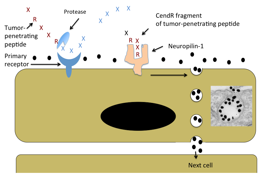
A few years ago the laboratory discovered peptides that not only home to tumor vessels, but are transported through the vascular wall and deep into tumor tissue. The key feature of these peptides is a R/KXXR/K sequence motif, named C-end Rule (CendR) motif or element. In tumor-penetrating peptides, the CendR element is cryptic. These peptides penetrate into tumor tissue in a 3-step process: (i) The peptide binds to a primary receptor on tumor endothelium. In iRGD, the RGD motif recognizes the avb3/avb5 integrins; the primary receptor for the LyP-1 family of peptides is cell surface p32/gC1qR. (ii) The peptide is then cleaved by a protease to expose the CendR element at the C-terminus of the peptide; and, (iii) the CendR element mediates binding to neuropilin-1 (NRP-1), to induce vascular and tissue penetration. The CendR transport pathway triggered through NRP-1 resembles macropinocytosis, but differs from it in being receptor-mediated. Importantly, the responsiveness of the pathway to triggering through NRP-1 is regulated by the nutrient status of cells and tissues. Its physiological function is likely to be to transport nutrients into tissues that lack them. Our ability to trigger the pathway specifically in tumors makes it useful in delivering drugs into tumors.
Targeting the Brain
The Ruoslahti laboratory has recently also applied phage screening to the identification of peptides that target brain diseases. So far, a peptide that specifically recognizes sites of brain injury, and a panel of peptides that are specific for Alzheimer’s brain have been obtained. This topic will be an expanding focus of the laboratory in the near future.
Nanomedicine
A major focus is to use homing peptides as targeting elements to deliver nanoparticles into tumors and other sites of disease. Nanoparticles are considered a promising new approach in medicine because they can be designed to perform more functions than a simple drug. The vasculature is an excellent target for nanoparticles because tumor vessels are readily available for circulating particles. In collaboration with chemistry and bioengineering laboratories, multifunctional nanoparticles for tumor targeting have been constructed. These particles can be directed into tumors in a highly selective manner as demonstrated by histology, non-invasive imaging, and tumor treatment results. The laboratory has constructed nanoparticles with the ability to amplify their own homing to tumors, and are currently working on nanoparticles coated with tumor-penetrating peptides. More recent work has dealt with nanoparticles that target brain diseases or atherosclerotic plaques. The general goal is to engineer nanoparticles with multiple functions. In addition to the specific targeting, such functions include avoidance of the reticuloendothelial system, self-amplification of the targeting, exit from vessels into tissue, ability to send signals for imaging, and controlled drug delivery.
Schematic Representation of the CendR Trans-tissue Transport Pathway
Note that CendR effect enhances the tissue penetration of molecules (depicted here as a black dots) that are co-administered with the peptide, as well as of cargo coupled to the peptide. The inset shows an electron microscopic image of a CendR endocytic vesicle that is budding from the cell surface into the cytoplasm and contains CendR peptide-coated gold nanoparticles (dark dots) See Ruoslahti, Adv. Drug Deliv. Rev. 2016.
Barbara Ranscht earned her PhD in Cell Biology/Developmental Neurobiology from the University of Tübingen, Germany in 1981. Her postdoctoral training was at King’s College in London, U.K., and the Massachusetts Institute of Technology in Cambridge, Massachusetts. Dr. Ranscht joined Sanford Burnham Prebys in 1987, and holds an adjunct professorship in the Department of Neurosciences at University of California, San Diego. From 1989 to 1992, Dr. Ranscht was the recipient of a McKnight Scholarship.
Education
1981: PhD, University of Tübingen, Germany Neurobiology
Related Disease
Attention-deficit hyperactivity disorder (ADHD), Cancer, Cardiovascular Diseases, Metabolic Syndrome, Multiple Sclerosis, Neurological and Psychiatric Disorders
Our lab studies cell surface interactions that regulate signaling networks in the nervous and cardiovascular systems and in cancer. With focus on the brain, we investigate membrane glycoproteins that enable crosstalk of neurons with the environment during circuitry development and disease. Our group has contributed seminal insights into the functions of Contactin-1 (Cntn1) and T-cadherin (Cdh13) in axon guidance, synapse formation and myelination using knock-out mouse models as well as biochemical, electrophysiological, histological and cellular approaches. Our current work explores the role of T-cadherin in brain circuitries enabling learning and memory formation, and determines how Contactin-1 functions in processes of myelination and remyelination. Exploiting our mouse genetic model, we uncovered a novel function for T-cadherin in protecting against stress-induced cardiac injury, and revealed disruptions of T-cadherin-mediated cellular interactions in cancer. In these latter contexts, our research identified T-cadherin as a major receptor for Adiponectin (Adipoq), a fat-secreted, circulating hormone that is of high clinical interest for its role in positively regulating energy balance and protecting against cellular insult. Current work in the lab aims to understand the molecular processes and signaling pathways regulated by T-cadherin or Contactin-1 in nervous system, cardiovascular and cancer functions with the goal to derive translational applications.
Barbara Ranscht’s Research Report
Functions of T-cadherin/Cdh13 in Neuronal Circuits Involved in Learning and Memory
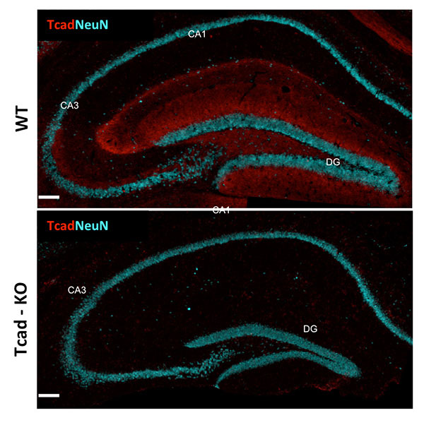
Cell adhesion molecules play seminal roles in synapse formation, stability and function in the central nervous system (CNS). T-cadherin (Cadherin-13; Cdh13), a glycosylphosphatidylinositol-linked cadherin-type cell adhesion molecule discovered in our laboratory, is prominent in diverse structures of the CNS. Mutations in the human CDH13 gene are linked to neuropsychiatric disorders, most prominently Attention Deficit Hyperactivity Disorders and its comorbidities. Our lab generated mice deficient for T-cadherin gene expression (Tcad-KO) mice to uncover the T-cadherin-dependent neuronal circuitries and probe its contribution to cognitive function. Tcad-KO mice show no overt developmental, neurological or sensory deficiencies, and are indistinguishable from wild type littermates under normal housing conditions. However, challenging the mice in behavioral tests revealed defects in performing select learning tasks. Investigating hippocampal circuitry as a central relay station for learning and memory, we identified reductions in CA1 synaptic transmission and long-term potentiation along with reduced spine densities and maturation of hippocampal pyramidal neurons. Current work aims to link T-cadherin to specific synaptic circuitry, and probe its function through conditional T-cadherin ablation in select neuron populations of the interconnected circuitry to contribute principal understanding to human cognitive disorders.
Figure 1: T-cadherin expression in adult hippocampus. T-cadherin staining delineates the dendritic layer of pyramidal neurons in CA1-CA3 subfields, and the molecular layer in the dentate gyrus. Staining is absent from the hippocampus of Tcad-KO mice. NeuN outlines the positions of pyramidal and granule neurons for orientation. Images by Hedie Badieh-Madhavi, PhD and Jinsha Liu, graduate student.
Mechanisms of Myelination and Remyelination
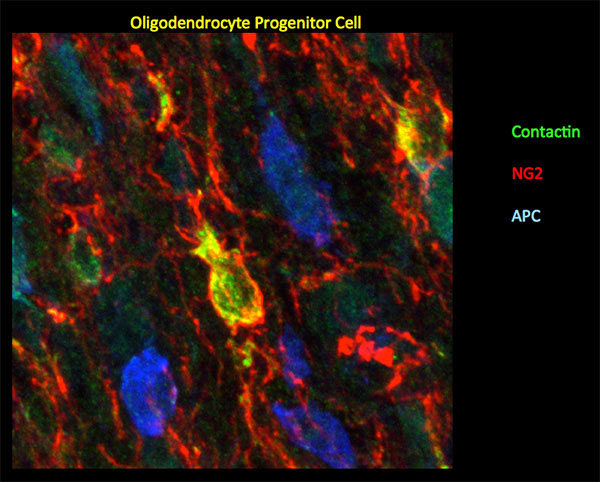
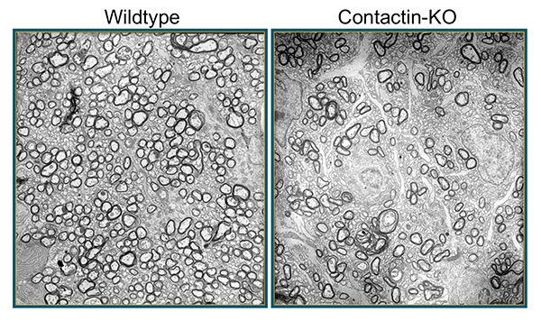
Myelin, a multilayered membrane sheath formed by oligodendrocytes around axons in the central nervous system (CNS) enables rapid nerve impulse conduction and sustains neuronal health and survival. Our lab is interested in the signals exchanged between axons oligodendrocytes during myelin formation and repair. Contactin-1, a GPI-linked cell surface glycoprotein, studied in our lab has emerged as one of the critical proteins to regulate axon-glia communication. In peripheral myelinated nerves, Contactin is solely expressed by neurons, and together with Caspr clusters at the paranodal axon domain to orchestrate formation of the septate-like axoglial junctions. In the CNS, Contactin delineates paranodes and nodes of Ranvier in mature myelin, and oligodendrocytes during the process of active myelination. We recently documented a vital dual role of Contactin-1 in central myelin formation: Contactin-mediated interactions between axons and oligodendrocytes regulate the extension and wrapping of myelin membranes, and loss of these interactions leads to hypomyelination in the mouse knockout model. The residual myelin is non-functional due to disrupted paranodal junctions that are characterized by loss of Contactin-associated paranodal Caspr, mislocalized potassium channels and disrupted transverse bands. Current work in the lab investigates the distinct contributions of Contactin in neurons and oligodendrocytes in mice with conditional Contactin-1 deletion, myelinating co-cultures and mouse models of myelin injury. If indicated, this work will form the basis for translational aspects of myelin repair.
Figure 2: In mouse P10 optic nerve, Ccntactin (green) delineates oligodendrocyte progenitor cells demarcated by NG2 (red). Staining for Contactin does not overlap with APC (blue), a marker for mature oligodendrocytes. Image by Gulsen Colakoglu, PhD
Figure 3: In mice deficient for Contactin-1, loss of axon-glia cross-talk leads to hypomyelination. Shown here are electron micropgraphs from P17 wildype and knockout (KO) optic nerves. Images Erik O. Berglund, MD, PhD
Adiponectin – T-cadherin/Cdh13 Signaling in Cardioprotection
Adiponectin is of significant clinical interest for its potency in counterbalancing the adverse effects of obesity-linked metabolic disease and cardiovascular dysfunctions. Research in the field is increasingly focusing on local actions of APN in its target tissues where binding to the cell surface leads to activation of intracellular signal transduction pathways that increase cellular energy and counteract adverse processes such as generation of reactive oxygen species, misbalanced cellular metabolism, and apoptotic cell death. Our lab identified T-cadherin as a major ligand-binding receptor for Adiponectin in the cardiovascular system (Denzel et al, 2010). T-cadherin effectively sequesters APN from serum and its absence leads to excessive APN concentrations in the circulation. Under cardiac stress, induced by pressure overload, T-cadherin is necessary to confer cardioprotection through APN. Downstream, APN engagement with T-cadherin activates the AMP-activated protein kinase (AMPK), a major downstream signaling target of APN. Since T-cadherin is a GPI-linked cell surface glycoprotein lacking the cytoplasmic region, it is our working model that T-cadherin connects through associated molecules to intracellular signaling pathways. We are exploring the heptahelical APN-receptors AdipR1/R2 as prime candidates while considering alternative possibilities.
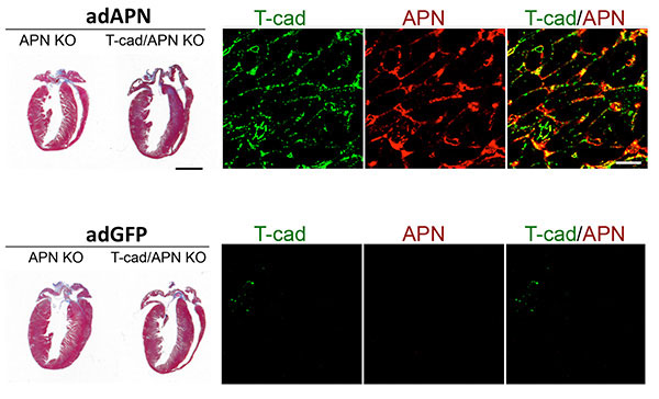
Figure 4: T-cadherin requirement in reverting exaggerated cardiac hypertrophy in APN-KO mice. Trichorme-stained hearts (left) show cardiac hypertrophy induced by pressure overload. Hypertrophy can be reverted in Adiponectin knockout (APN KO) mice by adenovirus-mediated expression of recombinant Adiponectin (adAPN; upper panel). In the absence of T-cadherin, APN-KO cardiac tissue does not respond (upper panel). Control virus expressing green fluorescent protein (adGFP) has no effect on the heart hypertrophic phenotype (lower panels). Mechanistically, as shown by immunohistochemistry, low T-cadherin expression in APN-KO hearts (Tcad, green, lower panel) is upreguated by adAPN (Tcad, green, upper panel) and binds adAPN which triggers downstream cardioprotective pathways. Data by Martin Denzel, PhD
Combining our cellular and functional analyses of T-cadherin with the extensive expertise in heart function at our Institute, we hope to make headway in determining APN-associated signaling pathways involving T-cadherin and AdipoR receptors. This work will fill the current knowledge gap of understanding APN interactions with its target cells in the cardiovascular system and may aid the development of novel molecular approaches for cardioprotection.
T-cadherin/Cdh13 in Cancer
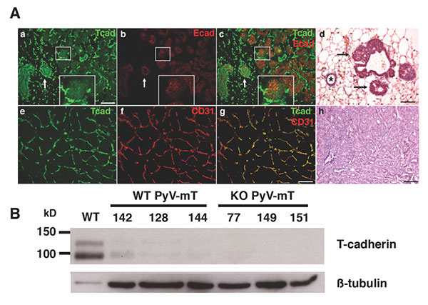
T-cadherin has also emerged as one of the key molecules altered in diverse types of cancer. Epithelial-mesenchymal transition (EMT) during neoplasia is characterized by the down-regulation of T-cadherin/CDH13, and this silencing is linked to increased proliferative and invasive potential of human cancers. We used the mouse genetic model to investigate functions of T-cadherin in tumors. In the mouse mammary tumor virus-driven polyoma middle T (MMTV-PyV-mT) model, T-cadherin is down-regulated from neoplastic epithelial cells while it is upregulated in the tumor vasculature. Under conditions of T-cadherin deficiency, MMTV-PyV-mT induced tumor growth occurs with delayed onset and at reduced growth rates that result in prolonged animal survival over non-mutant mice. Our analyses identified reduced tumor vascularization as the major underlying mechanism for this beneficial outcome. T-cadherin’s pro-angiogenic role is mediated by the association of Adiponectin: The APN-deficient MMTV-PyV-mT tumor phenotype mirrors the defects in T-cadherin knockout mice, and in vitro silencing of T-cadherin in endothelial cells abrogates the cellular responses to APN. Our lab is now interested in understanding the mechanism by which T-cadherin binding to Adiponectin affects endothelial cell function and develop strategies to control tumor angiogenesis.
Figure 5: T-cadherin in the neoplastic mammary gland. A. In early stage MMTV-PyV-mT mouse tumors, T-cadherin (green) and E-cadherin (red) are down-regulated from developing neoplasias (a-c; central white box magnified as insert) while both cadherins are expressed on the non-cancerous ductal epithelium (white arrows in a-c), Histology shows the pathology of a normal duct (asterisk) and developing neoplasias (d, black arrows; scale bars 50 mm). In developed MMTV-PyV-mT tumors, T-cadherin (green in e) is upregulated on the CD31-positive tumor vasculature (f and g). The pathology of a MMTV-PyV-mT tumor is shown in f. B, T-cadherin is detected as a protein doublet of 95 kD and 115kD by Western blotting of normal mouse mammary fat pads and is down-regulated in the neoplastic mammary gland. Beta tubulin Is used as a gel loading control. No T-cadherin protein is detected in the T-cadherin-deficient (KO) MMTV-PyV-mT tumors. Data by Lionel Hebbard, PhD
Dr. Levine is Emeritus Professor at Sanford Burnham Prebys. Prior to that, he was a Professor in the Department of Pediatrics at the University of California, San Diego School of Medicine, where he continues to see children with inherited metabolic diseases. Dr. Levine received his undergraduate degree in biochemistry from Harvard and his MD and PhD degree in genetics from the University of Washington. His clinical training as a pediatric geneticist was at the Children’s Hospital of Philadelphia. Dr. Levine has been working in the field of cell transplantation therapies for diabetes and b-cell biology for more than fifteen years. His laboratory was the first to develop immortalized cell lines from the human endocrine pancreas as models of beta-cell growth and differentiation. He has made insights into cellular senescence in the endocrine pancreas, finding that b-cells undergo rapid senescence when stimulated to proliferate. Most recently, he and his co-workers demonstrated the existence of endocrine stem cells in the adult human pancreas. The laboratory continues to pursue the development of cell therapies for diabetes using a variety of approaches, including high throughput screening.
Education
1979-86: PhD, University of Washington (Genetics) 1979-86: MD, University of Washington 1975-79: A.B., Harvard University (Biochemistry)
Postgraduate Training
1989-91: Genetics Fellow, Dept. of Pediatrics, UCSD School of Medicine 1988-89: Clinical Genetics Fellow, Children’s Hosp. of Philadelphia 1987-89: Pediatric Resident, Children’s Hosp. of Philadelphia 1986-87: Pediatric Intern, Children’s Hosp. of Philadelphia
Other Appointments
Health Sciences Clinical Professor of Pediatrics, UCSD School of Medicine Attending Physician, Rady Children’s Hospital
Related Disease
Cancer, Childhood Diseases, Metabolic Diseases, Obesity, Type 1 Diabetes, Type 2 Diabetes
Diabetes is a disease in which there is an insufficient amount of insulin, produced by the pancreatic b-cells, to control the blood glucose concentration. In type I diabetes, the b-cell are destroyed by an autoimmune response, while in type II diabetes, the b-cells are dysfunctional and are ultimately lost because of obesity-associated pathology. Our laboratory is interested in how b-cells regenerate under normal and pathophysiological conditions, with the goal of developing new therapies for diabetes that result in an increased number of those cells. Our studies of b-cell growth have also led us to develop lead compounds that are active in cancer, an interesting connection as both obesity and diabetes are now recognized as major risk factors for cancer.
Fred Levine’s Research Report
The major interest in the laboratory is pancreatic beta-cell biology and specifically the control of beta-cell growth and differentiation in the adult pancreas. In both type I and type II diabetes, β-cell mass decreases and is a major factor in the pathogenesis of diabetes. Thus, one major question in which we are interested is the existence and nature of adult endocrine stem/progenitor cells. Using both human and rodent models, we study cells in vitro and in vivo to determine the competence of various cell populations to undergo endocrine differentiation in response to defined stimuli. The goal is to achieve a sufficient understanding of the process of adult β-cell regeneration to allow us to enhance β-cell mass in diabetes.
A second major area of emphasis is to use high-throughput screening to discover compounds that modulate important aspects of β-cell biology. Our focus has been on modulation of insulin promoter activity, as that gene is acted upon by many diabetogenic stimuli. We have isolated and are characterizing a number of compounds and genes that were discovered in the screening process. Recently, we have discovered ligands for the orphan nuclear receptor HNF4a. HNF4a is involved in a number of disease processes, including diabetes, liver disease, and cancer.
Michiko N. Fukuda earned her PhD in biochemistry at the University of Tokyo in 1980. She did postdoctoral work at Fred Hutchinson Cancer Research Center in Seattle prior to her recruitment to Sanford-Burnham Medical Research Institute in 1982.
Education
1980: PhD, University of Tokyo, Biochemistry 1970: MS, University of Tokyo, Biochemistry 1968: BS, Tokyo University of Education, Botany
Related Disease
Breast Cancer, Cancer, Congenital Disorders of Glycosylation, Endometriosis, Glycosylation-Related Disorders, Inherited Disorders, Ovarian Cancer, Prostate Cancer, Testicular Cancer
Michiko Fukuda’s Research Report
Identification of Peptide that Delivers Drugs to Tumors
Chemotherapy effectiveness is often limited by drug toxicity in healthy tissues, although methods that spare normal cells by delivering drugs specifically to tumors may help to overcome this constraint. We have identified a promising tumor-targeted drug delivery vehicle known as the IF7 peptide. Using in vitro assays, we found that the IF7 peptide bound to the protein annexin 1 (Anxa1), which is known from previous studies by others to be enriched on the surface of tumor vasculature in several tumor types. When we injected a fluorescently labeled IF7 peptide into mice with tumors, fluorescent signals appeared in the tumors within one minute of injection. By contrast the tumors showed no fluorescence when mice were pre-injected with anti-Anxa1 antibodies that inhibits IF7-Anxa1 binding, suggesting that the peptide targets tumor by homing to Anxa1.
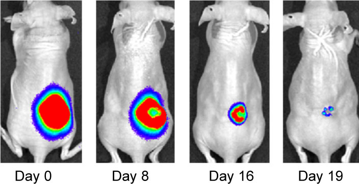
Effect of IF7-conjugated anti-cancer drug SN-38 on colon cancer mouse model. Activa cancer cells in a large tumor produce chemiluminescence which is captured by imaging. (Hatakeyama et al, 2011).
When IF7 peptide was conjugated with potent anti-cancer drug SN-38 and IF7-SN38 conjugate was injected intravenously to mice with tumors, IF7 could deliver SN-38 to tumors. We found that daily injections of an IF7-SN38 conjugate reduced a large tumor in the mouse without apparent side effects, whereas non-homing peptide-SN38 conjugate or with SN-38 alone did not reduce the tumors (Hatakeyama et al, 2011). The findings suggest that IF7 peptide may represent a clinically relevant vehicle for anti-cancer drugs.
Role of Trophinin in Human Embryo Implantation and Cancer
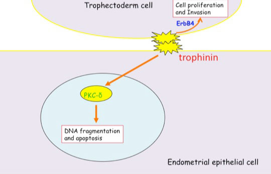
Invasion of the trophoblast into the endometrium, an essential element of embryo implantation, resembles invasion of malignant tumors. At the initial phase of implantation, the trophoblast and the uterine epithelium establish their first contact via their respective apical cell membranes. We have identified new molecules, trophinin, tastin, and bystin that mediate cell adhesion between trophoblastic cells and endometrial epithelial cells at the respective apical cell membranes. Trophinin is an intrinsic membrane protein, and tastin and bystin are cytoplasmic proteins. All of these molecules are strongly expressed in cells involved in embryo implantation in humans. However, trophinin is not expressed in human endometrial epithelia throughout the hormonal cycle, except only those cells located close to the implanting blastocyst. Trophinin expression by endometrial epithelia is induced by human chorionic gonadotrophin (hCG) derived from the implanting embryo (Sugihara et al, 2008). While embryos invade maternal cells (Sugihara et al, 2007), maternal tissue accepts embryos. We asked what happens in the maternal epithelia when trophinin-mediated adhesion takes place, and found that trophinin-mediated cell adhesion triggers an apoptotic signal in maternal epithelial cells (Tamura et al, 2011).
Distinct signal transduction of trophinin-mediated cell adhesion in trophectoderm cell and endometrial epithelial cells. In trophoblastic cells, ErbB4 (receptor tyrosine kinase) is arrested by bystin/trophinin complex. When trophinin-mediated cell adhesion takes palce, ErbB4 is released from bystin. This allows ErbB4 to be activated by phosphorylation. In endometrial epithelial cells, trophinin-mediated cell adhesion releases PKCd from trophinin. PKCd was then translocated to the nucleus, where it activates caspase 3 for apoptosis (Tamura et al., 2011).
References Cited
Hatakeyama S, Sugihara K, Shibata TK, Nakayama J, Akama TO, Tamura N, Wong SM, Bobkov AA, Takano Y, Ohyama C, Fukuda M, Fukuda MN (2011) Targeted drug delivery to tumor vasculature by a carbohydrate mimetic peptide. Proc Natl Acad Sci U S A 108: 19587-19592
Sugihara K, Kabir-Salmani M, Byrne J, Wolf DP, Lessey B, Iwashita M, Aoki D, Nakayama J, Fukuda MN (2008) Induction of trophinin in human endometrial surface epithelia by CGbeta and IL-1beta. FEBS Lett 582: 197-202
Sugihara K, Sugiyama D, Byrne J, Wolf DP, Lowitz KP, Kobayashi Y, Kabir-Salmani M, Nadano D, Aoki D, Nozawa S, Nakayama J, Mustelin T, Ruoslahti E, Yamaguchi N, Fukuda MN (2007) Trophoblast cell activation by trophinin ligation is implicated in human embryo implantation. Proc Natl Acad Sci U S A 104: 3799-3804
Tamura N, Sugihara K, Akama TO, Fukuda MN (2011) Trophinin-mediated cell adhesion induces apoptosis of human endometrial epithelial cells through PKC-delta. Cell Cycle 10 : 135-143
Dr. Tautz develops novel drugs targeting protein tyrosine phosphatases that are implicated in cancer, thrombosis, and Alzheimer’s disease.
Dr. Tautz earned his PhD in Organic Chemistry and Biochemistry from the University of Karlsruhe (Germany) with Dr. Janos Retey in 2002. He continued his research at the Burnham Institute with Dr. Tomas Mustelin, first as a postdoc and later as a staff scientist. In 2009 Dr. Tautz joined the faculty of the Sanford Burnham Prebys Medical Discovery Institute.
Education
2002: PhD, University of Karlsruhe, Germany, Chemistry, Biochemistry 1997: MS, University of Karlsruhe, Germany, Chemistry
Funding Awards and Collaborative Grants
American Heart Association Innovative Research Grant
Honors and Recognition
2014: Semifinalist, Stadtman Investigator Search, National Institutes of Health 2008/2006 : Society for Biomolecular Sciences Travel Awards 2006: American Chemical Society Travel Award 2006: William and Lillian Fishman Award for Exceptional Postdoctoral Research 2002: PhD in Chemistry magna cum laude, University of Karlsruhe, Germany
Related Disease
Alzheimer’s Disease, Cancer, Cardiovascular Diseases, Inflammatory/Autoimmune Disease, Leukemia/Lymphoma, Neurodegenerative and Neuromuscular Diseases
Many human diseases stem from abnormalities in the activities of protein kinases and protein phosphatases. While efficacious therapeutics targeting protein kinases have been successfully used in the clinic (e.g., Gleevec), effective strategies to target specific protein phosphatases are still elusive. Dr. Tautz’ laboratory works on novel, more effective approaches to target these important enzymes. He focuses on protein tyrosine phosphatases implicated in cancer, thrombosis, autoimmunity, and Alzheimer’s disease.
Lutz Tautz’s Research Report
1. Discovery of a Novel Drug Target in Arterial Thrombosis
Arterial thrombosis is the primary cause of most cases of myocardial infarction and stroke, the leading causes of death in the developed world. Platelets, highly specialized cells of the circulatory system, are key contributors to thrombotic events. Antiplatelet drugs, which prevent platelets from aggregating, have been very effective in reducing the mortality and morbidity of these conditions. However, approved antiplatelet therapies have adverse side effects, most notably the increased risk of bleeding. In collaboration with researchers at the University of Liege in Belgium (Drs. Souad Rahmouni and Cecile Oury), we recently identified DUSP3 (also known as VHR) as a major regulator in platelet signaling and thrombosis. Intriguingly, bleeding was not affected by DUSP3 deficiency in mice, suggesting that DUSP3 plays a key role in arterial thrombosis, but is dispensable for primary hemostasis. We develop a specific small-molecule inhibitor of DUSP3 that effectively inhibited platelet aggregation in human platelets, thereby phenocopying the effect of DUSP3 deficiency in murine platelets. We are now poised to optimize this compound for in vivo studies in order to provide proof-of-concept for a novel and potentially safer antiplatelet strategy based on DUSP3.
2. Discovery of a Novel Mechanism in T Cell Activation
PTPs are crucial for maintaining the homeostasis of the immune system, including the regulation of antigen receptor-mediated lymphocyte activation and cytokine-induced differentiation. The lymphoid tyrosine phosphatase (LYP, PTPN22) is a critical negative regulator of T cell antigen receptor signaling. A single-nucleotide polymorphism (SNP) in PTPN22 was shown to correlate with the incidence of various autoimmune diseases, including type 1 diabetes and rheumatoid arthritis. First, we helped to demonstrate that the disease-associated allele is a gain-of-function mutant, i.e. a better inhibitor of T cell receptor activation. Then, we developed a specific chemical probe of LYP which we utilized to identify the associated mechanism that leads to increased LYP activity. In contrast to what was known from work in mice, we showed that in human cells LYP needs to dissociate from CSK in order to inhibit T cell activation. We also showed that our LYP inhibitor acts by stabilizing a unique inactive conformation of LYP.
We have been working on chemical probes for cancer targets for several years (e.g., VHR, HePTP). Currently, we focus on the role of SHP2 in leukemia and breast cancer. PTPN11, the gene encoding SHP2, has been widely recognized as an oncogene. Germline mutations in PTPN11 were first observed in ~50% of cases of Noonan syndrome, an autosomal dominant developmental disorder with increased risk of malignancy. Numerous somatic gain-of-function mutations in PTPN11 have been identified in various leukemias. Hyperactivated SHP2 was also found in several types of solid tumors, including breast cancer. Previously reported SHP2 inhibitors lack efficacy in cancer cells and/or selectivity over related homologs. Novel SHP2 antagonists are needed for proof-of-principle studies that support a therapeutic approach based on SHP2. We have identified novel SHP2 lead compounds with significantly improved efficacy and selectivity. Currently, we optimize these compounds to make them suitable for in vivo studies.
Alzheimer’s disease (AD) is characterized by a progressive loss of cognitive function. The FDA has approved four drugs to treat the cognitive deficits in AD (donepezil, galantamine, rivastigmine, and memantine). However, none of these drugs halt disease progression. Our collaborator Dr. Paul Lombroso (Yale) identified the STriatal-Enriched Phosphatase (STEP) as a novel therapeutic target involved in the initial synaptic dysfunction that occurs prior to loss of neurons. His work suggests that inhibition of STEP could provide a disease-modifying strategy and early treatment option for AD. We recently received funding from the Alzheimer’s Association to develop a high-throughput assay to screen large chemical libraries for compounds that inhibit STEP function in neuronal cells. Once such compounds are identified, we will test their potential to reverse the biochemical and cognitive defects in AD animal models.