Manuel Perucho earned his PhD in biological sciences at the University of Madrid, Spain in 1976. He did postdoctoral work at the Max-Planck-Institut für Molekulare Genetik, Berlin and at Cold Spring Harbor Laboratory, where he was subsequently appointed to staff in 1981. Following appointments at SUNY Stony Brook as Assistant and Associate Professor in 1982 and 1987, respectively, Dr. Perucho joined the California Institute for Biological Research in La Jolla, serving as Research Program Director from 1993 to 1995. Dr. Perucho was recruited to Sanford Burnham Prebys in 1995.
Other Appointments
Adjunct Professor, Pathology Department, University of California, San Diego
Related Disease
Colorectal Cancer, Endometrial Cancer, Gastric Cancer, Ovarian Cancer
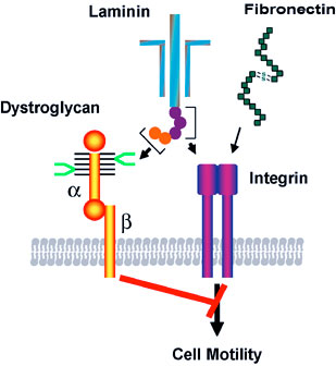
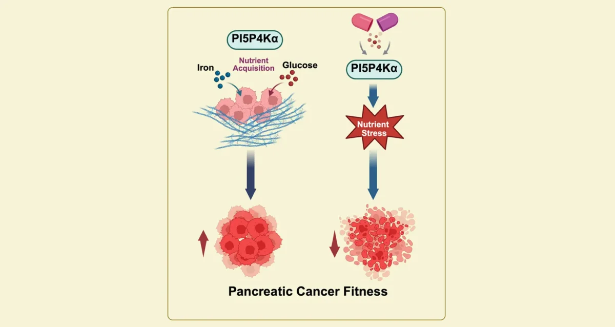
Dr. Perucho studies tumors from the intestinal tract that sometimes develop when the cellular machinery preserving the integrity of the genome – like computer spell-check programs that detect errors and correct them – is not working properly. When these corrector genes (mutators) are inactivated, the mutations that occur in all normal cells accumulate in large numbers because they are not repaired. This sparks genomic instability and cancer eventually develops when mutations occur in some cancer genes, such as oncogenes and tumor suppressor genes. However, some mutator genes are not inactivated by mutations, but by epigenetic silencing. This results from the disintegration of the epigenetic code, an unexplored process that is strongly associated with aging. This is important because many hereditary colon tumors originate by mutations in mutator genes that are transmitted from generation to generation. Molecular diagnosis of the deficient mutator genes determines which members of these families will be affected in the future. Identification of tumors with this kind of genomic instability is also useful for detecting familial cancer patients and predicting survival.
Manuel Perucho’s Research Report
Genomic Instability in Cancer Pathways
Our research efforts focus on the analysis of the genomic instability underlying two alternative pathways for oncogenesis (see figure below). Most neoplasms lose the chromosomal balance of the diploid normal cell following a pathway for cancer that involves the mutational inactivation of critical tumor suppressor genes. A minority of cancers manifest another type of genomic instability – the accumulation of hundreds of thousands of mutations, including insertions and deletions of a few base pairs in simple repeated sequences or microsatellites.
We study the aneuploidy of the tumor cell of the suppressor pathway for cancer by unbiased Arbitrarily Primed PCR DNA fingerprinting. Gains and losses of sequences from defined chromosomal regions can be simultaneously identified in multiple tumors generating a molecular karyotype or “amplotype.” Amplotyping offers useful applications for cancer diagnosis and prognosis and maps chromosomal regions harboring cancer genes with positive and negative roles in cell growth or survival. These cancer genes are under positive and negative selection pressure during tumorigenesis and are detected by the frequent gains and losses of specific chromosomal regions, respectively.
Understanding Colon Cancer
In colon cancer, the initial event in this carcinogenic pathway is the inactivation of the APC tumor suppressor. In hereditary cases of the familial polyposis (FAP) cancer syndrome, one mutated allele is transmitted in the germline, while in sporadic cases both alleles are inactivated by somatic mutations. Usually one allele is inactivated by a nonsense or frameshift mutation and the other allele is inactivated by the deletion of the chromosomal region (loss of heterozygosity), typical of the aneuploid cancer cell.
The first event in the Microsatellite Mutator Phenotype (MMP) pathway for colon cancer is the inactivation of a gene involved in genome stability, such as the hMLH1 DNA mismatch repair gene. In sporadic cases, the inactivation of the mutator gene usually occurs by somatic mutations or by epigenetic silencing. In many familial cases, including a majority of the Hereditary Non-Polyposis Colorectal Cancer (HNPCC) syndrome, one allele is inactivated by a germline mutation and the other by any of the other mechanisms (mutation, LOH, epigenetic silencing, etc.).
The MMP pathway for gastrointestinal cancer presents two distinctive features that seem paradoxical at first sight. First, despite accumulating hundreds of thousands of clonal somatic mutations in simple repeated sequences, these tumors exhibit a low mutation incidence in APC, K-ras and p53, prototypical cancer genes in colorectal carcinogenesis. Second, these tumors harbor ubiquitous biallelic mutations in non-functional poly (A)n sequences, such as the poly A tails of the Alu repeats. However, they also accumulate many monoallelic (i.e., heterozygous) mutations in functional sequences, such as the coding regions of mutator (hMSH3, hMSH6), suppressor (TGFbRII, p53) and apoptotic (Bax) genes.
The first paradox may be explained by the existence within some genes of simple repeats that are preferred targets for the MMP. Thus, in the presence of the mutator phenotype, mutations in these genes (i.e., Bax) occur sooner than in other genes of the same oncogenic signaling pathways that do not have these repeats (i.e., p53). The second paradox can also be explained by another peculiar feature of these MMP tumors. Due to their exacerbated mutator phenotype, the disruption of the homeostatic controls for cell growth and survival may also occur by the accumulation of heterozygous mutations in multiple genes whose products play redundant but synergistic roles at different points of the cell proliferation and apoptotic networks. The occurrence of multiple heterozygous mutations presumably reduces the threshold amounts of the corresponding gene products. This accumulative haploinsufficiency model is not restricted to cell proliferation and apoptotic pathways, but also applies to other networks involved in the control of genome integrity.
Robert Oshima graduated from the University of California Santa Barbara in Cellular Biology. He earned his PhD from the University of California at San Diego in 1973 with Paul Price. He joined Dr. Jerry Schneider’s laboratory in the UCSD Medical School to work on the biochemistry of cystinosis, a genetic lysosomal storage disease. During that time, he contributed to the development of a treatment that extends the life of patients greatly.
He acquired expertise in developmental biology and stem cells in the laboratories of Drs. Boris Ephrussi and Mary Weiss at the Centre National Recherche Scientifique, Gif-sur-Yvette, France in 1975. He continued those studies upon returning to UCSD and then moved to MIT in 1979 where he purified two markers of mouse stem cell differentiation that are widely used in the cancer pathology and developmental studies.
He joined the Sanford Burnham Prebys (formerly known as the La Jolla Cancer Research Foundation) in 1982 where he acted as Associate Scientific Director, a Program Director in the NCI designated Cancer Center, Postdoctoral Training Program Director, started the Tumor Analysis Shared Service and directed research on stem cells and cancer that resulted in over 100 publications. He also served as a reviewer for multiple cancer research granting agencies and taught at UC San Diego as an Adjunct Professor of Pathology from 1997.
He is currently Professor Emeritus (2015) and continues to advise and consult in cancer research. His particular cancer research interest is in methods of directing premalignant cancer cells to adopt a normal benign cell fate instead of becoming invasive malignant cancer.
Related Disease
Breast Cancer, Colorectal Cancer, Crohn’s Disease (Colitis), Preeclampsia
Cancer and development are closely related processes. The Oshima laboratory investigates the differences between normal stem cells and cancer stem cells. Furthermore, we are searching for genes and chemical compounds that control the differentiation of cancer cells and persuading them to adopt a normal developmental fate. Differentiation therapy has the promise of causing fewer deleterious side effects than killing cancer cells.
Robert Oshima’s Research Report
Stem Cells in Breast Cancer and Placental Development
Ets2 is one of a family of transcription factors that all utilize a conserved Ets protein domain for DNA binding. Like its Drosophila homologs, pointed and yan, Ets2 is regulated by growth factors and oncogenes that use Ras signaling pathways. The phosphorylation of a single threonine residue in an evolutionarily conserved protein-docking domain of Ets2 results in transcriptional activation and the induction of Ets2-dependent genes.
Early mouse placental development is dependent on the function of the Ets2 transcription factor. Ets2 regulates trophoblast stem (TS) cell self-renewal and thus placental development. One of the genes regulated by Ets2 in TS cells is the Cdx2 homeobox transcription factor. In collaboration with Dr. Mana Parast at UCSD, we extended our interest in placental development by developing a model system of human placental development. Human embryonic stem cells (ESC) and induced pluripotent stem cells (IPSC) can differentiate in culture to a trophoblast like derivative. We are screening chemical libraries by high content antibody staining methods for chemicals that improve the yield of trophoblast progenitors and direct their differentiation to either extra villous, invasive trophoblast cells or synchiotrophoblast layers.
Recently, we developed methods for the selective propagation of mouse mammary cancer stem cells in culture (Castro et al. Stem Cells 2013). These cells were shown to be capable of differentiation to a benign luminal epithelial-like fate both in culture and in animals. Inhibition of ROCK1 kinase inhibited both their spontaneous differentiation to luminal epithelial cells and their adoption of a mesenchymal tumorigenic phenotype.
Previously, in collaboration with Dr. Alexey Terskikh, we investigated the role of the Maternal Embyronic Leucine Zipper Kinase (MELK) in normal mammary epithelial stem cells and mouse mammary cancer. Using a transgenic reporter gene for Melk expression, we found that Melk expression is preferentially expressed in proliferative mammary epithelial progenitor cells and tumor cells. Both tumorsphere formation in culture and tumor formation in vivo is suppressed by knocking down Melk expression with Lentiviral-mediated shRNA in MMTV-Wnt1 tumor cells. These results have recently been confirmed in human triple negative breast cancer cells.
Furthermore, the development of differentiation therapy came from a collaborative project with Pfizer. Administration of bosutinib, a Src family inhibitor, to mice with mammary tumors caused by the MMTV-PyMT oncogene greatly restricted tumor progression by inducing differentiation of the tumor to epidermal and lactational cell fates without widespread cell death. This is an example of the possibility of restricting cancer by inducing differentiation.
Figure 1. Regulation of cancer stem cell differentiation by low oxygen atmosphere. Single cancer stem cells were plated in two different concentrations of O2 and stained for markers of luminal (K8) or basal (K14) epithelial cells. In 20 percent O2 differentiated cells expressing only K8 or K14 are observed. (Castro et al. 2013)
Figure 2. Presumptive trophoblast derivatives on the top of an hES cell colony, identified by keratin 7 staining (red). Nuclei stained by DAPI (blue). (Maurer et al. 2008)
Minoru Fukuda earned his PhD in biochemistry from the University of Tokyo in 1973 and did his postdoctoral training at the Yale University School of Medicine. Following a period with joint appointments at University of Washington and Fred Hutchinson Cancer Research Center in Seattle, he was recruited to Sanford-Burnham Medical Research Institute in 1982 as Director of the Glycobiology Program. Dr. Fukuda directs the program project grant, which consolidates the research efforts of the members of the Glycobiology Program.
Dr. Fukuda is a recipient of a Merit Award from the National Cancer Institute and the 1997 recipient of the Karl Meyer Award from the Society of Glycobiology. He served as an Executive Editor for Biochimica et Biophysica Acta, as an Associate Editor for Cancer Research and Editorial Member for Journal of Biological Chemistry. He also has edited 11 books including three books from Oxford University Press and three volumes of Methods in Enzymology and holds an Adjunct Professor appointment at the University of California, San Diego.
Education
1973: PhD, University of Tokyo, Biochemistry 1970: MS, University of Tokyo, Biochemistry 1968: BS, University of Tokyo, Biochemistry
Related Disease
Brain Cancer, Colorectal Cancer, Gastric Cancer, Helicobacter pylori, Prostate Cancer
The cell surface is heavily coated with carbohydrates. The structure of those cell surface carbohydrates displays a dramatic change during development, and mature cells express cell surface carbohydrates specific to different organs and tissues. Cell surface carbohydrates thus serve as a zip code for different organs and tissues. Sialyl Lewis X represents such an oligosaccharide. After discovery of sialyl Lewis X in neutrophils by Dr. Minoru Fukuda, his laboratory demonstrated that sulfated form of sialyl Lewis X is essential for lymphocyte homing and recruitment of natural killer cells in preventing tumor metastasis to the peripheral lymph node. With colleagues from Japan, Dr. Fukuda discovered that certain carbohydrates function as antibiotics against Helicobacter pylori infection, which is a leading cause for peptic ulcer and gastric carcinoma. Most recent studies in Dr. Fukuda’s laboratory revealed that decrease of the laminin-binding glycans on α-dystroglycan in carcinoma cells leads to tumor cell migration, invasion, and metastasis. The restoration of the unique glycans by the expression of distinct β3-N-acetylglucosaminyltransferase renders these cells act like normal cells. The results indicate that certain carbohydrates on normal cells and enzymes that synthesize those glycans, such as β3-N-acetylglucosaminyltransferase, function as tumor suppressors These findings will be useful in developing carbohydrate-based therapy for the treatment of inflammation and tumor metastasis.
Minoru Fukuda’s Research Report
Cell Surface Carbohydrates as Tumor Suppressor
Many studies have focused on carbohydrates that increase in cancer cells, but only a few have looked at carbohydrates that appear in normal cells but decrease or disappear in cancer cells. A specific mucin-type O-glycans (core 3 O-glycans) is one of such glycans, and we found core 3 O-glycans suppress tumor formation and metastasis. When core 3 O-glycans were forced to express on human prostate cancer cell lines, those prostate cancer cells produced much smaller tumors and almost no metastasis. By contrast, the parent cancer cells, which did not express core 3 O-glycans, produced robust primary and metastatic tumors. We showed that the expression of core 3 O-glycans decreases a formation of α2β1-integrin complex, receptors that mediate cell adhesion, diminishing cancer cell migration.
We also revealed tumor suppressor function in the unique laminin-binding glycans on dystrophin complex, — carbohydrates located on α-dystroglycan, which is also associated with cell adhesion. We discovered that the unique glycans play a critical role in epithelial-basement membrane interaction in normal cells, and the decrease or loss of the glycans, due to downregulation of β3-N-acetylglucosaminyltransferase, leads to increased malignancy by invasive carcinoma cells. Restoration of the laminin-binding glycans by forced expression of β3-N-acetylglucosaminyltransferase, on the other hand, results in reduced cell migration, thus dramatic decrease in tumor formation and metastasis. We demonstrated that interaction of laminin with the unique glycans on α-dystroglycan counteracts the cell migration signals that are mediated by integrin binding to its ligands, thereby decreasing tumor formation and metastasis. These findings also suggest that the laminin-binding glycans can be an excellent marker for epithelial-mesenchymal transitions.
These results indicate that certain carbohydrates on normal cells and enzymes that synthesize those glycans, such as β3-N-acetylglucosaminyltransferase, function as tumor suppressors. Upregulation of those key enzymes may become a novel way to treat cancer.
Neural Cell-specific Glycans in Development and Cancer
Polysialic acid and HNK-1 glycan represent carbohydrates enriched in neural cells. Polysialic acid is mainly attached to NCAM in embryos, while the majority of NCAM in adults lack this carbohydrate. To understand the roles of these glycans in neural development, we have cloned cDNAs encoding human polysialyltransferase, PST, and HNK-1 sulfotransferase, HNK-1ST, that are responsible for the synthesis of polysialic acid and HNK-1 glycan, respectively. By using these cloned cDNAs, we demonstrated that polysialic acid facilitates the invasion of glioma, the most common form of adult brain tumor. Our studies also showed that mutant mice with deficient STX, another polysialyltransferase, exhibit reduced behavioral response to fear conditioning, apparently due to anomalies in mossy fibers of the hippocampus. Our studies also demonstrated that neural development is significantly impaired in mutant mice that entirely lack polysialic acid due to inactivation of two polysialyltransferases. We found that this defect is caused by impairment of neural cell migration.
Mucin-type O-glycans in Immune Cell Interactions
Previously, we found that an increase of core 2-branched oligosaccharides is associated with leukemia and immunodeficiency, such as in Wiskott-Aldrich syndrome and AIDS. To determine the roles of core 2-branched O-glycans in immune cell interactions, the enzyme (C2GnT-1) responsible for the core 2-branched oligosaccharide was knocked-out by gene targeting. Compared to wild-type mice, leukocytes from the gene knockout mice exhibited a reduced binding to L-, E- and P-selectin in this order. In contrast, homing of lymphocytes was moderately reduced. Lymphocyte homing is mediated by binding of L-selectin on lymphocytes to sulfated L-selectin oligosaccharide ligands, 6-sulfo sialyl Lewis X in high endothelial venules (HEV) of secondary lymphoid organs. By analyzing remaining L-selectin ligands in C2GnT-1 knockout mice, we discovered novel L-selectin ligands that are based on extended core 1 oligosaccharides. The core portion of this novel L-selectin ligand is also an epitope for MECA-79 antibody that inhibits lymphocyte homing in vivo. Moreover, crossbreeding between mutant mice with deficient L-selectin ligand sulfotransferase and another sulfotransferase led to our findings that these two enzymes in cooperation synthesize L-selectin ligands. These mutant mice lack 6-sulfate group in L-selectin ligands that results in impaired inflammatory response. More recently, mice deficient in L-selectin ligands on mucin-type O-glycans were generated. The studies on the mutant mice revealed novel functions of N-glycan-based L-selectin ligand, which supports both lymphocyte homing and inflammatory response. This finding brought a new paradigm in selectin-carbohydrate interaction.
Carbohydrate-dependent Adhesion in Tumor Metastasis
Previously we found that the amount of core 2 O-glycans is significantly increased in colon and lung carcinomas and the increase of core 2 O-glycans is highly correlated to vessel invasion and lymph node metastasis. More recently, we discovered that forced expression of core 2 O-glycans by transfecting C2GnT-1 in prostate cancer cell lines resulted in increased tumor formation.
In parallel, we discovered that forced expression of selectin ligands, sialyl Lewis X on B16 melanoma cells leads to increased lung tumor formation. We also showed that tumor formation in lymph node is suppressed by natural killer (NK) cells, which are recruited by L-selectin mediated homing of NK cells to lymph nodes.
Roles of Carbohydrates in Helicobacter pylori-mediated Inflammation and Cancer
Helicobacter pylori is a leading cause of peptic ulcer and gastric cancer. Previously it was shown by others that H. pylori adhere to gastric mucosa in a carbohydrate-dependent manner. The infection of H. pylori leads to chronic inflammation, which apparently leads to peptic ulcer and gastric cancer.
Our recent studies showed that H. pylori-induced inflammation is associated with the formation of peripheral lymph node addressin (PNAd) characterized by binding to MECA-79 antibody and L-selectin. The number of HEV-like vessels expressing PNAd increases as H. pylori-induced inflammation progresses. Moreover, PNAd disappears once H. pyloriis eradicated by antibiotic treatment. These findings indicate that H. pylori-induced inflammation is facilitated by de novo formation of PNAd thereby recruiting lymphocytes. It may be possible to attenuate or prevent the formation of peptic ulcers or gastric cancer by inhibiting L-selectin ligand synthesis, for example by inhibiting the sulfotransferases.
Binding of laminin to the specific carbohydrate (shown in bright green) on α-dystroglycan counteracts the migration signals initiated by integrin binding to extracellular matrix proteins such as laminin and fibronectin. The synthesis of this specific carbohydrate requires a unique β3-N-cetylglucosaminyltransferase, and the downregulation of the glycosyltransferase in carcinoma cells leads to increased cell migration, thereby increased tumor formation and metastasis. Thus the specific carbohydrate structure at cell surface functions as a tumor suppressor, which is controlled by the unique β3-N-acetylglucosaminyltransferase.
While over half of the world’s population is infected with H. pylori, only a fraction of those individuals progress to peptic ulcer and gastric cancer. In relation to these observations, α1,4-N-acteylglucosaminyl capping structure (α4GlcNAc) is present in deeper portions of the gastric mucosa, where H. pylori rarely colonizes. We discovered that α4GlcNAc capping structure functions as an antibiotic against H. pylori infection by inhibition of the synthesis of α-glucosyl cholesterol, a major component of the H. pyloricell wall. This unprecedented discovery should be useful in developing drugs to inhibit H. pylori colonization, through inhibition of cholesterol α-glucosyltransferase. Such drugs lead to a novel treatment for prevention and potential treatment of peptic ulcer and gastric carcinoma.
After receiving his early training in clinical chemistry/biochemistry at the University of Buenos Aires, Argentina, Dr. Millán first joined the La Jolla Cancer Research Foundation (LJCRF) in 1977, the predecessor of Sanford Burnham Prebys, as a trainee in clinical enzymology. He completed his PhD studies in Medical Biochemistry at the University of Umeå, Sweden and after post-doctoral stints in Copenhagen and LJCRF he was appointed to the faculty at SBP in 1986. He served as Professor of Medical Genetics in the Department of Medical Biosciences at his alma mater, Umeå University, Sweden, from 1995-2000. He was appointed Sanford Investigator at the Sanford Children’s Health Research Center at Sanford Burnham Prebys in 2008.
Honors and Recognition
2018: ASBMR Lawrence G. Raisz Award for Pre-clinical Research. 2001: Gold Medal of the Royal Academy of Medicine and Surgery, Murcia, Spain 1992: Honorary title of AcadémicoCorresponsal at the Royal Academy of Medicine and Surgery, Murcia, Spain.
Related Disease
Arthritis, Bone Mineralization Disorders, Cardiovascular Diseases, Colorectal Cancer, Crohn’s Disease (Colitis), Heart Disease, Inherited Disorders, Metabolic Syndrome, Peripheral Vascular Disease, Testicular Cancer
Phenomena or Processes
Cardiovascular Biology, Disease Therapies, Extracellular Matrix, Protein Structure-Function Relationships
Anatomical Systems and Sites
Cardiovascular System, Musculoskeletal System, Vasculature
Research Models
Mouse
The Millán laboratory works on understanding the mechanisms that control normal skeletal and dental mineralization and elucidating the pathophysiological abnormalities that lead to heritable soft bones conditions such as Hypophosphatasia (HPP) and to soft-tissue calcification, including vascular calcification, that is a hallmark in patients affected by a variety of rare genetic diseases as well as in chronic kidney disease. Dr. Millán’s research has already contributed to the implementation of a novel therapy for HPP, a genetic disease caused by deficiency in tissue-nonspecific alkaline phosphatase (TNAP) function, that leads to accumulation in the extracellular space of inorganic pyrophosphate (PPi), a potent inhibitor of mineralization. HPP is characterized by defective mineralization of bones (rickets or osteomalacia), and teeth that display a lack of acellular cementum, hypomineralized dentin and enamel, and periodontal defects. Dr. Millán’s team has demonstrated the effectiveness of enzyme replacement therapy using mineral-targeted recombinant TNAP (asfotase alfa) to prevent the skeletal and dental defects in the TNAP knockout mouse model of infantile HPP. This therapy was approved in 2015 for the treatment of patients with pediatric-onset HPP.
Current efforts, in collaboration with Professor Miyake’s group in Japan (https://www.nms-gt.org/en/members), focus on developing gene therapy as an alternative approach to treat HPP. Dr. Millán’s group has also identified key pathophysiological changes that lead to calcification of the arteries in animal models of generalized arterial calcification of infancy, pseudoxanthoma elasticum and related genetic diseases as well as in animal models of chronic kidney disease. His group, in collaboration with scientists at the Conrad Prebys Center for Chemical Genomics at Sanford Burnham Prebys, has developed proprietary compounds able to ameliorate the soft-tissue calcification in these conditions and clinical trials are now underway using these first-in-class small molecule inhibitors.
Dr. Commisso obtained his PhD at the University of Toronto in the Department of Molecular Genetics and completed his postdoctoral training at New York University School of Medicine. Dr. Commisso made the seminal discovery that Ras-mutant cancer cells use macropinocytosis as an amino acid supply pathway. His laboratory’s research interests are centered on elucidating the underlying mechanisms of how metabolic stress influences the tumor ecosystem in pancreatic cancer. The lab is particularly interested in identifying metabolic adaptations and dependencies that contribute to tumor progression and can be targeted to develop novel therapeutic modalities for this disease.
Related Disease
Cancer, Colorectal Cancer, Lung Cancer, Pancreatic Cancer
Research in the Commisso lab is focused on biological discoveries that have the potential to lead to novel therapeutic strategies for cancer. Of particular interest to our laboratory are Ras-driven cancers, such as pancreatic cancer, which are extremely aggressive and are in urgent need of new and innovative therapies. The biological process that we study in the lab is called macropinocytosis, a fluid-phase form of bulk endocytic uptake, which we have linked to cancer cell metabolism in Ras-mutated tumors.
Cosimo Commisso’s Research Report
Macropinocytosis is an endocytic mechanism of fluid-phase uptake that produces large intracellular vesicles known as macropinosomes. Macropinosomes are heterogeneous in size and shape and serve to internalize large volumes of extracellular fluid along with the associated membrane. In transformed cells, macropinocytosis is stimulated by oncogenes, such as Ras. Ras proteins are small, membrane-localized GTPases that are activated in response to growth factors and they regulate a variety of outputs, including cell proliferation, survival and invasion. Gain-of-function mutations in Ras-encoding genes cause Ras proteins to be trapped in their active state, leading to the constitutive activation of downstream pathways. The functional consequences of macropinocytosis stimulation in mutant Ras-expressing cells were unknown prior to our work. We have linked macropinocytic uptake in Ras-transformed cells to nutrient delivery and amino acid supply (Commisso et al., 2013). We demonstrated that the inhibition of this nutrient delivery pathway selectively compromises growth of Ras-driven tumors. With the long-term goal of specifically targeting such tumors, we have recently developed stream-lined methodology to detect and grade macropinocytosis in tumor tissue (Commisso et al., 2014). Our work was important for two main reasons. First, cancer cells are dependent on amino acids, such as glutamine, for their growth and survival. Therefore, the targeting of these amino acid supply pathways, such as macropinocytosis, represents a promising strategy in developing anti-cancer therapeutics. Second, macropinocytosis is emerging as a mechanism of entry for a variety of therapeutic agents, such as nanoparticles. Hence, identifying that this uptake pathway is active in Ras-driven tumors may have an impact on how these tumors are treated. The complete understanding of the functional significance of Ras-induced macropinocytosis to carcinogenesis and treatment ultimately depends on having a firm grasp of how this process is regulated and on the ability to specifically control it. To this end, a major research focus of the lab is to advance our understanding of the molecular pathways that drive macropinocytosis, which could lead to the identification of new molecular targets whose inhibition would restrain tumor growth and enhancers that could be manipulated to dial-up the uptake process in drug delivery strategies. Additional research interests in the lab include nutrient sensing pathways that are active in the context of macropinocytic uptake and macropinosome maturation, the process that leads to active protein catabolism within the tumor cell.
Dr. Adams most recently led the Epigenetics Unit at the Beatson Institute for Cancer Research and the University of Glasgow, Institute of Cancer Sciences, in Scotland. He has also held positions at Wistar Institute (University of Pennsylvania), Drexel University and Fox Chase Cancer Center in Philadelphia.
Peter D. Adams obtained his BA in biochemistry at the University of Oxford, England and his PhD at Imperial Cancer Research Fund (now CR-UK). He did postdoctoral work with Dr. William G. Kaelin, Jr. at Dana-Farber Cancer Institute. Peter D. Adams is co-Editor-in-Chief of the journal Aging Cell.
Education
1993: PhD, Signal Transduction, Imperial Cancer Research Fund (CRUK), London, UK (Dr. Peter Parker, advisor) 1989: BA, Biochemistry, Oxford University, England
Honors and Recognition
2003-2008: Leukemia and Lymphoma Society Scholar 1999-2001: W.W. Smith Charitable Trust Fellowship 1999-2001: V Foundation Scholar 1995-1996: Cancer Research Foundation of America Fellowship 1993-1995: SERC/NATO Fellowship 1989: B.A. with Honors in Biochemistry, Oxford University, UK 1986: Awarded a Distinction in Oxford University Preliminary Examinations 1984-1989: 1984-1989: Exhibition holder for Academic Achievement at Oxford University, UK 1983: Lane Scholarship for Academic Achievement at King Henry VIII School, UK
Related Disease
Aging, Aging-Related Diseases, Colorectal Cancer, Leukemia/Lymphoma, Liver Cancer, Skin Cancer and Melanoma
Phenomena or Processes
Epigenetics
Dr. Adams’ lab investigates the impact of chromatin and epigenetics on cellular senescence, aging and cancer. In particular, the lab hypothesizes that age-associated changes in chromatin and epigenetic programming contribute to the dramatic age-associated increase in incidence of cancer. While age is the biggest single risk factor for most cancers, the reason for this is current poorly understood.
Dr. Adams’ research focus is driving epigenetics as a cross-cutting scientific theme and promoting access to single cell technologies at the Institute.
Peter D. Adams’ Research Report
We have mapped the epigenetic landscape of senescent cells, and are defining the causes and consequences of this altered landscape. Cellular senescence is an irreversible proliferation arrest and pro-inflammatory phenotype triggered in primary cells by activated oncogenes and other molecular stresses. As a profound change in cell phenotype, the initiation and maintenance of senescence depends on reprogramming of chromatin, the epigenome and gene expression. Cellular senescence is a potent tumor suppressor mechanism. However, the accumulation of senescent cells with age also causes tissue aging, by blocking cell and tissue renewal and driving chronic inflammation. Indeed, in contrast to its acute tumor suppressive effects, chronic accumulation of inflammatory senescent cells is tumor promoting. In collaboration with Dr. Shelley Berger, we have mapped the distribution of several critical epigenetic regulators in proliferating and senescent cells, including DNA methylation, several histone modifications, histone variants and nuclear lamins. These collaborative studies have yielded critical insights into gene regulation in senescent cells (Cruickshanks et al., 2013; Rai et al., 2014; Shah et al., 2013), and the tumor suppressive and pro-aging effects of senescent cells (Cruickshanks et al., 2013; Nelson et al., 2016).
Landmark structure and functional studies on the HIRA histone chaperone complex and its role in senescence-mediated tumor suppression. In collaboration with Dr. Ronen Marmorstein in Philadelphia we have dissected the structure-function relationships between HIRA and its binding partners, UBN1, CABIN1 and ASF1a and substrate histone H3.3 (Zhang et al., 2005). This included a crystal structure of the HIRA/ASF1a interaction surface and more recently the UBN1/histone H3.3 interaction surface (Tang et al., 2006). We were the first to describe the distribution of the HIRA complex across the mammalian epigenome (Pchelintsev et al., 2013). In functional studies, we have demonstrated the role of this DNA replication independent histone chaperone complex in the control of chromatin in non-proliferating senescent cells (Rai et al., 2014). These studies have been facilitated by the mouse monoclonal and rabbit polyclonal antibodies that we have made to all subunits of the complex. More recently, we have generated the first conditional knock out mice of HIRA, UBN1 and CABIN1 and are using these to establish in vivo functions (Rai et al., 2014). Of particular note, we have revealed a function for HIRA in promoting healthy aging and the suppression of cancer (Rai et al., 2014).
We coined the term “chromostasis” to describe the presumptive homeostatic mechanisms that confer epigenetic stability over the lifecourse, and we were major contributors to the first demonstration of a DNA methylation clock in the mouse. Maintenance of cell phenotype and suppression of disease, including cancer, over the lifecourse depends on a high level of epigenetic stability. However, since chromatin is inherently dynamic (Rai et al., 2014), this steady state stability likely reflects a challenge for the cell. Therefore, presumptive “chromatin homeostasis” or “chromostasis” mechanisms are predicted to actively maintain an epigenetic steady state over the lifecourse, thereby suppressing age-associated disease (Rai et al., 2014). We have shown that histone chaperone HIRA is one such factor that contributes to epigenetic stability in non-proliferating cells (Ye et al., 2007). Recently, we reported the first DNA methylation clock in the mouse, and showed that diverse interventions – genetic, dietary and drug – that promote longevity of mice also suppress age-associated epigenetic changes and slow progression of this DNA methylation “clock”, i.e. enhance chromostasis (Cole et al., 2017; Wang et al., 2017).
We have demonstrated a “tug of war” between tumor suppressive oncogene-induced senescence and oncogenic activated Wnt signaling in melanocytic neoplasia (Adams and Enders, 2008; Ye et al., 2007). The balance between these tumor suppressive and oncogenic activities determines the efficiency of senescence-mediated tumor suppression. For example, we showed that in oncogene-expressing melanocytes a low level of activated Wnt signaling promotes benign nevus formation (Pawlikowski et al., 2013). However, a high level of activated Wnt signaling, caused by germline sequence variants, promotes giant congenital nevi in the form of congenital melanocytic nevus (CMN) syndrome (Pawlikowski et al., 2015). In a mouse model that closely recapitulates the human genetics, we showed that activated Wnt signaling and an activated Ras oncogene (NRasQ61K) cooperate to drive CMN syndrome, and that this is suppressed by acute post-natal treatment with MEK inhibitors (Pawlikowski et al., 2015). Based on these studies, our collaborator Dr. Veronica Kinsler is preparing to test MEK inhibitors in babies afflicted by CMN syndrome.
We first characterized Cytoplasmic Chromatin Fragments (CCF) in senescent cells and in collaboration defined a role for CCF as drivers of inflammation via the cGAS/STING cytoplasmic DNA sensing anti- viral pathway. Cellular senescence is a potent tumor suppressor mechanism by virtue of proliferation arrest and the senescence associated secretory phenotype (SASP) which promotes clearance of pre-malignant cells by the immune system. However, the mechanism responsible for initiation of SASP has been unknown. In 2013, our lab first characterized and named CCF as fragments of chromatin expelled from the nucleus of senescent cells into the cytoplasm (Ivanov et al., 2013). Then, in 2015, in collaboration with Shelley Berger’s laboratory, we showed that formation of CCF depends on interaction of lamin B1 and autophagy adaptor LC3 in the nucleus, and that lamin B1 is a nuclear substrate of autophagy (Dou et al., 2015). Most recently, again with Shelley Berger’s lab, we have shown that CCF are sensed by the cytoplasmic DNA sensing anti-viral apparatus, cGAS and STING, and this leads to activation of NFkB and SASP in senescent cells (Dou et al., 2017). The role of CCF as triggers of SASP, via cGAS and STING, has been confirmed by several other labs. Most recently, we showed that formation of CCF is triggered by a retrograde mitochondria-to-nucleus signalling pathway that constututes a target for candidate anti-SASP healthy aging interventions (Vizioli et al., 2020).
Lysosome-dependent processing of histones in senescent cells. Ivanov, A., Pawlikowski, J., Manoharan, I., van Tuyn, J., Nelson, D. M., Rai, T. S., Shah, P. P., Drotar, M., Wu, H., Berger, S. L., and Adams, P.D. J. Cell Biol. 2013 July 1; 202 (1): 129–143
Autophagy mediates degradation of nuclear lamina. Dou, Z., Capell, BC., Drake, AM., Shah, PP., Dorsey, J., Simola, D., Donahue, G., Zhu, Z., Sammons, M., Rai, TS., Natale, C., Ridky, T., Goldman, R., Adams, P.D., Berger, SL. Nature. 2015 Oct 28; 527:105-109
Cytoplasmic chromatin triggers inflammation in senescence and cancer. *Corresponding authors. Dou, D., Ghosh, K., Vizioli, MG., Zhu, J., Sen, P., Wangensteen, KJ., Simithy, J., Lan, Y., Lin, Y., Zhou, Z., Capell, BC., Xu, C., Xu, M., Kieckhaefer, JE., Jiang, T., Shoshkes-Carmel, M., Ahasan Al Tanim, KM., Barber, GN., Seykora, JT., Millar, SE., Kaestner, KH., Garcia, BA., Adams, P.D.*, Berger, SL*. Nature. 2017 Oct 4; 550:402-406