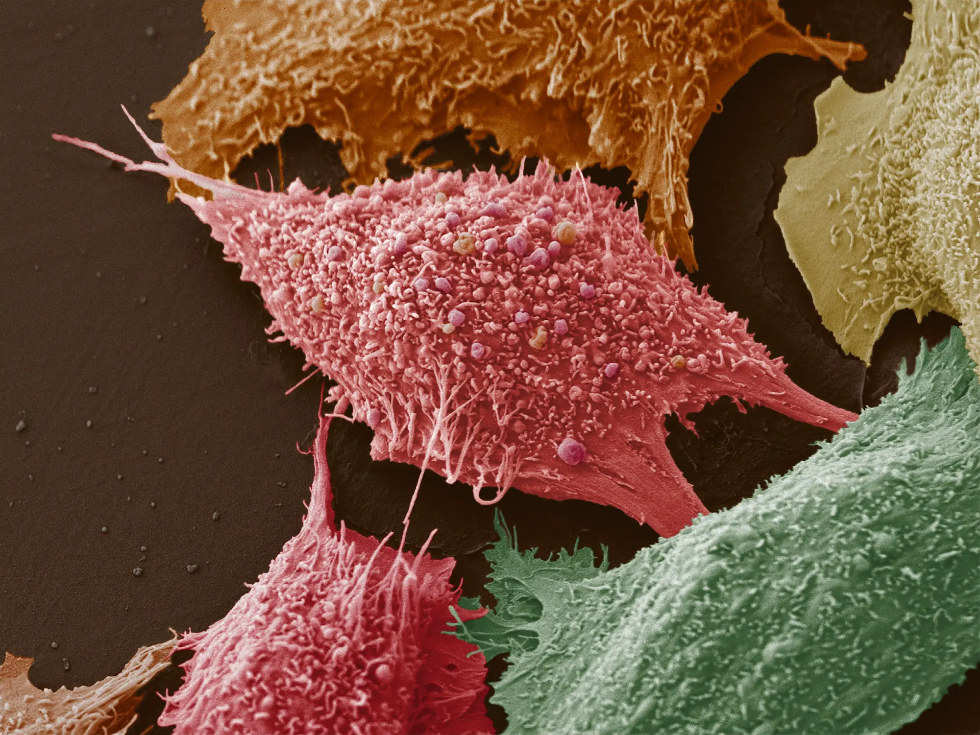
Overview
The Cellular Imaging Facility broadly supports research programs by providing access to sophisticated microscopes for digital imaging, as well as training, assistance and guidance. The core facility offers expertise, training and assistance in advanced biological microscopic imaging techniques and use of complex image processing software, use of well-maintained, aligned, and calibrated microscopic equipment, and troubleshooting of equipment and experimental problems.
Services
- Single and multiphoton confocal microscopy
- Live cell imaging
- Training and consultation
- Wide field microscopy:
- Bright field and dark field microscopy
- Phase and Nomarski differential interference contrast
- Polarization and epi-fluorescent microscopy
- Image analysis:
- Morphometry
- 3D and 4D rendering
- Deconvolution
Equipment & Resources
Confocal Microscopy
- Zeiss LSM 980 AiryScan Microscope System
The Zeiss LSM 980 Airyscan2 microscope is optimized for spectral multiplexing by simultaneous spectral detection of multiple labels. Adapted illumination and detection schemes allows imaging of most challenging three-dimensional samples with high framerates and beyond the diffraction limit, while still being gentle to sensitive samples- 4-line laser launch with 405nm, 488nm, 561nm, and 640nm lasers.
- light-efficient beam path with up to 36 simultaneous channels and full spectral flexibility from UV into the near infrared (NIR) range
- 34-channel spectral detector comprised of 1 UV + 32 Visible + 1 Far-red detectors and the Airyscan2 super-resolution detector system
- AirtScan2 part:
- 32 detector elements allowing for super resolution quantitative results
- optimized for spectral multiplexing
- shorter acquisition times (up to 10x faster)
- capture of larger view fields or dynamic processes
- Incubation chamber for live imaging (Co2/Temp/Humidity)
- Fully automated XY stage with piezo-Z stage for fast scans of slides, dishes, and well plates
- Nikon N-SIM E Super-Resolution / A1 ER Confocal Microscope System
- Structured illumination super-resolution microscope combined with enhanced resolution point scanning confocal
- Nikon’s N-SIM E is a super-resolution system that provides double the resolution of conventional optical microscopes. Combining N-SIM E and the A1 ER confocal microscope allows you the flexibility to select a location in the confocal image, and easily switch to view it in super-resolution, enabling the acquisition of more detail
- The new system has 10X, 20X, 40X oil, 60X oil, and Super-Res 100X oil objective lenses. N-SIM-E laser lines include 488nm, 561nm, and 640nm. The A1R Hybrid confocal has a 6-line laser launch with 405nm, 445/488/515nm, 561nm, and 640nm lasers
- LSM 710 NLO Zeiss Multiphoton Laser Point Scanning Confocal Microscope
- Four Single Photon Lasers are generating six excitation lines: Ar-ion gas laser produces 457, 488, 514 nm lines and three solid state lasers produce 561, 594 & 633 nm
- Multi-photon Mai-Tai Laser HB- DeepSee complex system (690-1024nm) dedicated for excitation of multiple fluorophores, including UV excited dyes, such as DAPI
- CO2 & Thermo Controlled Time lapse system
- The Microscope operates by ZEN-2011 acquisition/processing package
- Fluoview-1000 Olympus Laser Point Scanning Confocal Microscope
- Four Single Photon Lasers provide six excitation lines: diode laser emits 405nm line, Ar-ion laser produces 457, 488, 514 nm, DPSS laser – 561 nm and He-Ne laser – 633 nm
- Dual SIM Scanner for simultaneous stimulation & registration fluorophores
- Olympus software provides modules for Spectral Deconvolution; FRAP, FRET and Photo-activation applications
- CO2 & Thermo Controlled Time lapse system
- Yokogawa Spinning Disk Laser Confocal Microscope
- XY and Piezo-Z object stage (ASI 2000-Piezo)
- Single Photon Kr-Ar laser excites 488, 568 and 647 nm (Melles Griot)
- Cooled Monochrome CCD Quant EM Camera (Photometrics)
- MetaMorph (Mol. Devices) software (v.7.7.7)
- CO2 & Thermo Controlled Time lapse system
Wide Field Microscopy
- EVOS® FL Auto Imaging System
- Time-lapse imaging, Image stitching, and Automated cell counting
- Environmental chamber enabling precise control of temperature and humidity Automated X/Y scanning stage with interchangeable vessel holders for slides, multiwell plates, 35 mm dishes, 60 mm petri dishes, and T-25 flasks
- Dual cameras: Monochrome: high-sensitivity interline CCD; Color: high-sensitivity CMOS; 3.1 megapixels
- 4X, 10X, 20X, and 40X air objectives as well as a 60X oil lens
- Epifluorescence and transmitted light (bright-field and phase-contrast)
- 7 light cubes (DAPI, GFP, RFP, Texas Red, Cy5, Cy5.5, Cy7) for a wide range of fluorescent antibodies
- Inverted IX81 Olympus Wide Field and Fluorescence Microscope (FRET)
- DG4 UV source galvanometer equipped with 300W Xenon lamp, capable for sequential acquisition of 4 excitation channels & 10 position fast (30msec) Emission Filter Wheel (Sutter)
- FRET & Ca2+ Imaging, DIC
- XY-linear encoded objective stage ASI 2000(ASI Inc.); Z-IX81 step-motor
- ZDC ( Zero Drift focus Control, Olympus)) attachment
- Cooled Monochrome CCD Cascade 512B FT Camera. (Photometrics Inc.)
- MetaMorph software, v. 7.7.7 (Mol. Devices)
- CO2 & Thermo Controlled Time lapse system
- Inverted IX81 Olympus Wide Field and Fluorescence Microscope (TIRF)
- Excitation illumination system presented by combination of X-cite 120 Metal halide UV source (EXFO) for the epi-fluorescent microscopy and single photon lasers combiner containing three solid state lasers (488, 561 and 640 nm)
- XY and Piezo-Z object stage ASI 2000 (ASI Inc.)
- Excitation & Emission Filter Wheels (Sutter)
- Cooled, Monochrome CCD QuantEM Camera (Photometrics Inc.)
- CO2 & Thermo Controlled Time lapse system
- Inverted IX81 Olympus Wide Field and Fluorescence Microscope
- XY-Martzhauser Tango objective stage (Martzhauser Inc.); Z-IX81 step-motor
- Cooled, Color CCD SPOT RT-3 Camera (Diag.Instruments)
- X-cite 120 Metal Halide UV source (EXFO)
- Excitation & Emission Filter Wheels (Sutter)
- CO2 & Thermo Controlled Time lapse system
- Inverted TE300 Nikon Wide Field, Fluorescence Microscope #1
- Multiple Fluorescence; Phase and Nomarski DIC contrast.
- Cooled, Color CCD SPOT RT Camera (Diagnostic Instruments Inc.)
- Inverted TE300 Nikon Wide Field, Fluorescence Microscope #2
- Multiple Fluorescence; Nomarski (DIC) contrast
- XYZ object stage ASI 2000 (ASI Inc.)
- Cooled Color CCD SPOT RT Camera(Diagnostic Instruments Inc)
- Thermo Controlled Time lapse system
- Upright BX50 Olympus Wide Field, Fluorescence Microscope
- Transmitted light and Triple Fluorescent imaging
- Cooled Monochrome ORCA ER (Hi Res) camera (Hamamatsu)
- Software packages
- Image Pro Plus (Media Cybernetics)
- Volocity 4D Rendering (Improvision, PerkinElmer)
- MetaMorph, MetaFluor packages (Mol. Devices)
- Spot RT Acquisition / Processing Software (Diagnostic Instruments Inc.)
- ImageJ and IrfanView (free sources imaging packages)
Price List
For a Price List, please call (858) 795-5206 or email us.
Leadership
Brooke Emerling, PhD
Scientific Director
Leslie Boyd, BS
Facility Manager
lboyd@sbpdiscovery.org
Contact
Please call (858) 795-5206 or use the button below to send us an email.
