CODEX
Overview
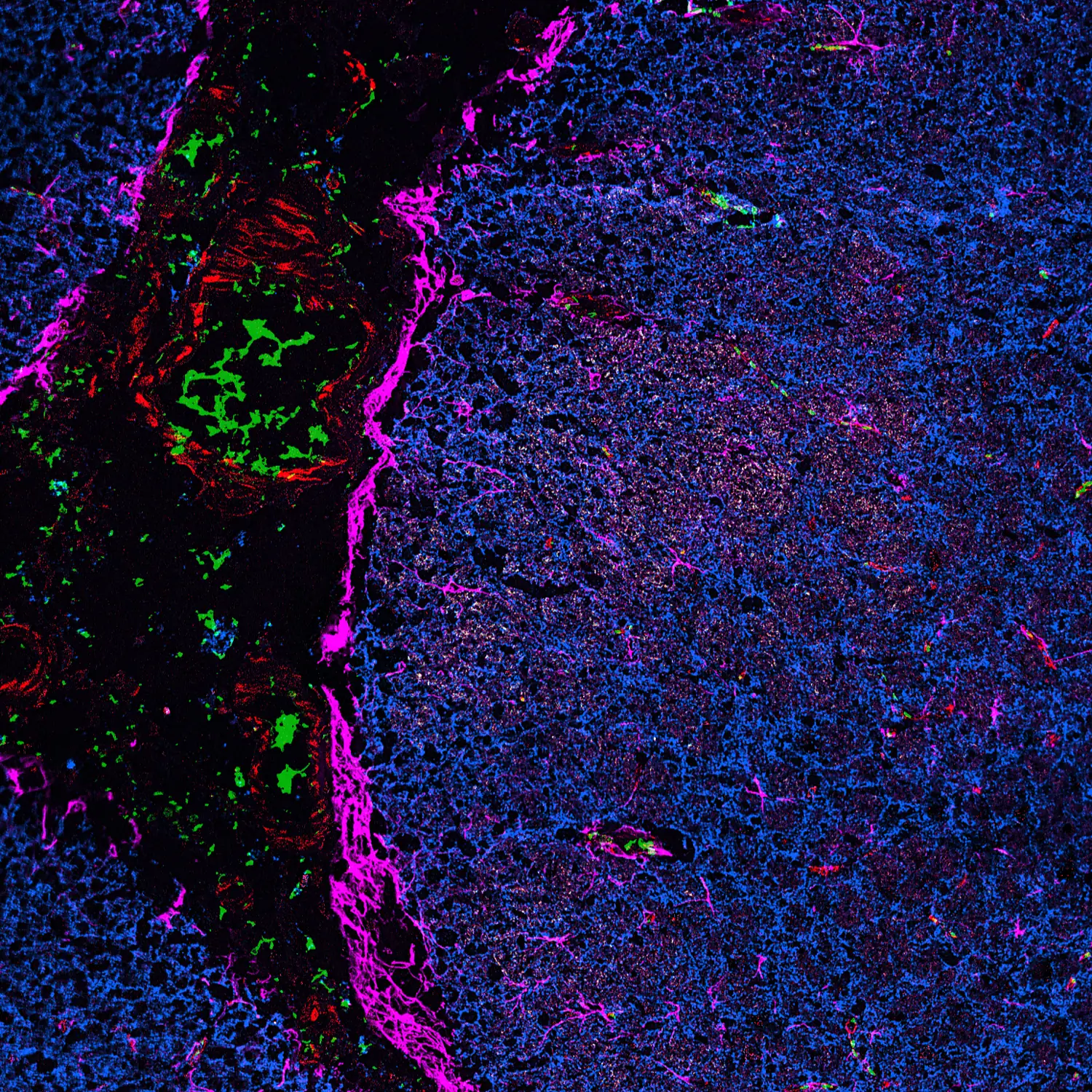
CODEX is a highly multiplexed spatial proteomics technology that enables simultaneous detection of dozens of protein markers within intact tissue while preserving their spatial context. By combining multiplex antibody imaging with high-resolution microscopy, CODEX allows researchers to characterize tissue architecture, cellular composition, and cell-to-cell interactions at single-cell resolution.
Compared with conventional immunofluorescence and lower-plex imaging approaches, CODEX enables the analysis of many biomarkers within the same tissue section, providing a more comprehensive view of complex tissue microenvironments while preserving spatial information.
The CODEX Core provides end-to-end spatial proteomics services, including experimental design, antibody panel development and conjugation, multiplex imaging, and comprehensive downstream data analysis. Trained users may also access selected instruments for independent use, including self-operated imaging on the Keyence microscope and cryostat sectioning following completion of the required training.
Investigators are encouraged to consult with the Core early in the experimental design process for guidance on tissue preparation, biomarker selection, panel optimization, and imaging strategies. Early consultation can help ensure compatibility with the CODEX workflow and maximize the quality and biological value of the resulting spatial proteomics data.
Services
From study planning to data interpretation, the CODEX Core offers comprehensive support for spatial proteomics projects. Services include:
- Scientific consultation and project planning
- Tissue preparation and sectioning
- Antibody panel design and custom conjugation
- Multiplex tissue staining
- High-dimensional imaging using the PhenoCycler™ platform
- Image processing, spatial analysis, and biological interpretation
Additional Services
- Keyence microscope training
- Self-operated Keyence microscope imaging (available to trained users)
- Self-operated cryostat sectioning
Panels Available
We currently have established CODEX panels available for several tissues and biological systems, including:
- Liver
- Muscle
- Epigenetic panel
- Tumor tissue
- Immune tissues
- Brain
- Lectin-based panels
Equipment & Resources
The CODEX Core is equipped with:
2 × PhenoCycler™ Open Systems (Quanterix)
Each instrument is integrated with a high-resolution Keyence fluorescence microscope, enabling automated cyclic imaging of multiplex antibody panels and generation of spatially resolved protein expression datasets.
2 × Keyence BZ-X810 Automated Fluorescence Microscopes
Microscope capabilities include:
- Air objectives: 4×, 10×, 20×, and 40×
- Optional oil immersion objective: 63× / 1.40 NA
- Multi-channel fluorescence imaging capabilities, including:
- Blue: DAPI, Hoechst, BFP
- Green: GFP/YFP, FITC, Alexa Fluor™ 488
- Red: TRITC, Cy3, Rhodamine, Alexa Fluor™ 546
- Far-red: Cy5, APC, Alexa Fluor™ 647
- Near-infrared: Cy7, Alexa Fluor™ 750
3 × High-Performance Image Analysis Workstations
The Core provides dedicated computational resources for processing and analyzing large-scale spatial imaging datasets. Workstations are equipped with high-performance NVIDIA GPUs (24 GB VRAM) to support image processing, cell segmentation, spatial analysis, and downstream data interpretation.
250 TB Secure Network Storage
A dedicated high-speed data infrastructure, including a 10 Gigabit Ethernet network and QNAP Network Attached Storage (NAS) systems, enables efficient transfer, management, and secure storage of large multiplex imaging datasets.
Epredia CryoStar™ NX50 Cryostat
The Core provides cryostat sectioning capabilities using an Epredia CryoStar™ NX50 cryostat equipped with Vacutome technology and adjustable chamber height, supporting high-quality tissue preparation for spatial proteomics applications.
Price List
For a complete list of services, please call (858) 646-1000 ext. 4180 or email us.
| CODEX Services | Internal Subsidized | Internal | External Non-Profit | External For-Profit |
|---|---|---|---|---|
| Consultation | Free | Free | Free | Free |
| Coverslip Coating (Batch of 20) | $100 | $125 | $135 | $263 |
| Tissue Sectioning (per hour) | $100 | $125 | $135 | $263 |
| H&E Staining | $100 | $125 | $135 | $263 |
| Multiplex Staining | $730 | $913 | $968 | $1,920 |
| Antibody Mix | Variable (see formula below) | |||
| Plate Preparation – Small Panel | $150 | $188 | $203 | $395 |
| Plate Preparation – Large Panel | $180 | $225 | $243 | $473 |
| Antibody Panel Design (per hour) | $150 | $188 | $203 | $395 |
| Antibody Conjugation (Each) | $80 | $100 | $108 | $210 |
| Imaging Service – Validation Panel | $500 | $625 | $675 | $1,315 |
| Imaging Service – Regular Panel | Variable (see formula below) | |||
| Data Analysis (per hour) | $90 | $113 | $122 | $237 |
| Keyence Microscope Training (per hour) | $70 | $88 | $95 | $184 |
| Self-Operated Keyence Microscope Imaging (per hour) | $30 | $38 | $40 | $80 |
| Self-Operated Cryostat Sectioning (per hour) | $80 | $100 | $108 | $210 |
Antibody Mix Cost
Estimated Cost = Antibody Total Mix Volume (µL) × $3 per µL +$150 (prep fees)
Imaging Service – Regular Panel Pricing
Estimated Cost =(Number of Tiles×$0.40)×(Number of Regions×$0.70)×(Number of Cycles×$0.50)+$150
Note: The $150 surcharge applies only to experiments with 20 or more imaging cycles.
Leadership
Beatrice Silvestri, PhD
CODEX Core Manager
(858) 646-3135
bsilvestri@sbpdiscovery.org
Contact
Dr. Beatrice Silvestri is a neuromuscular biologist with expertise in advanced imaging technologies, spatial biology, and multiplexed tissue profiling. She received her PhD training at Sapienza University of Rome, Italy, and completed her postdoctoral training in the laboratory of Dr. Yu Xin Wang at Sanford Burnham Prebys.
Her research has focused on applying high-dimensional imaging approaches to understand complex tissue microenvironments, including immune regulation, tissue regeneration, and disease-associated changes. Dr. Silvestri has extensive experience with the Akoya CODEX/PhenoCycler platform, including the development and optimization of spatial proteomics workflows, multiplex antibody panel design and validation, tissue preparation strategies, image acquisition, and computational analysis of large-scale spatial datasets.
As Manager of the CODEX Core, Dr. Silvestri provides scientific guidance and technical support to investigators throughout all stages of their projects, from experimental design and assay development to spatial data interpretation. She works closely with researchers to develop customized solutions that enable robust and reproducible spatial profiling studies across a variety of biological applications.
Please call (858) 646-3135 or use the button below to send us an email.