Adam Godzik Archives - Sanford Burnham Prebys
Institute News

SBP brings science to San Diego community at STEM

AuthorSusan Gammon
Date

March 8, 2018

STEM Expo Day kicked off a week-long festival on Saturday, March 3, 2018 at Petco Park. The event marked the 10th annual San Diego Festival of Science and Engineering Expo Day—one of the largest STEM (Science, Technology, Engineering and Math) festivals in the U.S. The community outreach event provided learning and hands-on activities for students in grades kindergarten through 12th grade, as well as adults and families to ignite their passion for STEM education.

“STEM Expo is a great way to get our future researchers excited about science, and I believe that’s why SBP’s involvement continues to expand,” said Karen Ocorr, PhD, an SBP faculty member who organized a research demonstration for the exhibit booth.

About 40 SBP volunteers, including faculty, postdocs, staff and graduate students, worked in three shifts throughout the day to talk with visitors and guide them through two sets of interactive programs.

Ocorr set up a station where children received paper lab coats with personalized name tags to explore the food preferences of fruit flies and how it affects their heart health. Visitors used microscopes and high-powered LED magnifying glasses to look at fly abdomens to see if they had eaten sugar (dyed red) or fat (dyed blue). Not surprisingly, the fruit flies prefer a high sugar diet. Ocorr shared videos of beating fly hearts showing that both high sugar and high fat are bad for heart function.

The second station gave visitors an opportunity to interact with models of drugs and human proteins made by a 3D printer, courtesy of Adam Godzik, PhD, director of the Bioinformatics and Structural Biology Program at SBB. Godzik’s lab also brought glasses for viewing a virtual reality program showing how protein folding configurations are used to design drugs.

The event, which was free and open to the public, had more than 130 interactive exhibits with hands-on learning activities.

In spite of rain an estimated 25,000 people participated in EXPO Day at Petco Park.

Institute News

How RNA splicing can trigger cancer

AuthorSusan Gammon
Date

September 21, 2017

Cancer, which is one of the leading causes of death worldwide, arises from the disruption of essential mechanisms of the normal cell life cycle, such as replication control, DNA repair and cell death. Thanks to the advances in genome sequencing techniques, biomedical researchers have been able to identify many of the genetic alterations that occur in patients that are common among and between tumor types. But until recently, only mutations in DNA were thought to cause cancer. In a new study published in the journal Cell Reports, researchers show that alterations in a process known as alternative splicing may also trigger the disease.

Although DNA is the instruction manual for cell growth, maturation, division, and even death, it’s proteins that actually carry out the work. The production of proteins is a highly regulated and complex mechanism: cellular machinery reads the DNA fragment that makes up a gene, transcribes it into RNA and, from the RNA, makes proteins. However, each gene can lead to several RNA molecules through alternative splicing, an essential mechanism for multiple biological processes that can be altered in disease conditions.

Using data for more than 4,000 cancer patients from The Cancer Genome Atlas (TCGA project), an international team of scientists that included Adam Godzik, PhD, professor at Sanford Burnham Prebys Medical Discovery Institute (SBP), has analyzed the changes in alternative splicing that occur in each tumor patient and studied how these changes could impact the function of genes. The results of the study show that alternative splicing changes lead to a general loss of functional protein domains, and particularly those domains related to functions that are also affected by genetic mutations in cancer patients.

From previous work, the research team learned that tumor type and stage can be predicted by observing alterations in alternative splicing. With this new study, the team discovered that changes in alternative splicing that occur in cancer impact protein functions in a way that is similar to that previously described for genetic mutations.

All of these alterations in protein functions would cause changes in cells morphology and function, giving them the characteristics of tumor cells, such as a high proliferative potential or the ability to avoid programmed cell death.

According to Godzik, “These changes potentially have oncogenic power in cells, which means, the ability to turn a healthy cell into a cancer cell.” A novel aspect of the study is that these changes tend to occur in genes other than those often mutated in cancer, and in patients with a low number of mutated genes.

“Changes in alternative splicing provide cancer with new ways in which it can escape fine cellular regulation. Therefore, the study of alternative splicing opens new doors in the research to cure cancer and may provide new alternatives to the treatment of this disease.

Institute News

Battling infectious diseases with 3D structures

AuthorSusan Gammon, PhD
Date

April 25, 2017

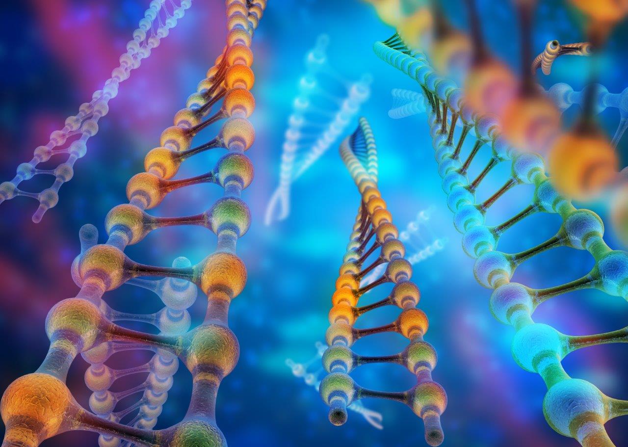
Sanford Burnham Prebys Medical Discovery Institute (SBP) scientists are part of an international team led by Northwestern University Feinberg School of Medicine that has determined the 3D atomic structure of more than 1,000 proteins that are potential drug and vaccine targets to combat some of the world’s most dangerous emerging and re-emerging infectious diseases.

These experimentally determined structures have been deposited into the World-Wide Protein Data Bank, an archive supported by the National Institutes of Health (NIH), and are freely available to the scientific community. The 3D structures help expedite drug and vaccine research and advance the understanding of pathogens and organisms causing infectious disease.

“Almost 50 percent of the structures that we have deposited in the Protein Data Bank are proteins that were requested by scientific investigators from around the world,” said Feinberg’s Wayne Anderson, PhD, director of the project. “The NIH has also requested us to work on proteins for potential drug targets or vaccine candidates for many diseases, such as the Ebola virus, the Zika virus and antibiotic-resistant bacteria. We have determined several key structures from these priority organisms and published the results in high-impact journals such as Nature and Cell.

Teamwork with an international consortium

This milestone effort, funded by two five-year contracts from the National Institute of Allergy and Infectious Diseases (NIAID), totaling a budget of $57.7 million, represents a decade of work by the Center for Structural Genomics of Infectious Diseases (CSGID) at Feinberg, led by Anderson in partnership with these institutions:

  • University of Chicago
  • University of Virginia School of Medicine
  • University of Calgary
  • University of Toronto
  • Washington University School of Medicine in St. Louis
  • UT Southwestern Medical Center
  • J. Craig Venter Institute
  • Sanford Burnham Prebys Medical Discovery Institute
  • University College London

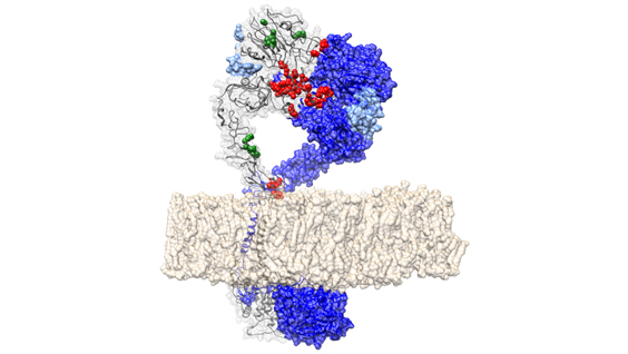
How the 3D structures are made

Before work begins on a targeted protein, a board appointed by the NIH examines each request. Once approved, the protein must be cloned, expressed and crystallized, and then X-ray diffraction data is collected at the Advanced Photon Source at Argonne National Laboratory. This data defines the location of each of the hundreds or even thousands of atoms to generate 3-D models of the structures that can be analyzed with graphics software. Each institution in the Center has an area of expertise it contributes to the project, working in parallel on many requests at once.

The bioinformatics group SBP, led by Adam Godzik, PhD, focuses on steps that have to be taken before the experimental work starts. Every protein suggested by the research community as a target for experimental structure determination is analyzed and an optimal procedure for its experimental determination is mapped out.

Experimental structure determination used to have a very high failure rate and the money and time spent on failed attempts is a major contributor to the total expense and time needed to solve protein structures. Both can be significantly improved using “Big Data” approaches, as researchers learn from thousands of successful and failed experiments in structural biology. The SBP bioinformatics group uses these approaches to improve success rates at CSGID, allowing our center to solve more structures at lower costs.

Until recently the process of determining the 3D structure of a protein took many months or even years to complete, but advances in technology, such as the Advanced Photon Source, and upgrades to computational hardware and software has dramatically accelerated the process. The Seattle Structural Genomics Center for Infectious Disease, a similar center funded by NIAID, is also on track to complete 1,000 3-D protein structures soon. Browse all of the structures deposited by the CSGID.

Anyone in the scientific community interested in requesting the determination of structures of proteins from pathogens in the NIAID Category A-C priority lists or organisms causing emerging and re-emerging infectious diseases, can submit requests to the Center’s web portal. As part of the services offered to the scientific community, the CSGID can also provide expression clones and purified proteins, free of charge.

This project has been supported by federal funds from the NIAID, NIH,  Department of Health and Human Services, under contract numbers HHSN272200700058C and HHSN272201200026C.

Institute News

Showing kids why mutants matter at the STEM Expo

AuthorJessica Moore
Date

March 7, 2017

Want to see some mutant worms? If you do, you’re like the hundreds—or possibly thousands—of children and their parents who visited the Sanford Burnham Prebys Medical Discovery Institute (SBP) booth at the STEM Expo, held Saturday, March 4 to kick off the San Diego Festival of Science and Engineering.

Those families got to watch live C. elegans—tiny worms used at SBP to study the biology of aging—move under a magnifying glass. By comparing normal worms to mutants that don’t move as well, they learned about how a single change to DNA can have a huge impact on how a whole body works. Scientists from the lab of Malene Hansen, PhD, associate professor at SBP, who provided the worms, explained how mutations in the same gene also cause human diseases like Ehlers-Danlos syndrome, which involves hyperflexible joints and skin.

They also had the chance to use virtual reality headsets to look at the shapes of proteins—the machines that make things happen inside cells—and how they can change as a result of a mutation. The 3D visualization tool was created by the lab of Adam Godzik, PhD, professor and director of the Bioinformatics and Structural Biology Program at SBP. Researchers from Godzik’s group told visitors about how they use similar computer modeling to find new mutations that could be important in cancer.

Kids left the SBP booth with temporary DNA tattoos on their hands (or sometimes their faces), mini lab coats personalized on the spot so they could pretend to be scientists at home and, maybe, a newfound interest in biology, thanks to the enthusiasm of the Institute’s many volunteers.

Institute News

Genetic drivers of immune response to cancer discovered through ‘big data’ analysis

AuthorJessica Moore
Date

July 18, 2016

Scientists at the Sanford Burnham Prebys Medical Discovery Institute (SBP) have identified over 100 new genetic regions that affect the immune response to cancer. The findings, published in Cancer Immunology Research, could inform the development of future immunotherapies—treatments that enhance the immune system’s ability to kill tumors.

“By analyzing a large public genomic database, we found 122 potential immune response drivers—genetic regions in which mutations correlate with the presence or absence of immune cells infiltrating the tumors,” said lead author Eduard Porta-Pardo, PhD, a postdoctoral fellow at SBP. “While several of these correspond to proteins with known roles in immune response, many others offer new directions for cancer immunology research, which could point to new targets for immunotherapy.”

Immunotherapy has been heralded as a turning point in cancer because it can treat even advanced cases that have spread to other organs. Several drugs in this class are now widely used and often lead to remarkable success, eradicating or dramatically shrinking tumors and preventing recurrence.

Most current immunotherapies rely on a similar strategy—releasing the brakes on the immune system. These treatments are powerful if the tumor is recognized by the immune system as a threat and allows immune cell infiltration, but some cancers remain undercover or block immune cell entry into the tumor in as yet unknown ways.

“To develop immunotherapies that are relevant to a wide range of cancers, we need to know a lot more about how the immune system interacts with tumors,” said Adam Godzik, PhD, professor and director of the Bioinformatics and Structural Biology Program and senior author of the study. “Our study provides many new leads for this endeavor.”

“We are exploring cancer mutations at fine resolution by accounting for the fact that mutations can affect the encoded protein in different ways depending on where the resulting change is located,” commented Porta-Pardo. “Our algorithm, domainXplorer, identifies correlations between a phenotype, in this case the amount of immune cells in the tumor, and mutations in individual protein domains—parts of a protein with distinct functions.

“This work emphasizes the value of open data,” Godzik added. “Because we could access genomic data from over 5,000 tumor samples from The Cancer Genome Atlas (TCGA), we could jump straight to analysis without having to set up a big collaborative network to gather and sequence so many samples.”

“Our plan for the next phase of this research is to use this algorithm to search for genetic regions correlating with the levels of specific immune cell types within the tumor, which will reveal further details of cancer immunology.”

Institute News

Symposium brings leaders in tumor immunology to SBP

Authorjmoore
Date

March 21, 2016

SBP’s La Jolla campus recently hosted a one-day conference on Cancer Immunology and the Tumor Microenvironment, one of the hottest topics in cancer research. The symposium on March 17 attracted approximately 160 attendees from across the La Jolla biomedical research mesa.

The symposium was organized by Carl Ware, PhD and Robert Rickert, PhD, directors of the Inflammatory and Infectious Disease Center and the Tumor Microenvironment and Cancer Immunology Program, respectively. They planned a scientific agenda that covered diverse aspects of research on harnessing the immune system to treat cancer, from improving current immunotherapies to identifying new immunological targets. Talks were given by prestigious scientists including:

Two SBP researchers also presented. Linda Bradley, PhD, professor in the Immunity and Pathogenesis Program, discussed her work on the immune cell surface protein PSGL-1, showing that it regulates PD-1, a so-called “immune checkpoint.” PD-1 is found on T cells and normally acts as on “off switch” to keep the immune system from attacking cells in the body. PD-1 regulators are a new approach to treating cancer that work by unleashing cytotoxic T cells to kill cancer cells.


Adam Godzik, PhD, professor and director of the Bioinformatics and Structural Biology Program, spoke about using bioinformatic analyses to search for new cancer drivers related to the immune response. Cancer drivers are genes that, when altered, are responsible for cancer progression. Combining cancer mutation and protein structure databases, his team has identified many genes involved in immune recognition of tumors.

Institute News

Upcoming symposium: Cancer Immunology and the Tumor Microenvironment

Authorjmoore
Date

March 10, 2016

On March 17, SBP La Jolla is hosting a symposium on the interactions between the immune system and tumors, including how they can be leveraged for cancer treatment. The symposium is organized by Carl Ware, PhD and Robert Rickert, PhD, the directors of the Inflammatory and Infectious Disease Center and the Tumor Microenvironment and Cancer Immunology Program, respectively, and features presentations by leaders in the field:

Crystal Mackall, MDStanford University

Yang-Xin Fu, MD, PhDUT Southwestern

Mikala Egeblad, PhDCold Spring Harbor Laboratory

Linda Bradley, PhDSanford Burnham Prebys Medical Discovery Institute

Jose Conejo-Garcia, MD, PhDWistar Institute

Jonathan Powell, MDJohns Hopkins School of Medicine

Shannon Turley, PhDGenentech

Karen Willard-Gallo, PhDInstitut Jules Bordet – Belgium

Sandip Patel, MDUC San Diego

Adam Godzik, PhDSanford Burnham Prebys Medical Discovery Institute

The symposium will be held from 9-4:30 in Fishman Auditorium (overflow seating in the Building 12 auditorium), with a reception to follow. If you plan to attend, please register here.

Institute News

‘Big Data’ used to identify new cancer driver genes

Authorsgammon
Date

October 20, 2015

In a collaborative study led by Sanford Burnham Prebys Medical Discovery Institute (SBP), researchers have combined two publicly available ‘omics’ databases to create a new catalogue of ‘cancer drivers’. Cancer drivers are genes that when altered, are responsible for cancer progression. The researchers used cancer mutation and protein structure databases to identify mutations in patient tumors that alter normal protein-protein interaction (PPI) interfaces. The study, published today in PLoS Computational Biology, identified more than 100 novel cancer driver genes and helps explain how tumors driven by the same gene may lead to different patient outcomes.

“This is the first time that three-dimensional protein features, such as PPIs, have been used to identify driver genes across large cancer datasets,” said lead author Eduard Porta-Pardo, PhD, a postdoctoral fellow at SBP. “We found 71 interfaces in proteins previously unrecognized as cancer drivers, representing potential new cancer predictive markers and/or drug targets. Our analysis also identified several driver interfaces in known cancer genes, such as TP53, HRAS, PI3KCA and EGFR, proving that our method can find relevant cancer driver genes and that alterations in protein interfaces are a common pathogenic mechanism of cancer.”

Cancer is caused by the accumulation of mutations to DNA. Until now, scientists have focused on finding alterations in individual genes and cell pathways that can lead to cancer. But the recent push by the National Institutes of Health (NIH) to encourage data sharing has led to an era of unprecedented ability to systematically analyze large scale genomic, clinical, and molecular data to better explain and predict patient outcomes, as well as finding new drug targets to prevent, treat, and potentially cure cancer.

“For this study we used an extended version of e-Driver, our proprietary computational method of identifying protein regions that drive cancer. We integrated tumor data from almost 6,000 patients in The Cancer Genome Atlas (TCGA) with more than 18,000 three-dimensional protein structures from the Protein Data Bank (PDB),” said Adam Godzik, PhD, director of the Bioinformatics and Structural Biology Program at SBP. “The algorithm analyzes whether structural alterations of PPI interfaces are enriched in cancer mutations, and can therefore identify candidate driver genes.”

“Genes are not monolithic black boxes. They have different regions that code for distinct protein domains that are usually responsible for different functions. It’s possible that a given protein only acts as a cancer driver when a specific region of the protein is mutated,” Godzik explained. “Our method helps identify novel cancer driver genes and propose molecular hypotheses to explain how tumors apparently driven by the same gene have different behaviors, including patient outcomes.”

“Interestingly, we identified some potential cancer drivers that are involved in the immune system. With the growing appreciation of the importance of the immune system in cancer progression, the immunity genes we identified in this study provide new insight regarding which interactions may be most affected,” Godzik added.

The study was performed in collaboration with the European Bioinformatics Institute (UK), Centro de Investigación Principe Felipe (Spain), and CIBER de Enfermedades Raras (Spain).

Institute News

Sanford-Burnham’s 36th Annual Symposium: The Microbiome and Human Health

Authorsgammon
Date

November 3, 2014

On Thursday, October 30, 2014, Sanford-Burnham hosted more than 250 attendees at its 36th annual symposium to hear opinion-leading scientists discuss their latest findings on the microbiome. The microbiome is a relatively new frontier for research scientists with aims to understand how the trillions of microbes—bacteria, viruses, fungi, and others—that live in our nose, mouth, gut, and skin interact with human cells to influence health and disease. Continue reading “Sanford-Burnham’s 36th Annual Symposium: The Microbiome and Human Health”