angiogenesis Archives - Sanford Burnham Prebys
Institute News

Scientists discover new survival strategy for oxygen-starved pancreatic cancer cells

AuthorMonica May
Date

October 23, 2019

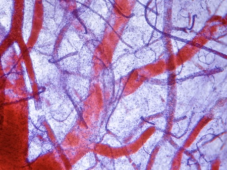
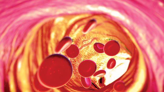
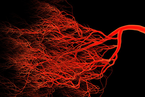
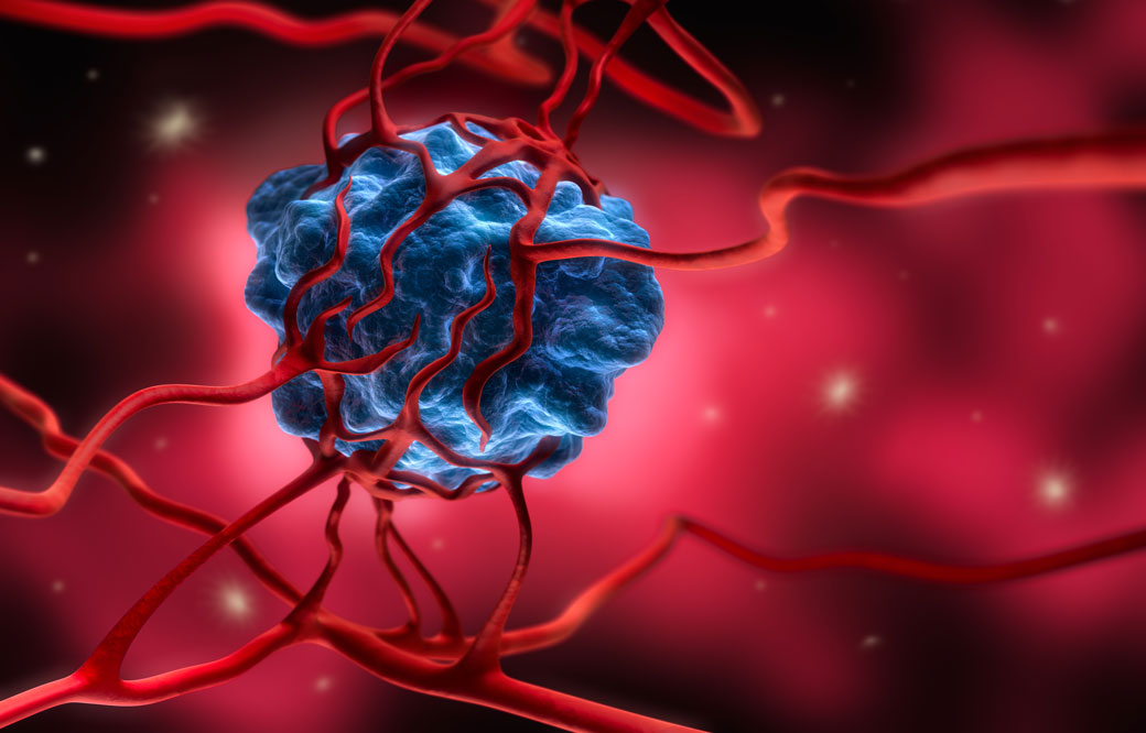
Oxygen is essential to life. When fast-growing tumor cells run out of oxygen, they quickly sprout new blood vessels to keep growing, a process called angiogenesis. 

By blocking pancreatic cancer’s oxygen-sensing machinery—the same field of research studied by the winners of the 2019 Nobel Prize in Medicine—Sanford Burnham Prebys scientists have uncovered a new way that tumors turn on angiogenesis in an animal model. The discovery, published in Cancer Research, could lead to a treatment that is given with an anti-angiogenetic medicine, thereby overcoming drug resistance. 

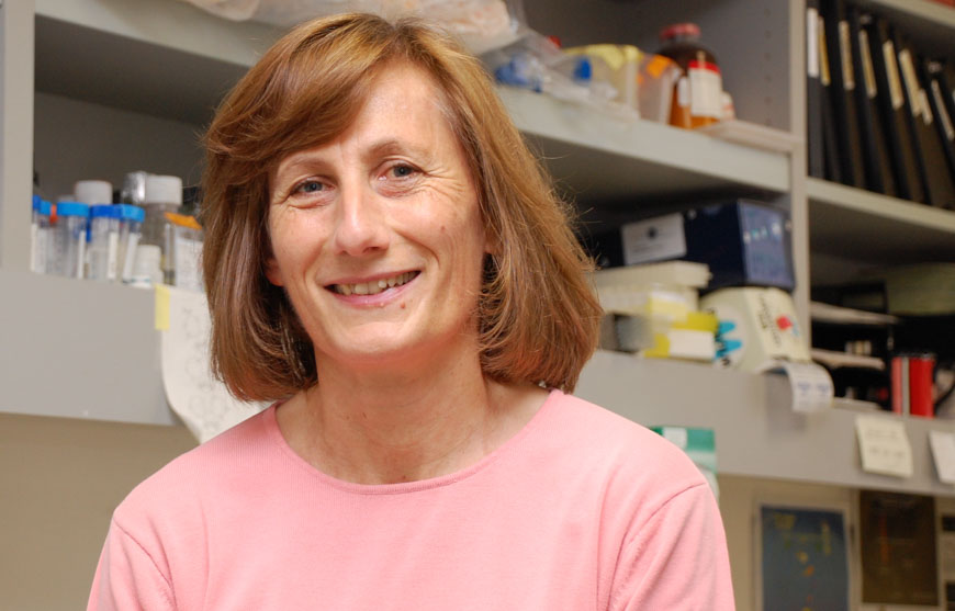
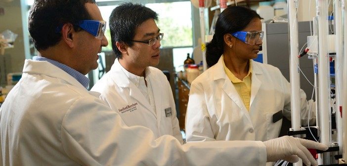
“Treatment resistance is a major challenge for cancer treatments that block blood vessel growth,” says Garth Powis, D.Phil., professor and director of Sanford Burnham Prebys’ National Cancer Institute (NCI)-designated Cancer Center and senior author of the study. “Our research identifies a new way angiogenesis is activated, opening new opportunities to find medicines that might make existing cancer treatments more effective.” 

Many cancer treatments work by blocking angiogenesis, which rarely occurs in healthy tissues. However, these medicines eventually stop working, and the cancer returns, sometimes in as little as two months. Scientists have been researching why this treatment resistance occurs so it can be stopped.

In this study, the scientists focused on pancreatic cancer, which is notoriously desperate for oxygen and also difficult to treat. Fewer than 10% of people diagnosed with pancreatic cancer are alive five years later. 

To see how a pancreatic tumor responds to a disruption in its oxygen supply, the Sanford Burnham Prebys researchers used a mouse model to block an oxygen-sensing protein called HIF1A—which should cripple the tumor’s growth. Instead of dying, however, after about a month the cells multiplied—indicating they had developed a new way to obtain oxygen. 

Further work revealed that the cancer cells were clear and swollen with the nutrient glycogen (a characteristic also seen in some ovarian and kidney cancers). In response to the excess glycogen, special immune system cells were summoned to the tumor, resulting in blood vessel formation and tumor survival. Each of these responses represents a new way scientists could stop pancreatic tumors from evolving resistance to treatment.

“Our team’s next step is to test tumor samples from people with pancreatic cancer to confirm this escape mechanism occurs in a clinical setting,” says Powis. “One day, perhaps we can create a second medicine that keeps anti-angiogenic drugs working and helps more people survive pancreatic cancer.”


Research reported in this press release was supported by the U.S. National Institutes of Health (NIH) (5F31CA203286, CA216424 and P30CA030199). The content is solely the responsibility of the authors and does not necessarily represent the official views of the NIH. The study’s DOI is 10.1158/0008-5472.CAN-18-2994. 

Institute News

Targeting long-sought EphA2 receptor becomes crystal clear

AuthorMonica May
Date

May 13, 2019

Scientists have long sought to target a cellular receptor called EphA2 because of its known role in many disorders, including cancer, inflammatory conditions, neurological disorders and infectious diseases. However, lack of information about the structure formed when EphA2 links to other molecules—ligands—has hindered drug development. 

Now, scientists at Sanford Burnham Prebys have crystallized EphA2 together with peptide ligands (short proteins) and used the structure to engineer more powerful compounds that activate or inactivate the receptor, paving the way for new therapies. The discovery was published in the Journal of Biological Chemistry.

“EphA2 plays a central role in a plethora of biological and disease processes,” says Elena Pasquale, PhD, professor in the Tumor Initiation and Maintenance Program at Sanford Burnham Prebys. “Our team’s identification of potent, highly selective peptides that regulate the receptor is a key step toward rational design of therapies for the numerous disorders that are driven by EphA2.” 

EphA2 is found in the cells that line the surfaces of our body, including our skin, blood vessels and other organs. The receptor is typically only present at high levels during disease states, making it a promising drug target. Activating the receptor could hinder tumor growth, while inhibiting it could reduce unwanted formation of blood vessels (angiogenesis), treat certain inflammation-driven disorders and block pathogens—such as malaria, chlamydia and the hepatitis C virus—from gaining entry into a cell through the receptor. Because EphA2 travels deep inside of the cell when activated, scientists could also harness it as a Trojan horse by attaching chemotherapies or imaging agents to the peptide ligands, which would subsequently be delivered to the desired cells. 

In the study, the scientists initially crystallized a weakly binding peptide in complex with EphA2, yielding a detailed picture of the binding features and providing clues to the receptor’s “sweet spot” or site of action. The researchers then used this information to repeat this process, engineering increasingly more powerful ligands. This work identified several peptides that strongly clasp the receptor and activate or inactivate it—which can be used to inform drug development.

Further quantitative Förster Resonance Energy Transfer (FRET) microscopy experiments, which measure receptor-receptor interactions, revealed that EphA2 receptors cluster together when activated by a peptide—an effect similar to that caused by its natural ligands—answering an unresolved question in the field. 

“In addition to helping guide therapeutic development paths, these peptides are also valuable research tools for scientists who are working to gain insights into this important receptor,” adds Pasquale. “Our hope is that with this new information, one day we can find targeted therapies to treat cancer, inflammatory disorders and infectious diseases that are regulated by EphA2.”


The co-first authors of the study are Maricel Gomez-Soler, PhD, and Marina Petersen Gehring, PhD, of Sanford Burnham Prebys; and Bernhard C. Lechtenberg, PhD, formerly of Sanford Burnham Prebys and currently of the Walter and Eliza Hall Institute of Medical Research. 

Additional authors include Elmer Zapata-Mercado and Kalina Hristova, PhD, of Johns Hopkins University. The study’s DOI is 10.1074/jbc.RA119.008213. 

This research was supported by the National Institutes of Health (NIH) (R01NS087070, R01GM131374 and P30CA030199). 

Institute News

Preventing leaky blood vessels could halt cancer

AuthorJosh Baxt
Date

June 12, 2018

We like to think blood vessels are solid, like a brand-new garden hose—nothing leaks in or out. But sometimes vessels lose integrity, a condition called vascular permeability. This can be a big problem, particularly in cancer, allowing tumor cells to get into the bloodstream and travel throughout the body. 

Researchers in the Komatsu laboratory at SBP’s Lake Nona campus in Orlando, Fla. have been studying permeability for some time, focusing on a protein called RRAS that’s known to stabilize blood vessels. Recently, Carole Perrot, PhD, a postdoc in the lab, was the first author on a paper in The FASEB Journal that showed RRAS is regulated by a molecule called cAMP. How that regulation played out was a big surprise. 

“I thought cAMP would increase RRAS expression,” says Perrot, “but it was completely the opposite.” 

Perrot’s expectations were built on previous research, which showed that cAMP reduces permeability. However, those studies weren’t long enough to fully understand what was happening inside the cells. Because cAMP is a signaling molecule that turns on several genes, it can take time before the results become apparent. 

“When cAMP is activated in the short term, it’s beneficial for vasculature,” says Perrot. “But if you activate the pathway for the long term, more than 48 or 72 hours, it does the opposite. You need more time to see which genes are triggered after the initial activation.” 

Over time, cAMP downregulates RRAS and destabilizes blood vessel cells’ contacts, making them more permeable. This fits with other evidence, which has shown that key proteins in the cAMP pathway are upregulated in tumor blood vessels. 

The ultimate goal is to better modulate RRAS and restore blood vessel functionality. Perrot and colleagues are continuing to identify the various pieces of this puzzle, and the next step will be to find the upstream molecules that trigger cAMP expression. This ongoing research could eventually provide new ways to combat metastasis. 

“If you suppress blood vessel leakage, it makes it easier for drugs to reach into the tumor during cancer therapy. Also, there is less opportunity for cancer cells to get into the blood vessels and disseminate throughout the body,” says Perrot. “If you restore RRAS expression in tumors, you normalize the vascular networks and potentially limit tumor progression.”

Institute News

Location matters, even for tumors

AuthorBill Stallcup, PhD
Date

September 7, 2017

Location, Location, Location! We often hear this in real estate, but it’s also true in biology. Cells need to be in the right place at the right time to help promote organ development and send the signals that keep our bodies working. But tumors rely on location as well, especially when it comes to angiogenesis—the process they use to recruit new blood vessels to “feed” their growth with oxygen and nutrients.

In the journal Cancers, William Stallcup, PhD, a professor in SBP’s NCI-Designated Cancer Center, describes the importance of location for vascular endothelial growth factor (VEGF), a protein that stimulates blood vessel formation.

“VEGF is a major stimulus for the formation of tumor blood vessels,” says Stallcup. “In fact, blocking VEGF has been one idea for slowing tumor growth by cutting off the tumor blood supply. This strategy has not been as successful as researchers had hoped, partly because blood vessel formation and the action of VEGF are both complex processes that we don’t fully understand.”

The vascular cells that form blood vessels are embedded in a fibrous meshwork called the vascular extracellular matrix (ECM). Some forms of VEGF bind tightly to this ECM, while other forms diffuse freely in tissues. The Stallcup lab’s studies reveal that the way VEGF and ECM interact is very important for tumor blood vessel formation.

According to Weon-Kyoo You, PhD, a former Stallcup postdoc and first author of the study, “When we looked at brain tumor growth in normal mice, we found that lots of VEGF was bound to the vascular ECM. But when we studied tumor growth in mutant mice that were deficient in ECM assembly, we saw that the VEGF mostly diffused away from tumor blood vessels and was dispersed in the tumor tissue.”

This change in VEGF location had large effects on the structure and function of tumor blood vessels. In normal mice, efficient binding of VEGF to the vascular ECM produced large diameter vessels that were not leaky—and brain tumors grew fast in these mice. In mice with deficient VEGF-ECM interactions, the blood vessels were thin and leaky—and brain tumors grew slowly due to lack of nutrients.

Stallcup concludes that, “We were very surprised at the correlation between healthy ECM, VEGF binding, and tumor growth. Apparently, having the VEGF located right there at the site where it can be readily used by vascular cells is a key factor in producing functional blood vessels.

“These studies add to our understanding of the tactics tumors use to grow, and also give us new clues about how we might be able to thwart cancer progression.”

Read the study here.

Institute News

Scientific breakthrough may limit damage caused by heart attacks

AuthorJessica Moore
Date

June 30, 2016

A research advance from the Sanford Burnham Prebys Medical Discovery Institute (SBP) and Stanford University could lead to new drugs that minimize the damage caused by heart attacks. The discovery, published in Nature Communications, reveals a key control point in the formation of new blood vessels in the heart, and offers a novel approach to treat heart disease patients.

“We found that a protein called RBPJ serves as the master controller of genes that regulate blood vessel growth in the adult heart,” said Mark Mercola, PhD, professor in SBP’s Development, Aging, and Regeneration Program and jointly appointed as professor of medicine at Stanford University, senior author of the study. “RBPJ acts as a brake on the formation of new blood vessels. Our findings suggest that drugs designed to block RBPJ may promote new blood supplies and improve heart attack outcomes.”

In the US, someone has a heart attack every 34 seconds. The ensuing loss of heart muscle, if it affects a large enough area, can severely reduce the heart’s pumping capacity, which causes labored breathing and makes day-to-day tasks difficult. This condition, called heart failure, arises within five years in at least one in four heart attack patients.

The reason heart muscle dies in a heart attack is that it becomes starved of oxygen—a heart attack is caused by blockage of an artery supplying the heart. If heart muscle had an alternative blood supply, more muscle would remain intact, and heart function would be preserved. Many researchers have therefore been searching for ways to promote the formation of additional blood vessels in the heart.

“Studies in animals have shown that having more blood vessels in the heart reduces the damage caused by ischemic injuries, but clinical trials of previous therapies haven’t succeeded,” said Ramon Díaz-Trelles, PhD, staff scientist at SBP and lead author of the study. “The likely reason they have failed is that these studies have evaluated single growth factors, but in fact building blood vessels requires the coordinated activity of numerous factors. Our data show that RBPJ controls the production of these factors in response to the demand for oxygen.

“We used mice that lack RBPJ to show that it plays a novel role in myocardial blood vessel formation (angiogenesis)—it acts as a master controller, repressing the genes needed to create new vessels,” added Diaz-Trelles. “What’s remarkable is that removing RBPJ in the heart muscle did not cause adverse effects—the heart remained structurally and functionally normal in mice without it, even into old age.”

“RBPJ is a promising therapeutic target. It’s druggable, and our findings suggest that blocking it could benefit patients with cardiovascular disease at risk of a heart attack. It may also be relevant to other diseases,” commented Pilar Ruiz-Lozano, PhD, associate professor of pediatrics at Stanford and adjunct professor at SBP, co-senior author. “Inhibitors of RBPJ might also be used to treat peripheral artery disease, and activators might be beneficial in cancer by inhibiting tumor angiogenesis.”

The paper is available online here.

Institute News

Scientists solve structure of important protein for tumor growth

Authorsgammon
Date

August 5, 2015

In a collaborative study between SBP and the Argonne National Laboratory, scientists have used a highly specialized X-ray crystallography technique to solve the protein structure of hypoxia-inducible factors (HIFs), important regulators of a tumor’s response to low oxygen (hyopoxia). The findings, published today in the journal Nature, open the door to search for new drugs to treat tumors by cutting off their supply of oxygen and nutrients. Continue reading “Scientists solve structure of important protein for tumor growth”

Institute News

Molecule that fixes “leaky” blood vessels can impact cancer, stroke, and blindness

Authorsgammon
Date

March 13, 2015

In a new study by Masanobu Komatsu, Ph.D., associate professor in the Cardiovascular Pathobiology Program and Tumor Microenvironment and Metastasis Programs, a cellular protein called R-Ras was found to suppress the effects of vascular endothelial growth factor (VEGF), a signaling molecule that helps create new blood vessels and is overexpressed in many tumors. The findings create a new route to treat cancer as well as certain causes of blindness and ischemic diseases. Continue reading “Molecule that fixes “leaky” blood vessels can impact cancer, stroke, and blindness”

Institute News

A nuclear receptor that binds more than 5,000 sites in the genome—and promotes angiogenesis.

Authorsgammon
Date

July 17, 2014

The retinoid X receptor (RXR) is a nuclear hormone receptor—meaning that it sits on various parts of the genome—and turns genes “on” and “off.” RXR is known to play an important role in many fundamental biological processes such as reproduction, cellular differentiation, bone development, and hematopoiesis. Continue reading “A nuclear receptor that binds more than 5,000 sites in the genome—and promotes angiogenesis.”