Anne Bang Archives - Sanford Burnham Prebys
Institute News

Using stem cells to study the biochemistry of learning

AuthorMiles Martin
Date

August 18, 2022

A method for studying human neurons could help researchers develop approaches for treating Alzheimer’s, schizophrenia and other neurological diseases

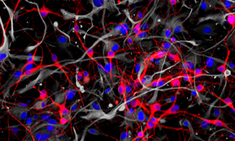
Researchers from the Conrad Prebys Center for Chemical Genomics have developed a procedure to use neurons derived from human stem cells to study the biological processes that control learning and memory. The method, described in Stem Cell Reports, uses electrodes to measure the activity of neuronal networks grown from human-induced pluripotent stem cells (iPSCs). The procedure tracks how synapses—the connections between neurons—strengthen over time, a process called long-term potentiation (LTP).

“Impaired long-term potentiation is thought to be central to many neurological diseases, including Alzheimer’s, addiction and schizophrenia,” says senior author Anne Bang, PhD, director of Cell Biology at the Prebys Center. “We’ve developed an approach to study this process in human cells much more efficiently than current methods, which could help trigger future breakthroughs for researchers working on these diseases.”

LTP helps our brain encode information, which is what makes it so critical for learning and memory. Impairment of LTP is thought to contribute to neurological diseases, but it has proven difficult to verify this hypothesis in human cells.
LTP helps our brain encode information, which is what makes it so critical for learning and memory. Impairment of LTP is thought to contribute to neurological diseases, but it has proven difficult to verify this hypothesis in human cells.

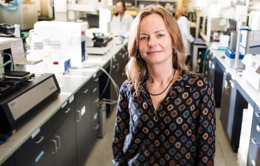
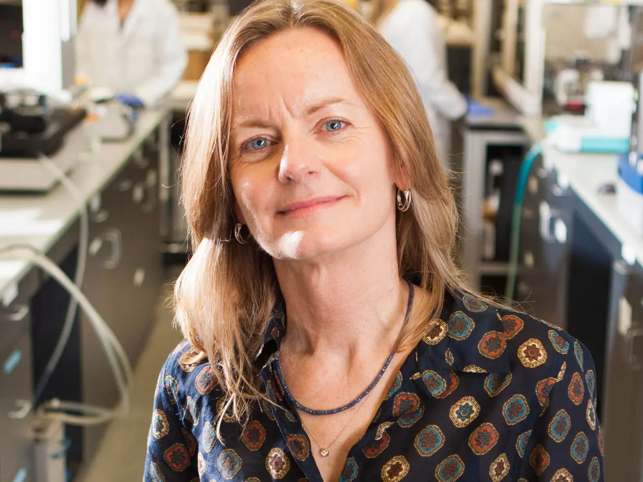
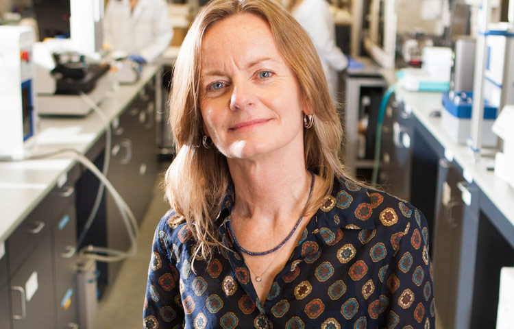
Anne Bang, PhD, director of Cell Biology at the Prebys Center.

“LTP is such a fundamental process,” says Bang. “But the human brain is hard to study directly because it’s so inaccessible. Using neurons derived from human stem cells helps us work around that.”

Although LTP can be studied in animals, these studies can’t easily account for some of the more human nuances of neurological diseases.

“A powerful aspect of human stem cell technology is that it allows us to study neurons produced from patient stem cells. Using human cells with human genetics is important in these types of tests because many neurological diseases have complex genetics underpinning them, and it’s rarely just one or two genes that influence a disease,” adds Bang.

To develop the method, first author and Prebys Center staff scientist Deborah Pré, PhD, grew networks of neurons from healthy human stem cells, added chemicals known to initiate LTP and then used electrodes to monitor changes in neuronal activity that occurred throughout the process.

The method can run 48 tests at once, and neurons continue to exhibit LTP up to 72 hours after the start of the experiment. These are distinct advantages over other approaches, which can often only observe parts of the process and are low throughput, which can make getting results more time consuming.

For this study, the researchers used neurons grown from healthy stem cells to establish a baseline understanding of LTP. The next step is to use the approach on neurons derived from patient-derived stem cells and compare these results to the baseline to see how neurological diseases influence the LTP process.

“This is an efficient method for interrogating human stem cell–derived neurons,” says Bang. “Doing these tests with patient cells could open doors for researchers to discover new ways of treating neurological diseases.”
Institute News

What scientists are learning about COVID-19 and the brain

AuthorMonica May
Date

December 8, 2020

We caught up with cell biologist Anne Bang, who recently teamed up with her husband to study how SARS-CoV-2 affects the brain

Brain fog. Memory loss. Dizziness and confusion. Although COVID-19 is primarily thought of as a lung disease, survivors continue to report lingering and highly concerning neurological effects—severe enough to impact their ability to work and live normal lives. Doctors are also seeing a worrisome increase in strokes in younger patients, among other observations.

To learn what scientists know so far about COVID-19 and its effect on the brain, we caught up with Anne Bang, PhD, director of Cell Biology at Sanford Burnham Prebys’ Conrad Prebys Center for Chemical Genomics. Bang recently teamed up with scientists at Penn Medicine and a virologist at Scripps Research—who also happens to be her husband—to investigate whether SARS-CoV-2 infects brain cells. Their findings were published in Cell Stem Cell.

What do scientists know about the brain and COVID-19 so far?

Unfortunately, information is still very limited. There are reports of viral replication in the brain and spinal cord fluid of people with COVID-19 who have neurological symptoms. But as you can imagine, taking brain biopsies from someone who has COVID-19 is not realistic. So we really don’t know a lot yet. For this reason, scientists are turning to systems that can model the human brain, such as brain cells created from induced pluripotent stem cells (iPSCs) and brain organoids, to study SARS-CoV-2’s impact on the brain.

What did you find in your study?

We created several types of brain cells using iPSCs and brain organoids, which we then infected with SARS-CoV-2. We found that SARS-CoV-2 primarily infects a brain cell type called choroid plexus cells—largely bypassing neurons and astrocytes. The choroid plexus is a specialized part of the blood-brain barrier, which controls what can enter your brain and produces cerebral spinal fluid. More research emerges every day, but so far, the consensus in the field seems to align with our findings.

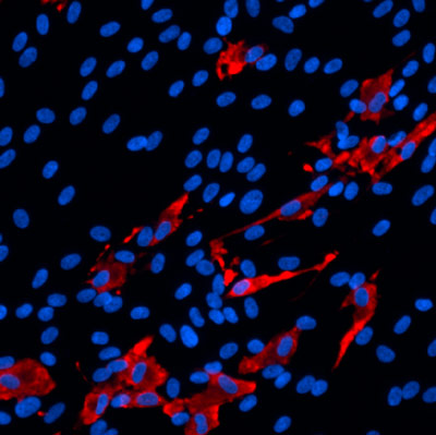
SARS_CoV2_ Infected human choroid plexus cells a type of brain cell

The scientists found that SARS-CoV-2 (red) primarily infects brain cells called choroid plexus cells (blue), which are part of the brain’s protective blood-brain barrier.

How might this finding translate to what we’re seeing in patients?

We know that choroid plexus cells produce high levels of ACE2, which is the receptor that SARS-CoV-2 uses to enter and infect cells. Because the choroid plexus is the “gatekeeper” to the brain, it’s possible that the virus enters the brain by infecting these cells. However, much more research is needed before we can give a definitive answer to this question.

We have more questions than answers right now about COVID-19. What is one question you wish we had the answer to?

How does the virus get from the nose and mouth and spread to other parts of the body? This is a big question for me and the scientific field. Once we know how the virus travels throughout the body, we can potentially stop its spread and control the dangerous symptoms.

What was it like working with your husband? Was this your first time working together?

It was really fun. I found out that he is great to work with. We’ve been together for 30 years, and incredibly, this was the first time we worked together.

Institute News

Drug screen conducted at Sanford Burnham Prebys identifies new therapeutic avenues for Alzheimer’s disease

AuthorMonica May
Date

February 7, 2019

A screen of more than 1,600 Food and Drug Administration (FDA)–approved drugs performed at SBP’s Conrad Prebys Center for Chemical Genomics (Prebys Center) has revealed new therapeutic avenues that could lead to an Alzheimer’s disease treatment. 

The findings come from a collaboration between SBP scientists and researchers at the University of California San Diego School of Medicine, Leiden University Medical Center and Utrecht University in the Netherlands and were published in Cell Stem Cell

The hunt is on for an effective treatment for Alzheimer’s, a memory-robbing disease that is nearing epidemic proportions as the world’s population ages. Nearly six million people in the U.S. are living with Alzheimer’s disease. This number is projected to rise to 14 million by 2060, according to the Centers for Disease Control and Prevention (CDC). 

Scientists have known for many years that a protein called tau accumulates and creates tangles in the brain during Alzheimer’s disease. Additional research is revealing that altered cholesterol metabolism in the brain is associated with Alzheimer’s. But the relationship between these two clues is unknown. 

By testing a library of FDA-approved drugs against induced pluripotent stem cells (iPSC) neurons created from people with Alzheimer’s disease, the scientists were able to identify 42 compounds that reduced the level of phosphorylated tau, a form of tau that contributes to tangle formation. The researchers further refined this group to only include cholesterol-targeting compounds. 

A detailed study of these drugs showed that their effect on tau was mediated by their ability to lower cholesteryl esters, a storage product of excess cholesterol. These results led them to an enzyme called CYP46A1, which normally reduces cholesterol. Activation of this enzyme by the drug efavirenz (brand names Sustiva® and Stocrin®) reduced cholesterol esters and phosphorylated tau in these neurons, making it a promising therapeutic target for Alzheimer’s disease. Further mapping of the enzyme’s action(s) within a cell could reveal even more therapeutic targets. 

“Our Prebys Center is designed to be a comprehensive resource that allows basic research—whether conducted at SBP, academic and nonprofit research institutions or industry—to be translated into medicines for diseases that urgently need better treatments,” says study author Anne Bang, PhD, director of Cell Biology at the Conrad Prebys Center for Chemical Genomics at SBP. “We are proud that the Prebys Centers’ drug discovery technologies helped reveal new paths that could lead to a potential treatment for Alzheimer’s, one of the most devastating diseases of our time.”


The senior author of the study is Lawrence S. B. Goldstein, PhD, distinguished professor at the University of California San Diego (UC San Diego) and scientific director of the Sanford Consortium for Regenerative Medicine. The co-first authors are Vanessa Langness, a PhD graduate student in Goldstein’s lab, and Rik van der Kant, PhD, a senior scientist at Vrije University in Amsterdam and former postdoctoral fellow in Goldstein’s lab. 

Additional study authors include Cheryl M. Herrera, Daniel Williams, Lauren K. Fong and Kevin D. Rynearson, UC San Diego; Yves Leestemaker, Huib Ovaa, Evelyne Steenvoorden and Martin Giera of Leiden University Medical Center; Jos F. Brouwers and J. Bernd Helms; Utrecht University; Steven L. Wagner, UC San Diego and Veterans Affairs San Diego Healthcare System.

Funding for this research came, in part, from the Alzheimer Netherlands Fellowship, ERC Marie Curie International Outgoing Fellowship, the National Institutes of Health (NIH) (5T32AG000216-24, IRF1AG048083-01) and the California Institute for Regenerative Medicine (RB5-07011).

Read more in UC San Diego’s press release. 

Institute News

Nanowire arrays allow electrical recording of neuronal networks

AuthorJessica Moore
Date

April 12, 2017

To examine a neuron’s health, activity and response to drugs, scientists record its electrical activity. Current methods of recording are destructive, so they can only be used to study a neuron for a brief period, and can only measure the activity of one cell at a time. But neurons don’t function individually—they act in networks, and commonly used systems for detecting the electrical activity of complex groups of neurons aren’t as sensitive as they could be.

A new technology developed through a collaboration between Anne Bang, PhD, director of Cell Biology in the Conrad Prebys Center for Chemical Genomics at the Sanford Burnham Medical Research Institute, and Shadi Dayeh, PhD, associate professor at UC San Diego, makes high-sensitivity recording possible in neuronal networks. Publishing in Nano Letters, the team describes nanowire arrays that could accelerate drug development for neurological and neuropsychiatric diseases.

“We envision that this nanowire technology could be used on stem-cell-derived brain models to identify the most effective drugs for disorders like bipolar disorder and Alzheimer’s,” says Bang.

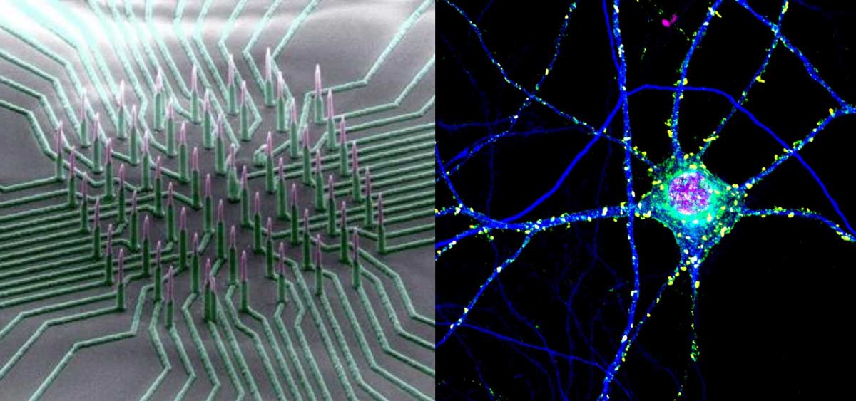
The nanowire technology developed in Dayeh’s laboratory is nondestructive and can simultaneously measure potential changes in multiple neurons — with the high sensitivity and resolution achieved by the current state of the art.

The device consists of an array of silicon nanowires densely packed on a small chip patterned with nickel electrode leads that are coated with silica. The nanowires poke inside cells without damaging them and are sensitive enough to measure small potential changes that are a fraction of or a few millivolts in magnitude. Neurons interfaced with the nanowire array survived and continued functioning for at least six weeks.

Another innovative feature of this technology is it can isolate the electrical signal measured by each individual nanowire. “This is different from existing nanowire technologies, where several wires are electrically shorted together and you cannot differentiate the signal from every single wire,” Dayeh says.

Dayeh noted that the technology needs further optimization for brain-on-chip drug screening. His team is working to adapt the arrays for heart-on-chip drug screening for cardiac diseases and in vivo brain mapping, which is still several years away. “Our ultimate goal is to translate this technology to a device that can be implanted in the brain.”

This story is based on a press release from UC San Diego.

Institute News

Consortium awarded $15 million to unravel bipolar disorder and schizophrenia

AuthorSusan Gammon
Date

August 31, 2016

Sanford Burnham Prebys Medical Discovery Institute (SBP), the Johns Hopkins University School of Medicine, the Salk Institute for Biological Studies, and the University of Michigan will embark on a $15.4 million effort to develop new systems for quickly screening libraries of drugs for potential effectiveness against schizophrenia and bipolar disorder, the National Institute of Mental Health (NIMH) has announced. The consortium, which includes two industry partners, will be led by Hongjun Song, PhD, of Johns Hopkins and Rusty Gage, PhD, of Salk.

Bipolar disorder affects more than 5 million Americans, and treatments often help only the depressive swings or the opposing manic swings, not both. And though schizophrenia is a devastating disease that affects about 3 million Americans and many more worldwide, scientists still know very little about its underlying causes — which cells in the brain are affected and how — and existing treatments target symptoms only.

With the recent advance of induced pluripotent stem cell (iPSC) technology, researchers are able to use donated cells, such as skin cells, from a patient and convert them into any other cell type, such as neurons. Generating human neurons in a dish that are genetically similar to patients offers researchers a potent tool for studying these diseases and developing much-needed new therapies.

“IPSCs are a powerful platform for studying the underlying mechanisms of disease,” says Gage, a professor of genetics at Salk. “Collaborations that bring together academic and industry partners, such as this one enabled by NIMH, will greatly facilitate the improvement of iPSC approaches for high-throughput diagnostic and drug discovery.”

A major aim of this collaboration is to improve the quality of iPSC technology, which has been limited in the past by a lack of standards in the field and inconsistent practices among different laboratories. “There has been a bottleneck in stem cell research,” says Gage, a professor of genetics at Salk. “Every lab uses different protocols and cells from different patients, so it’s really hard to compare results. This collaboration gathers the resources needed to create robust, reproducible tests that can be used to develop new drugs for mental health disorders.”

The teams will use iPSCs generated from more than 50 patients with schizophrenia or bipolar disorder so that a wide range of genetic differences is taken into account. By coaxing iPSCs to become four different types of brain cells, the teams will be able to see which types are most affected by specific genetic differences and when those effects may occur during development.

First the researchers must figure out, at the cellular level, what features characterize a given illness in a given brain cell type. To do that, they will assess the cells’ shapes, connections, energy use, division and other properties. They will then develop a way of measuring those characteristics that works on a large scale, such as recording the activity of cells under hundreds of different conditions simultaneously.

“SBP’s Conrad Prebys Center for Chemical Genomics will play a key role in this initiative,” says Anne Bang, PhD, a director at the Center. “We will be developing assays and testing prototype drug compounds to see if they induce the desired response in iPSC disease models from the consortium. Our goal is to establish assays suitable for high throughput drug screening, ultimately leading to discovery of drugs for preclinical and clinical studies.”

Once a reliable, scalable and reproducible test system has been developed, the industry partners will have the opportunity to use it to identify or develop drugs that might combat mental illness. “This exciting new research has great potential to expedite drug discovery by using human cells from individuals who suffer from these devastating illnesses. Starting with a deeper understanding of each disorder should enable the biopharmaceutical industry to design drug discovery strategies that are focused on molecular pathology,” says Husseini K. Manji, MD, F.R.C.P.C., global therapeutic area head of neuroscience for Janssen Research & Development.

The researchers also expect to develop a large body of data that will shed light on the molecular and genetic differences between bipolar disorder and schizophrenia. And, since other mental health disorders share some of the genetic variations found in schizophrenia and bipolar disorder, the data will likely inform the study of many illnesses.

The National Cooperative Reprogrammed Cell Research Groups program, which is funding the research, was introduced by the National Institute of Mental Health in 2013 to overcome barriers to collaboration by creating precompetitive agreements that harness the unique strengths of academic and industry research.

Institute News

LEAD San Diego visits Sanford-Burnham in La Jolla

Authorrbruni
Date

May 13, 2015

On Tuesday, May 5, Sanford-Burnham hosted LEAD San Diego, a local civic leadership organization that helps emerging and seasoned leaders in all sectors of the San Diego community expand their skills and enhance their knowledge of the local business enterprises, for a special breakfast and tour.

The visitors enjoyed a welcome by Institute CEO Dr. Perry Nisen before embarking on a tour of the state-of-the-art La Jolla, Calif., facilities.

Tour highlights included a stop at the Conrad Prebys Center for Chemical Genomics, where Dr. Anne Bang, Director, Cell Biology, gave a brief overview on  how the Institute utilizes cutting-edge technology to identify the precise small molecules that will become the building blocks of future medicines.

The visitors also had the opportunity to stop by the Center for Stem Cell Biology and Regeneration, where Dr. Yang Liu, a visiting researcher in the lab of Dr. Evan Synder, Professor, Human Genetics Program, gave them an overview of the Institute’s stem cell studies.

If you would like to arrange a visit to the Institute, please email Sandy Hanna at shanna@sanfordburnham.org or call (858)795-5056.

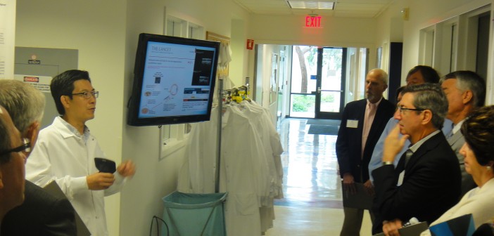
Dr. Yang Liu shares information on the Stem Cell Core with LEAD San Diego tour members.

Dr. Anne Bang with the LEAD San Diego group.LEAD San Diego Tour