Charles Spruck Archives - Sanford Burnham Prebys
Institute News

Three Sanford Burnham Prebys faculty receive promotions

AuthorMiles Martin
Date

June 30, 2022

Sanford Burnham Prebys is proud to announce the promotion of three of our faculty from assistant to associate professor. 

The promoted faculty, all from the Institute’s NCI-designated Cancer Center, include Ani Deshpande, PhD, Brooke Emerling, PhD and Charles Spruck, PhD

Ani Deshpande, PhD

Deshpande studies developmental processes in stem cells that get hijacked by cancer, focusing specifically on acute myeloid leukemia, one of the most common types of blood cancer. Earlier last year, Deshpande published a study with researchers at the National Institutes of Health (NIH) revealing that CRISPR gene editing can sometimes favor cells with cancer mutations, encouraging a cautious approach when using CRISPR therapies for certain cancers

Deshpande joined the Institute in 2015. Prior to that, he held positions at Memorial Sloan Kettering Cancer Center and Harvard Medical School.

Brooke Emerling, PhD

Emerling studies the metabolism of cancer cells, specifically how certain signaling proteins can contribute to the uninhibited growth typical of tumors. Emerling recently received a $2.3 million grant from the NIH to continue her work over the next four years.

Emerling joined the faculty at Sanford Burnham Prebys in 2016. Prior to that, she held positions at Weill Cornell Medicine and Harvard Medical School.

Charles Spruck, PhD 

Spruck develops new, effective, nontoxic treatments for patients with advanced cancers. Specifically, his recent studies have focused on the potential to treat cancer with viral mimicry, which tricks the body into thinking it has a viral infection, stimulating immune responses that can help the body fight cancer and improve the effects of other treatments. 

Spruck joined the Institute in 2010. Prior to that, he held positions at the Sidney Kimmel Cancer Center and Scripps Research.

Institute News

Boosting immunotherapy in aggressive brain cancer

AuthorMiles Martin
Date

November 3, 2021

Researchers from Sanford Burnham Prebys have collaborated the University of Pittsburgh Cancer Institute to reveal a new approach to enhance the effects of immunotherapy in glioblastoma, one of the most aggressive and treatment-resistant forms of brain cancer.

The study, published recently in Cancer Discovery, describes a novel method to ‘turn off’ cancer stem cells—the malignant cells that self-renew and sustain tumors—enabling the body’s own defense system to take charge and destroy tumors.

“Tumors are more than just masses of cells—each one is a complex system that relies on a vast network of chemical signals, proteins and different cell types to grow,” says senior author Charles Spruck, PhD, an assistant professor at Sanford Burnham Prebys. “This is part of why cancer is so difficult to treat, but it also presents us with opportunities to develop treatment strategies that target the machinery powering tumor cells rather than trying to destroy them outright.”

Glioblastoma is an extremely aggressive form of cancer that affects the brain and the spinal cord. Occurring more often in older adults and forming about half of all malignant brain tumors, glioblastoma causes worsening headaches, seizures and nausea. And unfortunately for the thousands of people who receive this diagnosis each year, glioblastoma is most often fatal.

“We haven’t been able to cure glioblastoma with existing treatment methods because it’s just too aggressive,” says Spruck. “Most therapies are palliative, more about reducing suffering than destroying the cancer. This is something we hope our work will change.”

Immune checkpoint inhibitors—which help prevent cancer cells from hiding from the immune system—can be effective for certain forms of cancer in the brain, but their results in glioblastoma have been disappointing. The researchers sought a way to improve the effects of these medications.

“Modern cancer treatment rarely relies on just one strategy at a time,” says Spruck. “Sometimes you have to mix and match, using treatments to complement one another.”

The researchers used genomic sequencing to investigate glioblastoma stem cells. These cells are the source of the rapid and consistent regeneration of glioblastoma tumors that make them so difficult to treat.

The team successfully identified a protein complex called YY1-CDK9 as essential to the cells’ ability to express genes and produce proteins. By modifying the activity of this protein complex in the lab, the team was able to improve the effectiveness of immune checkpoint inhibitors in these cells. 

“Knocking out this transcription machinery makes it much more difficult for the cells to multiply” says Spruck. “They start to respond to chemical signals from the immune system that they would otherwise evade, giving immunotherapy a chance to take effect.” 

While the approach will need to be tested in clinical settings, the researchers are optimistic that it may provide a way to improve treatment outcomes for people with glioblastoma. 

“What our results tell us is that these cells are targetable by drugs we already have, so for patients, improving their treatment may just be a matter of adding another medication,” adds Spruck. “For a cancer as treatment-resistant as glioblastoma, this is a great step forward.”

Institute News

Mining “junk DNA” reveals a new way to kill cancer cells

AuthorMonica May
Date

February 11, 2021

Scientists unearth a previously unknown vulnerability for cancer and a promising drug candidate that leverages the approach

Scientists at Sanford Burnham Prebys have uncovered a drug candidate, called F5446, that exposes ancient viruses buried in “junk DNA” to selectively kill cancer cells. Published in the journal Cell, the proof-of-concept study reveals a previously unknown Achilles’ heel for cancer that could lead to treatments for deadly breast, brain, colon and lung cancers.

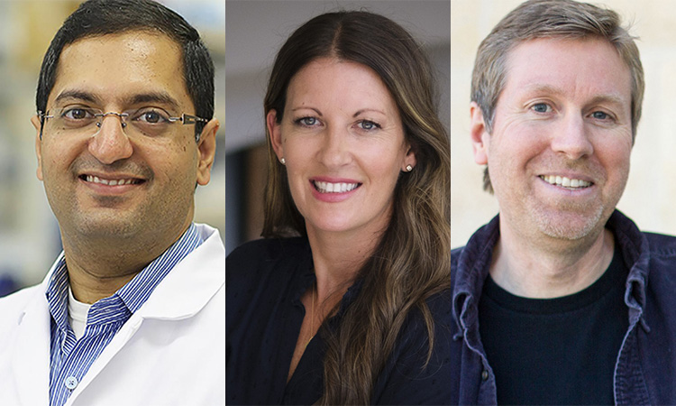
“We found within ‘junk DNA’ a mechanism to stimulate an immune response to cancer cells, while also causing tumor-specific DNA damage and cell death,” says Charles Spruck, PhD, assistant professor in the National Cancer Institute (NCI)-designated Cancer Center and senior author of the study. “This is a very new field of research, with only a handful of papers published, but this has the potential to be a game-changer in terms of how we treat cancer.”

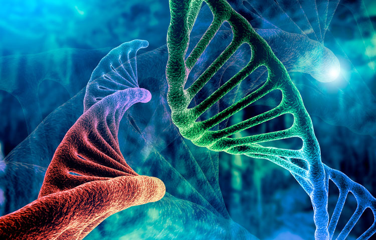
Since the human genome was fully sequenced in 2003, scientists have learned that our DNA is filled with some very strange stuff—including mysterious, noncoding regions dubbed “junk DNA.” These regions are silenced for a reason—they contain the genomes of ancient viruses and other destabilizing elements. An emerging area of cancer research called “viral mimicry” aims to activate these noncoding regions and expose the ancient viruses to make it appear that a cancer cell is infected. The hypothesis is that the immune system will then be triggered to destroy the tumor.

A one-two punch to cancer

In the study, Spruck and his team set out to find the molecular machinery that silences “junk DNA” in cancer cells. Using sophisticated molecular biology techniques, they found that a protein called FBXO44 is key to this process. Blocking this protein caused the noncoding sections of DNA to unwind—but not for long.

“When we revealed noncoding regions, which aren’t meant to be expressed, this caused DNA breakage. This told the cell that something is deeply wrong, and it committed suicide,” explains Spruck. “At the same time, the DNA of the ancient virus was exposed, so the immune system was recruited to the area and caused more cell death. So, we really delivered a one-two punch to cancer.”

The scientists then showed that a drug that targets the FBXO44 pathway, called F5446, shrank tumors in mice with breast cancer. The drug also improved the survival of mice with breast cancer that were resistant to anti-PD-1 treatment, an immunotherapy that is highly effective but often stops working over time. Additional studies in cells grown in a lab dish showed that the drug stops the growth of other tumors, including brain, colon and lung cancers.

The scientists also conducted many experiments to show that this silencing mechanism only occurs in cancer cells, not regular cells. Analysis of patient tumor databases confirmed that FBXO44 is overproduced in many cancers and correlated with worse outcomes—further indicating that a drug that inhibits this protein would be beneficial.

Moving the research toward people

As a next step, the scientists are working with the Conrad Prebys Center for Chemical Genomics to design an FBXO44 pathway-inhibiting drug that is more potent and selective than F5446. This state-of-the-art drug discovery facility is located at Sanford Burnham Prebys.

“Now that we have a compound that works, medicinal chemists can make modifications to the drug so we have a greater chance of success when we test it in people,” says Jia Zack Shen, PhD, staff scientist at Sanford Burnham Prebys and co-first author of the study. “Our greatest hope is that this approach will be a safe and effective pan-cancer drug, which maybe one day could even replace toxic chemotherapy.”


Institute News

Hearst Foundation’s new fellowship funds innovative research to fight breast cancer

AuthorJessica Moore
Date

October 20, 2016

Mark Goldberg, PhD, is working on a potential way to turn cancer stem cells into harmless cells. He and his advisor, Charles Spruck, PhD, assistant professor in the NCI-designated Cancer Center, are optimistic that they could turn this approach into new drugs that prevent breast cancer from returning.

Goldberg is supported by the first-ever research fellowship given by the David Whitmire Hearst Jr. Foundation. The funds were awarded specifically for this groundbreaking project.

“Breast cancer can spread to other organs very early, sometimes even before it’s detected,” said Spruck. “Those micrometastases—just one or a few cells—lie dormant for years, and are insensitive to anticancer drugs. Our goal is to switch those cells to a normal cell type that can’t generate a tumor.”

In as-yet unpublished research, Spruck’s lab recently discovered a protein that’s crucial for pre-cancerous cells to begin growing aggressively and out of control. Goldberg will use animal models of breast cancer to show that genetically inactivating this protein prevents secondary tumors from forming. The next step is to search for candidate drugs that inhibit the protein.

“If we find blockers of this protein that controls progression to malignancy, they could be given to breast cancer patients, after standard treatment has eradicated their primary tumor, to eliminate any remaining cancer stem cells,” added Spruck.

Goldberg’s background in bioengineering gives him a fresh perspective on cancer research. As a PhD student at Caltech, he designed implantable glucose and ion sensors using microfluidics and nanophotonics. That experience gives him a flexible, solutions-oriented approach to designing experiments.

“During Mark’s interview—the first time I met him—we came up with a really exciting way to apply what he was working on at Caltech to cancer research,” Spruck commented. “That kind of creative thinking and insight is invaluable—it’s why I hired him for this fellowship-supported spot.

“Because this research is so early-stage, it’s hard to get funded through traditional avenues. The Hearst Foundation fellowship allows us to get the evidence that this strategy works. That data will be key to getting the support we need for the drug discovery phase.” 

Institute News

New STRIVE awards announced

Authorsgammon
Date

October 8, 2015

“The STRIVE award is providing funds that allow us to accelerate the testing of a new hypothesis that connects the microbiome, epigenetics, and colorectal cancer.”        

          –Alex Strongin, PhD professor in the Bioinformatics and Structural Biology Program at SBP. Continue reading “New STRIVE awards announced”