colon cancer Archives - Sanford Burnham Prebys
Institute News

Mining “junk DNA” reveals a new way to kill cancer cells

AuthorMonica May
Date

February 11, 2021

Scientists unearth a previously unknown vulnerability for cancer and a promising drug candidate that leverages the approach

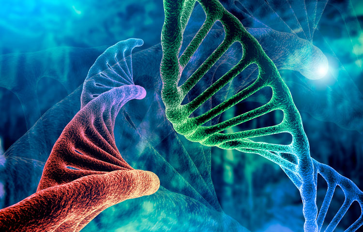
Scientists at Sanford Burnham Prebys have uncovered a drug candidate, called F5446, that exposes ancient viruses buried in “junk DNA” to selectively kill cancer cells. Published in the journal Cell, the proof-of-concept study reveals a previously unknown Achilles’ heel for cancer that could lead to treatments for deadly breast, brain, colon and lung cancers.

“We found within ‘junk DNA’ a mechanism to stimulate an immune response to cancer cells, while also causing tumor-specific DNA damage and cell death,” says Charles Spruck, PhD, assistant professor in the National Cancer Institute (NCI)-designated Cancer Center and senior author of the study. “This is a very new field of research, with only a handful of papers published, but this has the potential to be a game-changer in terms of how we treat cancer.”

Since the human genome was fully sequenced in 2003, scientists have learned that our DNA is filled with some very strange stuff—including mysterious, noncoding regions dubbed “junk DNA.” These regions are silenced for a reason—they contain the genomes of ancient viruses and other destabilizing elements. An emerging area of cancer research called “viral mimicry” aims to activate these noncoding regions and expose the ancient viruses to make it appear that a cancer cell is infected. The hypothesis is that the immune system will then be triggered to destroy the tumor.

A one-two punch to cancer

In the study, Spruck and his team set out to find the molecular machinery that silences “junk DNA” in cancer cells. Using sophisticated molecular biology techniques, they found that a protein called FBXO44 is key to this process. Blocking this protein caused the noncoding sections of DNA to unwind—but not for long.

“When we revealed noncoding regions, which aren’t meant to be expressed, this caused DNA breakage. This told the cell that something is deeply wrong, and it committed suicide,” explains Spruck. “At the same time, the DNA of the ancient virus was exposed, so the immune system was recruited to the area and caused more cell death. So, we really delivered a one-two punch to cancer.”

The scientists then showed that a drug that targets the FBXO44 pathway, called F5446, shrank tumors in mice with breast cancer. The drug also improved the survival of mice with breast cancer that were resistant to anti-PD-1 treatment, an immunotherapy that is highly effective but often stops working over time. Additional studies in cells grown in a lab dish showed that the drug stops the growth of other tumors, including brain, colon and lung cancers.

The scientists also conducted many experiments to show that this silencing mechanism only occurs in cancer cells, not regular cells. Analysis of patient tumor databases confirmed that FBXO44 is overproduced in many cancers and correlated with worse outcomes—further indicating that a drug that inhibits this protein would be beneficial.

Moving the research toward people

As a next step, the scientists are working with the Conrad Prebys Center for Chemical Genomics to design an FBXO44 pathway-inhibiting drug that is more potent and selective than F5446. This state-of-the-art drug discovery facility is located at Sanford Burnham Prebys.

“Now that we have a compound that works, medicinal chemists can make modifications to the drug so we have a greater chance of success when we test it in people,” says Jia Zack Shen, PhD, staff scientist at Sanford Burnham Prebys and co-first author of the study. “Our greatest hope is that this approach will be a safe and effective pan-cancer drug, which maybe one day could even replace toxic chemotherapy.”
Institute News

New therapeutic target for Crohn’s disease

AuthorJessica Moore
Date

September 20, 2016

Research from the Sanford Burnham Prebys Medical Discovery Institute (SBP) identifies a promising new target for future drugs to treat inflammatory bowel disease (IBD). The study, published in Cell Reports, also indicates that another protein, protein kinase C (PKC) λ/ι, may serve as a biomarker of IBD severity.

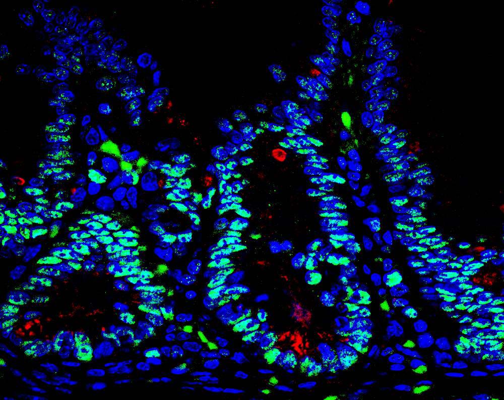
“The intestine is protected by specialized cells, called Paneth cells, that secrete antimicrobial peptides,” said Jorge Moscat, PhD, deputy director and professor in the NCI-designated Cancer Center and senior author of the paper. “We found that maintaining normal numbers of Paneth cells requires PKC λ/ι, and that the amount of PKC λ/ι decreases as IBD gets worse. We also discovered a way to prevent Paneth cell loss—inhibiting a protein called EZH2, which could be a new therapeutic strategy for IBD.”

IBD, which includes Crohn’s disease and ulcerative colitis, affects 1.4 million people in the U.S. These chronic conditions are often debilitating, as they cause unpredictable abdominal pain and diarrhea. Because current medications only help control symptoms and not the underlying disease, 70% of Crohn’s patients and 30% of those with colitis must eventually undergo surgery. In addition, IBD increases risk of intestinal cancer by as much as 60%.

“We also examined the effect of PKC λ/ι on tumor formation,” said Maria Diaz-Meco, PhD, also a professor in the Cancer Center and co-author of the paper. “In contrast to some previous studies indicating that it might promote cancer development, we demonstrate that in the intestine, PKC λ/ι is protective.”

“We inactivated the PKC λ/ι gene in the intestine of mice, which caused them to have very few Paneth cells,” added Diaz-Meco. “Without Paneth cells, the intestine is more susceptible to bacterial infiltration, which leads to inflammation. Since inflammation favors cancer, it makes sense that PKC λ/ι is a tumor suppressor in this setting.”

To find a way to boost Paneth cell numbers and possibly treat IBD, the team looked for what drives the deficit in these protector cells. The key link was overactive EZH2, which turns off genes needed to generate Paneth cells.

“We used an in vitro model—‘mini guts’ in a dish—to show that blocking EZH2 helps return the number of Paneth cells to normal,” said Yuki Nakanishi, MD, a postdoctoral fellow in the Moscat/ Diaz-Meco lab and lead author of the work. “This demonstrates that inhibiting EZH2 could be a new way to slow the progression of IBD.”

Importantly, the team verified the relevance of their findings in intestinal biopsy samples from 30 patients with Crohn’s disease. Disease progression correlated with lower levels of PKC λ/ι.

“EZH2 inhibitors are currently being developed by the pharmaceutical industry to treat other cancers, so they could be tested for IBD relatively soon,” said Moscat. “But first, we need to do preclinical studies to test whether they block progression of the disease.”

The paper is available online here.

Institute News

Making ERK work as a therapeutic target for colorectal cancer

AuthorJessica Moore
Date

June 3, 2016

Colorectal cancer is the third most common cancer in the US, affecting 1.2 million people. Despite extensive research, the five-year survival rate remains below 15%, underscoring the need for new treatments.

One-third of colorectal cancers are driven by over-activation of extracellular signal-regulated kinases 1 and 2 (ERK1/2), which regulates proliferation, metabolism, and cell movement. However, drugs targeting the ERK1/2 pathway are not widely used to treat colorectal cancer because they don’t appreciably slow cancer growth. New research co-led by Petrus de Jong, MD, PhD, postdoctoral associate at SBP, points to a possible reason for this lack of effect, as well as a solution.

“We genetically deleted the ERK1/2 pathway in the lining of the mouse intestine, and we expected to see less cell proliferation,” said de Jong, a co-first author on the paper. “Instead, the opposite occurred. There was more cell growth and the cells were less organized.” de Jong works in the laboratory of Garth Powis, D.Phil., professor and director of SBP’s NCI-Designated Cancer Center, who also contributed to the investigation.

The new study, published in Nature Communications, shows that the increased cell growth caused by disabling ERK1/2 results from increased activity of a related kinase, ERK5. The team went on to show that inhibiting both pathways suppresses proliferation of human colorectal cancer cell lines and slows growth of tumor-like structures in vitro.

“Therapies aimed at targeting ERK1/2 likely fail because ERK5 compensates,” said Eyal Raz, MD, senior author and professor at the UC San Diego School of Medicine. “Previously, ERK5 didn’t seem important in colorectal cancer. This is an underappreciated escape pathway for tumor cells. Hence, the combination of ERK1/2 and ERK5 inhibitors may lead to more effective treatments for colorectal cancer patients.”

“If you block one pathway, cancer cells usually mutate and find another pathway that ultimately allows for a recurrence of cancer growth,” said Koji Taniguchi, MD, PhD, assistant project scientist at UC San Diego and the other co-first author. “Usually, mutations occur over weeks or months. But other times, as in this case, the tumor does not need to develop mutations to find an escape route from targeted therapy. When you find the compensatory pathway and block both, there is no more escape.”

The scientists suggested that other inhibitors of the ERK1/2 pathway should be tested with ERK5 inhibitors in both human colorectal cancer cells and mouse models to identify the most effective combination that could advance to clinical trials.

This post is a modified version of the press release from UC San Diego. Photo from Ed Uthman via Flickr.

The paper is available online here.