Infectious Diseases Archives - Sanford Burnham Prebys
Institute News

A strange research ecosystem: Discussing Lyme disease with Victoria Blaho

AuthorMiles Martin
Date

December 22, 2021

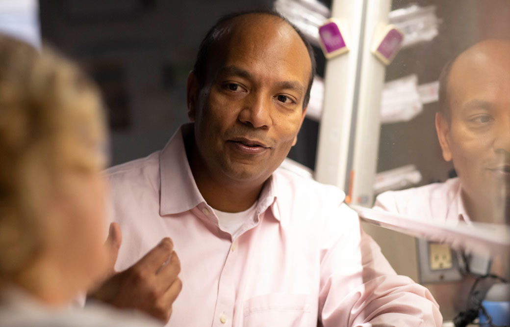
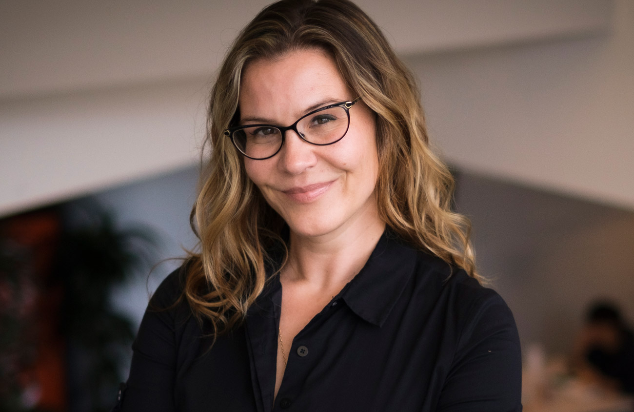
As an infectious disease immunologist studying Lyme disease, Victoria Blaho is one of a rare breed.

Sanford Burnham Prebys assistant professor Victoria Blaho, PhD, investigates the biochemical signals of the immune system and how they impact our bodies’ abilities to fight pathogenic infections, a branch of immunology that has become much less popular since the advent of antibiotics in the early 20th century.

Blaho’s disease of choice is Lyme disease, an unusual tick-borne bacterial infection that affects some 476,000 people in America each year, a number that is on the rise.

We caught up with Blaho to talk about why Lyme disease research is important, the progress being made and the work that remains in studying this strange and burdensome disease.

Why is Lyme disease research important?
Blaho: Lyme research is a very small field for a disease that is becoming bigger and bigger every year. Case counts are increasing for Lyme disease all over the world, and people get very sick from it. Some people are infected, take antibiotics and that’s the end of it. But others have chronic symptoms like arthritis or carditis that can last for years and become completely debilitating.

What makes Lyme difficult to study?
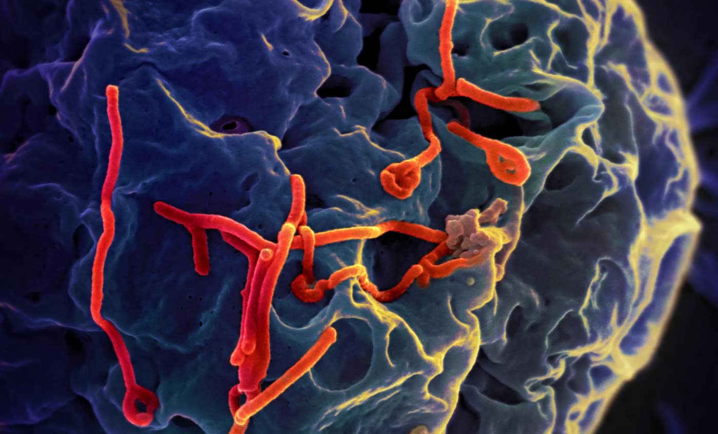
Blaho: One reason is that Lyme is an unusual infection from a microbiological standpoint. In the early days of Lyme research, there were studies showing that the bacteria that caused the disease, Borrelia burgdorferi, could change its physical form from a corkscrew shape to dormant blobs—and the blobs could be causing extended disease. This is a problem because scientists haven’t agreed on the true cause of chronic Lyme disease.

To make matters worse, a lot of the medical field still believes Lyme is easily curable with antibiotics, and if people are still having problems, then it’s psychosomatic. This makes it harder to get support for research into the longer-term inflammatory effects of Lyme. These politics make Lyme disease research a strange ecosystem of patients, physicians, researchers and funding agencies, and this is a barrier to learning more about the disease and helping people find relief.

How does your work enter the picture?
Blaho: I’ve been working on Lyme disease for over 15 years, since I was PhD student. It started because Celebrex was hugely popular at the time to treat arthritis, but nobody had ever studied it in the arthritis that emerges in Lyme disease. Celebrex inhibits an enzyme of the immune system that triggers inflammation, so we figured that Celebrex might work just as well in Lyme arthritis as in other types. But research on mice didn’t bear this out.
Inflammation doesn’t just peter out when an infections clears. The immune system has to clean up the mess. We discovered in mice that Celebrex inhibits the resolution of inflammation after Lyme disease has resolved, so the arthritis never went away.

Since then, my career has focused on exploring the signaling molecules that regulate inflammation and its resolution. These molecules affect all parts of the immune system and provide us with a whole host of different potential therapeutic targets for inflammatory diseases like chronic Lyme.

What are the next steps for your Lyme research and for the field at large? 
Blaho: My own immediate next step is to take the work I’ve been doing here at Sanford Burnham Prebys and connect it directly back to my original work with Lyme. My team here is currently working on a signaling molecule called S1P, and while we haven’t studied it in Lyme yet, we think there are connections between it and the immune mediators we first found through those Lyme studies.

Our next steps are to look for the protein that carries S1P in mice with Lyme disease. This protein is associated with disease susceptibility in other inflammatory illnesses like diabetes and cardiovascular disease, and we think it has a role to play in Lyme as well. We’re also planning to partner with the Bay Area Lyme Foundation to see if we can find changes in this protein in their collection of human samples.

More broadly, I think this field is hungry for innovation because there have been a small number of scientists focusing on it. If older ideas about Lyme being simple to treat were the complete picture, we’d already be able to better diagnose and treat patients. But we’re just not there yet.

Lyme may be a lot cleverer than we originally thought, but if we’re able to embrace new technologies and ideas and continue to push forward with new work, we’ll be able to find innovative approaches to fight Lyme and, ultimately, to help people suffering from this horrible disease.

Institute News

Ebola expert weighs in on news of a potential cure

AuthorMonica May
Date

August 13, 2019

Scientists recently reported that two treatments saved the lives of people infected with the Ebola virus—with the New York Times reporting that roughly 90% of newly infected patients were saved—suggesting we are ever so close to a cure. 

To place this news in context, we caught up with Ebola expert Sumit Chanda, PhD, whose team at Sanford Burnham Prebys is working to find a pill-based treatment for the deadly virus.

Tell us a bit more about Ebola and the recent outbreaks. 
Ebola is a virus responsible for severe, often fatal, hemorrhagic fevers in humans—meaning it damages blood vessels and can cause internal bleeding, among other symptoms. The mortality rate varies between 50% and 90%. The 2014 to 2016 West Africa epidemic has been of unprecedented scope, with more than 28,000 reported cases and more than 11,000 deaths. Exported cases were also documented in the U.S. and Europe. Since August 2018, a new outbreak is ongoing in the Democratic Republic of the Congo, with more than 2,800 total cases reported and more than 1,800 deaths. So far, no medication can treat people already sickened by Ebola (an experimental vaccine has shown effectiveness).

Describe the study and key findings for us. 
Last November, several potential treatments were evaluated in clinical trials in the outbreak area. Two of these treatments, mAb114 (Ridgeback Biotherapeutics) and REGN-EB3 (Regeneron Pharmaceuticals), were found to be highly effective in reducing Ebola-related deaths. These drugs are monoclonal antibodies, which are protein-based therapies—the same kind that are currently being used to treat cancers, autoimmune and other diseases. 

What is your reaction? Is this big news? Or is more research needed?
This is a very important result. For the first time, a clinical therapy significantly reduced mortality after Ebola exposure—especially when given early after infection. While it cannot be called a “cure,” since not everyone taking the therapy survived, it represents a hugely important advance by the scientific community and brings hope to people exposed to this virus and in the outbreak regions. 

What does this advance mean for people infected with the Ebola virus?
People impacted by Ebola have so far been skeptical about medical treatment, especially considering the low success rate of previous treatments. We expect that the high survival rate associated with these two treatments will encourage infected individuals to go to Ebola treatment centers. This will increase the number of people receiving the treatments, reducing the total amount of deaths and helping contain the spread of the virus.

What does this news mean for the quest to find an Ebola treatment?
This remarkable achievement gives me hope that a cure is possible, potentially by combining these therapies with additional drugs. There is more work to be done, however. These antibody-based treatments require administration by a medical professional in a specialized Ebola treatment center and can be expensive. An Ebola therapy that comes in the form of a low-cost pill—the focus of my lab’s work—will be easier to deploy to patients, especially in areas that do not have access to advanced facilities. Since it appears that early treatment is important, easy availability to a medicine will benefit rural patients who are usually at the epicenter of an outbreak—and will help prevent an epidemic from taking root in the first place. 

Anything else you’d like to add? 
We are now, more than ever, hugely optimistic that efforts to develop an Ebola antiviral drug, especially one that is low cost and can be easily distributed in affected regions, will be part of a complete cure regimen for Ebola.

Sumit Chanda, PhD, is the professor and director of the Immunity and Pathogenesis Program at Sanford Burnham Prebys. His team works to find new treatments for infectious diseases, including influenza A (flu), human immunodeficiency virus (HIV) and Ebola virus, by unraveling the cellular machinery that allows these viruses to thrive.

Institute News

Battling infectious diseases with 3D structures

AuthorSusan Gammon, PhD
Date

April 25, 2017

Sanford Burnham Prebys Medical Discovery Institute (SBP) scientists are part of an international team led by Northwestern University Feinberg School of Medicine that has determined the 3D atomic structure of more than 1,000 proteins that are potential drug and vaccine targets to combat some of the world’s most dangerous emerging and re-emerging infectious diseases.

These experimentally determined structures have been deposited into the World-Wide Protein Data Bank, an archive supported by the National Institutes of Health (NIH), and are freely available to the scientific community. The 3D structures help expedite drug and vaccine research and advance the understanding of pathogens and organisms causing infectious disease.

“Almost 50 percent of the structures that we have deposited in the Protein Data Bank are proteins that were requested by scientific investigators from around the world,” said Feinberg’s Wayne Anderson, PhD, director of the project. “The NIH has also requested us to work on proteins for potential drug targets or vaccine candidates for many diseases, such as the Ebola virus, the Zika virus and antibiotic-resistant bacteria. We have determined several key structures from these priority organisms and published the results in high-impact journals such as Nature and Cell.

Teamwork with an international consortium

This milestone effort, funded by two five-year contracts from the National Institute of Allergy and Infectious Diseases (NIAID), totaling a budget of $57.7 million, represents a decade of work by the Center for Structural Genomics of Infectious Diseases (CSGID) at Feinberg, led by Anderson in partnership with these institutions:

  • University of Chicago
  • University of Virginia School of Medicine
  • University of Calgary
  • University of Toronto
  • Washington University School of Medicine in St. Louis
  • UT Southwestern Medical Center
  • J. Craig Venter Institute
  • Sanford Burnham Prebys Medical Discovery Institute
  • University College London

How the 3D structures are made

Before work begins on a targeted protein, a board appointed by the NIH examines each request. Once approved, the protein must be cloned, expressed and crystallized, and then X-ray diffraction data is collected at the Advanced Photon Source at Argonne National Laboratory. This data defines the location of each of the hundreds or even thousands of atoms to generate 3-D models of the structures that can be analyzed with graphics software. Each institution in the Center has an area of expertise it contributes to the project, working in parallel on many requests at once.

The bioinformatics group SBP, led by Adam Godzik, PhD, focuses on steps that have to be taken before the experimental work starts. Every protein suggested by the research community as a target for experimental structure determination is analyzed and an optimal procedure for its experimental determination is mapped out.

Experimental structure determination used to have a very high failure rate and the money and time spent on failed attempts is a major contributor to the total expense and time needed to solve protein structures. Both can be significantly improved using “Big Data” approaches, as researchers learn from thousands of successful and failed experiments in structural biology. The SBP bioinformatics group uses these approaches to improve success rates at CSGID, allowing our center to solve more structures at lower costs.

Until recently the process of determining the 3D structure of a protein took many months or even years to complete, but advances in technology, such as the Advanced Photon Source, and upgrades to computational hardware and software has dramatically accelerated the process. The Seattle Structural Genomics Center for Infectious Disease, a similar center funded by NIAID, is also on track to complete 1,000 3-D protein structures soon. Browse all of the structures deposited by the CSGID.

Anyone in the scientific community interested in requesting the determination of structures of proteins from pathogens in the NIAID Category A-C priority lists or organisms causing emerging and re-emerging infectious diseases, can submit requests to the Center’s web portal. As part of the services offered to the scientific community, the CSGID can also provide expression clones and purified proteins, free of charge.

This project has been supported by federal funds from the NIAID, NIH,  Department of Health and Human Services, under contract numbers HHSN272200700058C and HHSN272201200026C.

Institute News

New grant supports Ebola drug discovery

AuthorJessica Moore
Date

March 24, 2017

Ebola’s reputation as a killer virus is well deserved—the most recent outbreak, in West Africa from 2014-2016, caused more than 11,000 deaths among 28,000 infections, according to the World Health Organization. Outbreaks have occurred regularly since 1976, so another is likely, but the timing is hard to predict. While an effective vaccine against the virus has been developed and will likely be approved, there are no drugs available to treat Ebola infections.

Ebola is not just deadly. It also causes an awful disease—sudden fever, fatigue, muscle pain, and headache that last for days, followed by vomiting and diarrhea that lead to severe dehydration, requiring IV fluids. Many Ebola patients also bleed internally and externally, from IV insertion sites, the nose and eyes.

A new $4.1 million grant from the National Institute of Allergy and Infectious Diseases supports research to find compounds that block the growth of Ebola virus, which could lead to new antiviral drugs. Sumit Chanda, PhD, professor and director of the Immunity and Pathogenesis Program at Sanford Burnham Prebys Medical Discovery Institute (SBP), and Anthony Pinkerton, PhD, director of medicinal chemistry at SBP’s Conrad Prebys Center for Chemical Genomics, are collaborators on the effort. Christopher Basler, PhD, professor and director of the Center for Microbial Pathogenesis at Georgia State University, is directing the project.

“Drugs for Ebola are still urgently needed,” says Chanda. “Even with a vaccine, there’s still the possibility that someone who hasn’t been vaccinated might be exposed and carry it to an area where it’s not endemic.”

The SBP investigators will not be working with the disease-causing Ebola virus itself. Instead, they’ll be using non-infectious components, avoiding the need for special containment facilities.

The scientific team, which also includes Megan Shaw, PhD, associate professor at the Icahn School of Medicine at Mount Sinai, and Robert Davey, PhD, scientist at Texas Biomedical Research Institute, will first identify inhibitors of the viral machinery. Later phases of the project will confirm efficacy against live Ebola virus, determine how the drug candidates block the viral machinery and develop additional tests to identify drug candidates that will inhibit not only Ebola virus, but also the related Marburg virus.

“Marburg is also highly lethal,” says Pinkerton. “Drugs that work against the whole virus family would provide an even greater benefit to public health.”

This story was based in part on a press release from Georgia State University.

Institute News

Research to combat biothreat of “Black Death”

AuthorJessica Moore
Date

March 7, 2017

The plague, also known as the Black Death, wiped out a third of Europe’s population in the 14th century and has a long history of exploitation as a biological weapon. Even today, outbreaks of the disease, caused by the bacterium Yersinia pestis, persist in Asia and Africa and the southwestern US.

Bubonic or septicemic plague result when bacteria are transmitted by contact with infected fluid (e.g. a flea bite), spread through the lymphatic system and enter the bloodstream. Both carry very high (40%–60%) mortality. Pneumonic plague results when bacteria are transmitted through aerosolized droplets and colonize the lungs—this form is highly contagious, spreads rapidly and causes extremely high (~100%) mortality.

Although most plague is treatable if detected within hours of infection, the limited number of effective antibiotics, the emergence of antibiotic-resistant strains, the lack of an effective vaccine, and the potential weaponization of aerosolized bacteria with bio-engineered antibiotic resistance all underscore the need to develop medical countermeasures. These factors have led the U.S. Department of Health and Human Services to designate Y. pestis as a Tier 1 Select Agent—the class reserved for pathogens that can be weaponized to kill millions of people.

“My lab is working on developing new ways to combat Y. pestis,” says Francesca Marassi, PhD, professor at SBP. “Understanding the basic mechanism of bacterial infection is the key first step.”

Finding new defenses against the Y. pestis microbe is important enough that Marassi’s research is being supported by a prestigious five-year grant from the National Institutes of Health.

“We want to determine the architecture of the outer membrane surface of the bacterium because this is the first line of contact with the human host upon infection,” Marassi explains. “We’re studying a protein called Ail (adhesion invasion locus), which is exposed on the bacterial surface and interacts with human proteins in ways that help Y. pestis survive in blood—without Ail, bacterial virulence is highly attenuated.”

Marassi’s lab just made a key advance – developing methods to determine the structure of Ail in the membrane. The results are now published in the Journal of Biomolecular NMR.

“These findings set the stage for studying the interactions of Ail with its protein partners on human host cells,” adds Marassi. “Being able to see the structure of Ail gives us vital insight for the development of drugs to fight the disease.

“Moving forward, we will look at the structural basis of Ail interactions with human host proteins to find sites that interact—that could potentially be interrupted with new drugs.”

Institute News

The search for new anthrax treatments isn’t over

AuthorJessica Moore
Date

October 5, 2016

A bioterror attack using virulent anthrax would be nearly as deadly today as it was in 2001, when anthrax spores sent through the mail killed five people. Even with aggressive treatment, only about half of those who breathe anthrax spores survive because the bacterium rapidly produces huge amounts of deadly toxins.

To inform future therapies, the lab of Robert Liddington, PhD, professor in the Bioinformatics and Structural Biology Program, examined how toxins enter cells. Their new study, published in the Journal of General Physiology, shows that the bacterial toxin is remarkably efficient at getting across cell membranes.

“When we pushed the system to its limit, we found that the pore formed by the toxin is incredibly robust,” said Liddington. “It acts like a ‘conveyer belt,’ continuously feeding toxic enzymes across the membrane.”

During anthrax infections, the bacterium Bacillus anthracis secretes a three-unit toxin: two enzymes named lethal factor (LF) and edema factor (EF) and a third protein called protective antigen (PA). After they’re engulfed by cells, they’re contained within membrane-bound acid baths (vesicles) that they have to escape to avoid being broken down. To do that, the PA proteins link together to form a pore across the membrane, allowing LF and EF to be transported into the interior of the cell. There, they wreck signals that would alert other cells to the presence of the bacteria, eventually causing the cell to die.

Liddington’s lab teamed with Isabelle Rouiller, PhD, and her group at McGill University, using a cutting-edge high-resolution imaging technique called cryo-electron microscopy to build a three-dimensional map of the “pre-pore” complex. The map showed seven PA proteins surrounding a narrow pore, with three LF molecules perched at the rim, ready to be moved across. As well as binding to the PA subunits, each LF molecule also bound to its neighbor.

The team hopes that these new findings will aid the development of better treatments for anthrax infection. “By identifying new interactions between different parts of the toxin, our findings suggest new ways to thwart toxin entry,” explained Liddington. “That might allow us to design new antitoxins that work better than or in combination with the two that have been approved by the FDA.”

This post is based in part on a press release from the Journal of General Physiology.

The paper is available online here.