NIH funding Archives - Page 5 of 7 - Sanford Burnham Prebys
Institute News

Targeting long-sought EphA2 receptor becomes crystal clear

AuthorMonica May
Date

May 13, 2019

Scientists have long sought to target a cellular receptor called EphA2 because of its known role in many disorders, including cancer, inflammatory conditions, neurological disorders and infectious diseases. However, lack of information about the structure formed when EphA2 links to other molecules—ligands—has hindered drug development. 

Now, scientists at Sanford Burnham Prebys have crystallized EphA2 together with peptide ligands (short proteins) and used the structure to engineer more powerful compounds that activate or inactivate the receptor, paving the way for new therapies. The discovery was published in the Journal of Biological Chemistry.

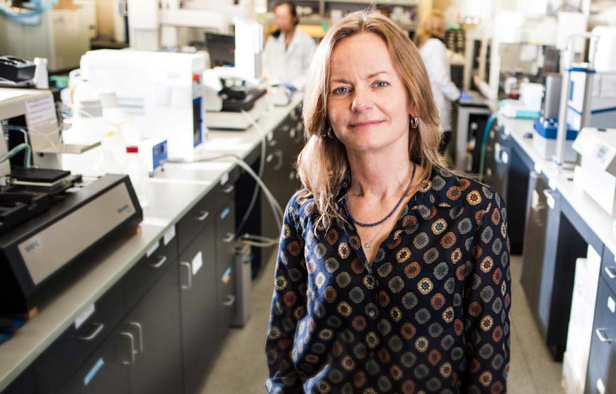
“EphA2 plays a central role in a plethora of biological and disease processes,” says Elena Pasquale, PhD, professor in the Tumor Initiation and Maintenance Program at Sanford Burnham Prebys. “Our team’s identification of potent, highly selective peptides that regulate the receptor is a key step toward rational design of therapies for the numerous disorders that are driven by EphA2.” 

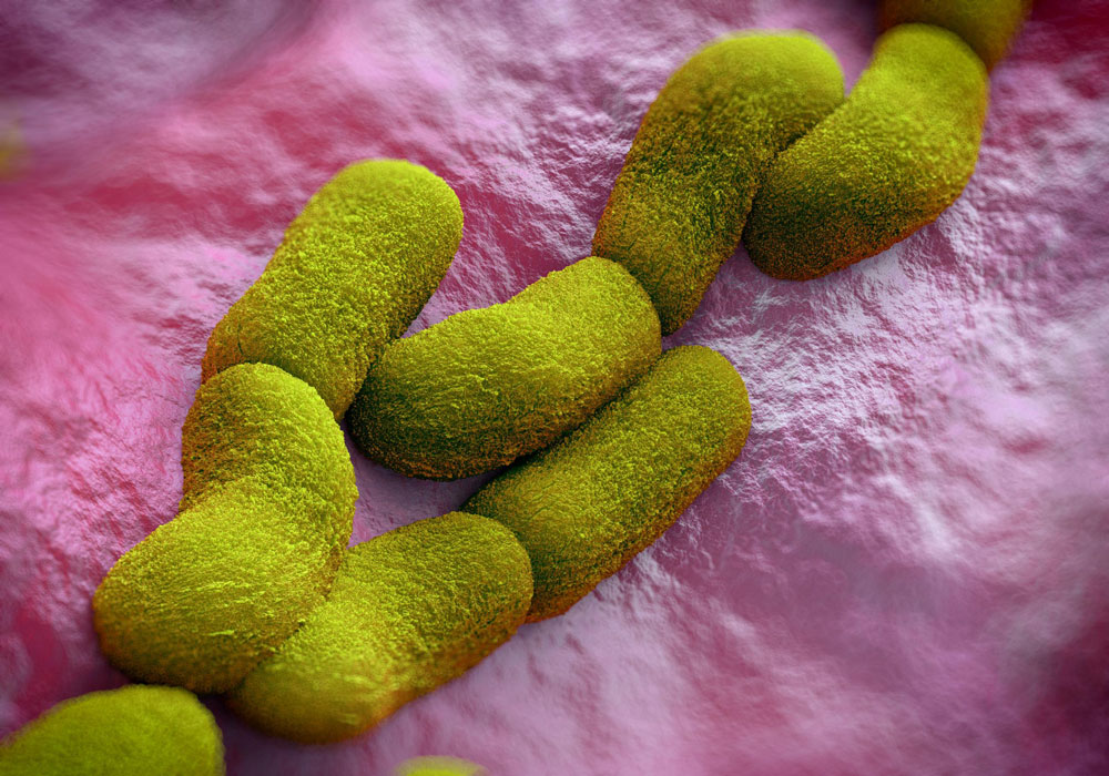
EphA2 is found in the cells that line the surfaces of our body, including our skin, blood vessels and other organs. The receptor is typically only present at high levels during disease states, making it a promising drug target. Activating the receptor could hinder tumor growth, while inhibiting it could reduce unwanted formation of blood vessels (angiogenesis), treat certain inflammation-driven disorders and block pathogens—such as malaria, chlamydia and the hepatitis C virus—from gaining entry into a cell through the receptor. Because EphA2 travels deep inside of the cell when activated, scientists could also harness it as a Trojan horse by attaching chemotherapies or imaging agents to the peptide ligands, which would subsequently be delivered to the desired cells. 

In the study, the scientists initially crystallized a weakly binding peptide in complex with EphA2, yielding a detailed picture of the binding features and providing clues to the receptor’s “sweet spot” or site of action. The researchers then used this information to repeat this process, engineering increasingly more powerful ligands. This work identified several peptides that strongly clasp the receptor and activate or inactivate it—which can be used to inform drug development.

Further quantitative Förster Resonance Energy Transfer (FRET) microscopy experiments, which measure receptor-receptor interactions, revealed that EphA2 receptors cluster together when activated by a peptide—an effect similar to that caused by its natural ligands—answering an unresolved question in the field. 

“In addition to helping guide therapeutic development paths, these peptides are also valuable research tools for scientists who are working to gain insights into this important receptor,” adds Pasquale. “Our hope is that with this new information, one day we can find targeted therapies to treat cancer, inflammatory disorders and infectious diseases that are regulated by EphA2.”


The co-first authors of the study are Maricel Gomez-Soler, PhD, and Marina Petersen Gehring, PhD, of Sanford Burnham Prebys; and Bernhard C. Lechtenberg, PhD, formerly of Sanford Burnham Prebys and currently of the Walter and Eliza Hall Institute of Medical Research. 

Additional authors include Elmer Zapata-Mercado and Kalina Hristova, PhD, of Johns Hopkins University. The study’s DOI is 10.1074/jbc.RA119.008213. 

This research was supported by the National Institutes of Health (NIH) (R01NS087070, R01GM131374 and P30CA030199). 

Institute News

Drug screen conducted at Sanford Burnham Prebys identifies new therapeutic avenues for Alzheimer’s disease

AuthorMonica May
Date

February 7, 2019

A screen of more than 1,600 Food and Drug Administration (FDA)–approved drugs performed at SBP’s Conrad Prebys Center for Chemical Genomics (Prebys Center) has revealed new therapeutic avenues that could lead to an Alzheimer’s disease treatment. 

The findings come from a collaboration between SBP scientists and researchers at the University of California San Diego School of Medicine, Leiden University Medical Center and Utrecht University in the Netherlands and were published in Cell Stem Cell

The hunt is on for an effective treatment for Alzheimer’s, a memory-robbing disease that is nearing epidemic proportions as the world’s population ages. Nearly six million people in the U.S. are living with Alzheimer’s disease. This number is projected to rise to 14 million by 2060, according to the Centers for Disease Control and Prevention (CDC). 

Scientists have known for many years that a protein called tau accumulates and creates tangles in the brain during Alzheimer’s disease. Additional research is revealing that altered cholesterol metabolism in the brain is associated with Alzheimer’s. But the relationship between these two clues is unknown. 

By testing a library of FDA-approved drugs against induced pluripotent stem cells (iPSC) neurons created from people with Alzheimer’s disease, the scientists were able to identify 42 compounds that reduced the level of phosphorylated tau, a form of tau that contributes to tangle formation. The researchers further refined this group to only include cholesterol-targeting compounds. 

A detailed study of these drugs showed that their effect on tau was mediated by their ability to lower cholesteryl esters, a storage product of excess cholesterol. These results led them to an enzyme called CYP46A1, which normally reduces cholesterol. Activation of this enzyme by the drug efavirenz (brand names Sustiva® and Stocrin®) reduced cholesterol esters and phosphorylated tau in these neurons, making it a promising therapeutic target for Alzheimer’s disease. Further mapping of the enzyme’s action(s) within a cell could reveal even more therapeutic targets. 

“Our Prebys Center is designed to be a comprehensive resource that allows basic research—whether conducted at SBP, academic and nonprofit research institutions or industry—to be translated into medicines for diseases that urgently need better treatments,” says study author Anne Bang, PhD, director of Cell Biology at the Conrad Prebys Center for Chemical Genomics at SBP. “We are proud that the Prebys Centers’ drug discovery technologies helped reveal new paths that could lead to a potential treatment for Alzheimer’s, one of the most devastating diseases of our time.”


The senior author of the study is Lawrence S. B. Goldstein, PhD, distinguished professor at the University of California San Diego (UC San Diego) and scientific director of the Sanford Consortium for Regenerative Medicine. The co-first authors are Vanessa Langness, a PhD graduate student in Goldstein’s lab, and Rik van der Kant, PhD, a senior scientist at Vrije University in Amsterdam and former postdoctoral fellow in Goldstein’s lab. 

Additional study authors include Cheryl M. Herrera, Daniel Williams, Lauren K. Fong and Kevin D. Rynearson, UC San Diego; Yves Leestemaker, Huib Ovaa, Evelyne Steenvoorden and Martin Giera of Leiden University Medical Center; Jos F. Brouwers and J. Bernd Helms; Utrecht University; Steven L. Wagner, UC San Diego and Veterans Affairs San Diego Healthcare System.

Funding for this research came, in part, from the Alzheimer Netherlands Fellowship, ERC Marie Curie International Outgoing Fellowship, the National Institutes of Health (NIH) (5T32AG000216-24, IRF1AG048083-01) and the California Institute for Regenerative Medicine (RB5-07011).

Read more in UC San Diego’s press release. 

Institute News

Parents gain answers about their child’s mysterious condition, thanks to SBP scientists

AuthorMonica May
Date

December 11, 2018

For the parents of a six-year-old Hispanic boy and a seven-year-old Qatari girl, answers remained elusive. Both children had alarming symptoms, including developmental delays, uncontrollable seizures and “floppy baby syndrome” (hypotonia). But despite doctors’ best efforts, the origin of the disease remained unknown. 

Now, these two children are linked by rare mutations in a gene called FUK—providing their families and doctors a better understanding of the cause of their medical conditions. Using biochemical techniques to analyze the boy’s cells, Sanford Burnham Prebys Medical Research Institute (SBP) scientists determined that a malfunctioning enzyme called fucokinase is to blame—caused by a mutation in the FUK gene. Because cells from the girl weren’t available, computer modeling was used—and indicated this same mutation likely caused the disease. The study published in the American Journal of Human Genetics.

Like a molecular spark plug, the fucokinase enzyme ignites one step in a cellular communication cascade—which culminates in the linkage of a sugar, fucose, to another carbohydrate. This final fucose-carbohydrate product is important for immune system regulation, tissue development, cell adhesion (“stickiness” to the environment) and more. 

Based on these findings, the scientists now know the condition is a congenital disorder of glycosylation (CDG), an umbrella term for disorders caused by abnormal linking of sugars to cellular building blocks, including proteins, fats (lipids) and carbohydrates. Although more than 130 types of CDGs exist, the boy and girl are the only known living individuals who have this mutation. 

“Our hope is that by reporting this information, we will help doctors grant more answers to patients and their loved ones,” says Hudson Freeze, PhD, senior author of the paper and director and professor of the Human Genetics Program at SBP. “Based on our findings, genetic databases around the world will now note this mutation causes disease—a potentially life-changing shortcut in the quest for answers.” 

The researchers analyzed skin and immune cells that were collected from the boy. They observed reductions in the amount of the fucokinase enzyme—as much as 80 percent in skin cells and more than half in immune cells, compared to a control protein. Consistent with these findings, downstream products typically created by fucokinase weren’t incorporated into the final fucose-carbohydrate product—indicating the enzyme was not working.

Because cells from the girl were not available, the scientists used computer modeling to predict the impact of her FUK gene mutation. This approach indicated the mutation occurs at an important site on the enzyme that would likely cause disease.

“We know that dampening down the activity of the FUK gene is linked to metastatic cancer—a deadly event that occurs when tumors gain the ability to travel throughout the body,” says Freeze. “In addition to providing long-awaited answers to these families, these findings could help us understand how certain cancers spread throughout the body, including liver, colorectal and skin cancers (melanoma).” 

Both children were identified through the National Institutes of Health’s Undiagnosed Diseases Network, which is designed to accelerate discovery and innovation in the way patients with previously undiagnosed diseases are diagnosed and treated. 

Additional study authors include: Jill Rosenfeld, Lisa Emrick, MD, Lindsay Burrage, MD, PhD, Brendan Lee, MD, PhD, William Craigen, MD, PhD, Baylor College of Medicine; Mahim Jain, MD, PhD, Johns Hopkins School of Medicine; David Bearden, MD, University of Rochester School of Medicine; and Brett Graham, MD, PhD, Baylor College of Medicine and Indiana University School of Medicine. The study’s DOI is https://doi.org/10.1016/j.ajhg.2018.10.021

Research reported in this story was supported by National Institutes of Health (NIH) grants R01DK099551, U01HG007709, and K08DK106453; Baylor College of Medicine Intellectual and Developmental Disabilities Research Center (U54 HD083092), Diana & Gabriel Wisdom and the Rocket Fund. The content is solely the responsibility of the authors and does not necessarily represent the official views of the NIH. 

Interested in keeping up with SBP’s latest discoveries, upcoming events and more? Subscribe to our monthly newsletter, Discoveries.

Institute News

SBP scientist awarded Susan G. Komen® and NIH grants to advance breast cancer research

AuthorMonica May
Date

October 25, 2018

Breast cancer remains the second most common cancer for American women. While treatment advances are being made, more research is needed. Current treatments don’t work for every woman.
 
Now, breast cancer researcher Svasti Haricharan, PhD, assistant professor at Sanford Burnham Prebys Medical Discovery Institute (SBP), has been awarded more than half a million dollars in combined grants from Susan G. Komen® and the National Institutes of Health (NIH). 

This funding will advance Haricharan’s breast cancer research—including developing a diagnostic test that could guide therapeutic options—and allow her to apply lessons from breast cancer to additional cancers. 

Susan G. Komen grant

The majority of women diagnosed with breast cancer have the estrogen-positive (ER-positive) form, meaning the tumor grows in response to estrogen. Hormone therapies (anti-estrogen drugs) that block estrogen—and thus stop the tumor from growing—are available. However, this treatment doesn’t work for 40 percent of women with ER-positive breast cancer. 

“Currently, doctors are unable to predict which ER-positive patients will respond to treatment—so an estrogen-blocking medicine is given, and a ‘wait and see’ approach is taken to see if the treatment will work,” says Haricharan. “However, if a woman doesn’t respond to treatment, during this time the tumor is instead still growing and may metastasize—when it becomes deadlier and even harder to treat. Knowing upfront if an individual will respond to treatment allows doctors to skip a treatment that won’t work and move immediately to prescribing a medicine that may be effective.” 

Haricharan’s previous work found that about one-third of women with ER-positive breast cancer who were treatment resistant had a mutation in DNA damage-repair genes—providing a potential biomarker that could predict who would respond to treatment. 

Luckily, an FDA-approved test that detects defects in DNA damage repair is currently available for colorectal cancer patients. The grant from Susan G. Komen enables Haricharan to evaluate whether this same test can be used to predict response to anti-estrogen drugs in ER-positive breast cancer patients. 

Additionally, research from Haricharan’s previous lab identified a medicine that is FDA approved for advanced or metastatic breast cancer patients and holds potential as a frontline breast cancer treatment (the first treatment prescribed by a doctor). The grant will allow her to bring these pieces of the puzzle together—developing a predictive test and evaluating a potential alternative treatment. 

“Because an FDA-approved test is already on the market, development of a breast cancer test to predict response to hormone therapy may be accelerated. I’d estimate my work could enable a commercially available test in less than five years—though of course a real-world assessment will be needed to obtain doctor and insurance-company approval,” says Haricharan. “Pairing a new test that can guide therapeutic options with a potential treatment would be an important advance for ER- positive breast cancer. I want to express my greatest thanks to Susan G. Komen for funding this important work.” 

NIH grant

Haricharan was also awarded a K22 grant from the NIH, which helps early-career scientists transition to independent research careers. This grant will allow her to apply insights from her breast cancer research to additional cancers. 

Studies have indicated there are links between the growth of colorectal and bladder tumors and estrogen response. While women are less frequently diagnosed with bladder cancer, they tend to have a greater risk of dying from the disease. In contrast, estrogen may have a protective effect on the development of colorectal cancers. 

The NIH grant will enable Haricharan to work to better understand the role DNA damage-repair mutations may play in response to standard-of-care treatment for ER-positive breast, colorectal and bladder cancers. Once this role has been established, the grant will help fund a search for effective targeted treatments.

“Both bladder and colorectal cancers are often caught at a late stage, when the cancer is harder to treat,” says Haricharan. “I hope that this research will ultimately yield tests that can predict response to treatment and guide treatment options for these deadly cancers.” 

Link to the NIH grant: A pan-cancer role for MUTL loss in inducing treatment resistance 

More information about the Susan G. Komen grant: Susan G. Komen Announces $26 Million Investment in New Research to Find Solutions for Aggressive and Metastatic Breast Cancers, and to Help Communities Most at Risk
 

Interested in keeping up with SBP’s latest discoveries, upcoming events and more? Subscribe to our monthly newsletter, Discoveries.

Institute News

SBP scientist honored by the American Society for Bone and Mineral Research

AuthorMonica May
Date

September 28, 2018

José Luis Millán, PhD, professor in the Human Genetics Program at Sanford Burnham Prebys Medical Discovery Institute (SBP), has received the 2018 American Society for Bone and Mineral Research (ASBMR) Lawrence G. Raisz Award for his outstanding achievements in pre-clinical and translational research. 

Millán has dedicated his career to understanding the mechanism of initiation of skeletal and dental mineralization. His pioneering research has led to the first-ever FDA-approved drug for a rare soft bone disease, hypophosphatasia (HPP); and a second drug candidate developed through a research collaboration with Daiichi Sankyo Company, Limited (Daiichi Sankyo) that entered a Phase 1, first-in-human clinical trial in 2017

ASBMR’s award is named in honor of Lawrence G. Raisz, MD, a prominent scientist, mentor, teacher and clinician in the field of bone and mineral metabolism. Raisz was a founding member of ASBMR and the first editor-in-chief of the Journal of Bone and Mineral Research. 

“Lawrence G. Raisz deeply influenced the skeletal mineralization field, so it is a true honor to receive an award in his memory,” says Millán. “In accepting this award, I want to thank the many individuals who enabled our achievements—from the National Institutes of Health (NIH), which has generously funded our research since the 1980s, to the collaborators and lab members who were instrumental in our scientific advances. I also want to thank SBP for providing state-of-the-art technologies that were invaluable to our research.”

Millán was presented the award onstage today at the ASBMR 2018 Annual Meeting in Montreal. 
 

Interested in keeping up with SBP’s latest discoveries, upcoming events and more? Subscribe to our monthly newsletter, Discoveries.

Institute News

Battling infectious diseases with 3D structures

AuthorSusan Gammon, PhD
Date

April 25, 2017

Sanford Burnham Prebys Medical Discovery Institute (SBP) scientists are part of an international team led by Northwestern University Feinberg School of Medicine that has determined the 3D atomic structure of more than 1,000 proteins that are potential drug and vaccine targets to combat some of the world’s most dangerous emerging and re-emerging infectious diseases.

These experimentally determined structures have been deposited into the World-Wide Protein Data Bank, an archive supported by the National Institutes of Health (NIH), and are freely available to the scientific community. The 3D structures help expedite drug and vaccine research and advance the understanding of pathogens and organisms causing infectious disease.

“Almost 50 percent of the structures that we have deposited in the Protein Data Bank are proteins that were requested by scientific investigators from around the world,” said Feinberg’s Wayne Anderson, PhD, director of the project. “The NIH has also requested us to work on proteins for potential drug targets or vaccine candidates for many diseases, such as the Ebola virus, the Zika virus and antibiotic-resistant bacteria. We have determined several key structures from these priority organisms and published the results in high-impact journals such as Nature and Cell.

Teamwork with an international consortium

This milestone effort, funded by two five-year contracts from the National Institute of Allergy and Infectious Diseases (NIAID), totaling a budget of $57.7 million, represents a decade of work by the Center for Structural Genomics of Infectious Diseases (CSGID) at Feinberg, led by Anderson in partnership with these institutions:

  • University of Chicago
  • University of Virginia School of Medicine
  • University of Calgary
  • University of Toronto
  • Washington University School of Medicine in St. Louis
  • UT Southwestern Medical Center
  • J. Craig Venter Institute
  • Sanford Burnham Prebys Medical Discovery Institute
  • University College London

How the 3D structures are made

Before work begins on a targeted protein, a board appointed by the NIH examines each request. Once approved, the protein must be cloned, expressed and crystallized, and then X-ray diffraction data is collected at the Advanced Photon Source at Argonne National Laboratory. This data defines the location of each of the hundreds or even thousands of atoms to generate 3-D models of the structures that can be analyzed with graphics software. Each institution in the Center has an area of expertise it contributes to the project, working in parallel on many requests at once.

The bioinformatics group SBP, led by Adam Godzik, PhD, focuses on steps that have to be taken before the experimental work starts. Every protein suggested by the research community as a target for experimental structure determination is analyzed and an optimal procedure for its experimental determination is mapped out.

Experimental structure determination used to have a very high failure rate and the money and time spent on failed attempts is a major contributor to the total expense and time needed to solve protein structures. Both can be significantly improved using “Big Data” approaches, as researchers learn from thousands of successful and failed experiments in structural biology. The SBP bioinformatics group uses these approaches to improve success rates at CSGID, allowing our center to solve more structures at lower costs.

Until recently the process of determining the 3D structure of a protein took many months or even years to complete, but advances in technology, such as the Advanced Photon Source, and upgrades to computational hardware and software has dramatically accelerated the process. The Seattle Structural Genomics Center for Infectious Disease, a similar center funded by NIAID, is also on track to complete 1,000 3-D protein structures soon. Browse all of the structures deposited by the CSGID.

Anyone in the scientific community interested in requesting the determination of structures of proteins from pathogens in the NIAID Category A-C priority lists or organisms causing emerging and re-emerging infectious diseases, can submit requests to the Center’s web portal. As part of the services offered to the scientific community, the CSGID can also provide expression clones and purified proteins, free of charge.

This project has been supported by federal funds from the NIAID, NIH,  Department of Health and Human Services, under contract numbers HHSN272200700058C and HHSN272201200026C.

Institute News

Research to combat biothreat of “Black Death”

AuthorJessica Moore
Date

March 7, 2017

The plague, also known as the Black Death, wiped out a third of Europe’s population in the 14th century and has a long history of exploitation as a biological weapon. Even today, outbreaks of the disease, caused by the bacterium Yersinia pestis, persist in Asia and Africa and the southwestern US.

Bubonic or septicemic plague result when bacteria are transmitted by contact with infected fluid (e.g. a flea bite), spread through the lymphatic system and enter the bloodstream. Both carry very high (40%–60%) mortality. Pneumonic plague results when bacteria are transmitted through aerosolized droplets and colonize the lungs—this form is highly contagious, spreads rapidly and causes extremely high (~100%) mortality.

Although most plague is treatable if detected within hours of infection, the limited number of effective antibiotics, the emergence of antibiotic-resistant strains, the lack of an effective vaccine, and the potential weaponization of aerosolized bacteria with bio-engineered antibiotic resistance all underscore the need to develop medical countermeasures. These factors have led the U.S. Department of Health and Human Services to designate Y. pestis as a Tier 1 Select Agent—the class reserved for pathogens that can be weaponized to kill millions of people.

“My lab is working on developing new ways to combat Y. pestis,” says Francesca Marassi, PhD, professor at SBP. “Understanding the basic mechanism of bacterial infection is the key first step.”

Finding new defenses against the Y. pestis microbe is important enough that Marassi’s research is being supported by a prestigious five-year grant from the National Institutes of Health.

“We want to determine the architecture of the outer membrane surface of the bacterium because this is the first line of contact with the human host upon infection,” Marassi explains. “We’re studying a protein called Ail (adhesion invasion locus), which is exposed on the bacterial surface and interacts with human proteins in ways that help Y. pestis survive in blood—without Ail, bacterial virulence is highly attenuated.”

Marassi’s lab just made a key advance – developing methods to determine the structure of Ail in the membrane. The results are now published in the Journal of Biomolecular NMR.

“These findings set the stage for studying the interactions of Ail with its protein partners on human host cells,” adds Marassi. “Being able to see the structure of Ail gives us vital insight for the development of drugs to fight the disease.

“Moving forward, we will look at the structural basis of Ail interactions with human host proteins to find sites that interact—that could potentially be interrupted with new drugs.”

Institute News

Bret Goodpaster to lead part of $170M NIH effort to understand the molecular effects of exercise

AuthorJessica Moore
Date

January 9, 2017

Everyone knows that physical activity is good for your health, but we’re far from understanding all the details of how exercise improves the function of the cells and organs of the body. To make up for this gap in knowledge, the National Institutes of Health (NIH) has funded a nationwide consortium that aims to construct a molecular map of the changes caused by exercise. Bret Goodpaster, PhD, professor at Sanford Burnham Prebys Medical Discovery Institute (SBP), and director of the Exercise and Metabolism Core at the Translational Research Institute for Metabolism and Diabetes (TRI-MD) at Florida Hospital, will co-lead one of the seven clinical centers involved in the consortium, called Molecular Transducers of Physical Activity, or MoTrPAC.

“This is a huge undertaking that’s guaranteed to advance our insight into how physical activity improves and preserves health,” says Goodpaster. “It’s great that the NIH is making this a priority. Along with research on how diseases develop, there should be just as much work looking at the other side—prevention.”

To build the molecular map, clinical investigators will recruit thousands of active and sedentary volunteers with a range of fitness levels and body compositions, who will perform resistance and aerobic exercises. Biological samples will be collected before and after physical activity, allowing scientists to analyze changes in thousands of molecules.

“We know from epidemiology that exercise has a myriad of health benefits—from diabetes and cardiovascular disease prevention to mitigating cancer and Alzheimer’s disease risk, but those kinds of studies provide limited information,” adds Goodpaster. “To really draw conclusions about cause and effect, we have to do experiments in humans, and that requires a large number of study participants because people are so diverse. This investment is exactly what we need, especially now that sequencing and ‘omics’ technologies can be used efficiently on a big scale.”

Institute News

Team led by Jamey Marth awarded $12.8M to develop new ways to prevent sepsis

AuthorJessica Moore
Date

July 26, 2016

A multidisciplinary team of scientists led by Jamey Marth, PhD, professor in the NCI-designated Cancer Center and director of UC Santa Barbara’s Center for Nanomedicine, is poised to undertake a major biomedical research initiative focused on the escalating problem of sepsis, the body’s abnormal response to severe infections.

The multi-investigator program will be supported by a five-year, $12.8 million research grant from the National Institutes of Health (NIH).

“Millions of people are diagnosed with sepsis each year worldwide, and on average 30 percent die from the complications of sepsis. No new effective treatments have been developed in decades,” said Marth.

Playing a lead role in the translational component is Jeffrey Fried, MD, an acute care physician at Santa Barbara Cottage Hospital and an expert in sepsis. Fried and Marth have collaborated over the past four years.

“With Dr. Fried’s expertise, we have already made unexpected discoveries pertaining to human sepsis,” Marth said.

“While we have made great strides at our hospital in reducing the mortality of sepsis by two-thirds over the past 11 years, we have reached a plateau of what we can accomplish without new treatments,” Fried explained. “Marth and his co-investigators have done seminal work in investigating the molecular basis of sepsis. This work should translate into the development of radically different and more effective approaches to treating sepsis in the future.”

Additional contributing biomedical scientists and clinicians include UC San Diego faculty member Jeffrey Esko, PhD, an expert in the mechanisms of blood-based diseases, and Dzung Le, MD, PhD, head of the clinical hematology and coagulation laboratory at UC San Diego’s Hillcrest and Thornton hospitals. Jeffrey Smith, PhD, also a professor in SBP’s NCI-designated Cancer Center, brings leading expertise in mass spectrometry methods applied to blood systems.

“I look forward to contributing to this potentially transformative research,” said Smith. “The proteomics analyses at SBP will link regulation of specific blood proteins to disease states, which should point to targets for future therapeutic development.”

The program will also benefit from the involvement of renowned scientists and clinicians on its advisory board. “Sepsis remains the leading killer of patients in intensive care units and there are no approved medications,” said advisory board member Victor Nizet, MD, PhD, chief of the Division of Host-Microbe Systems and Therapeutics at UC San Diego’s School of Medicine. “The highly innovative discoveries by Jamey Marth and his team have inspired a rethinking of how blood components respond to severe infection and suggest new ways to restore normal function and protect vital organs from injury.”

Marth noted the program’s potential to reduce the frequency of disability and death in patients diagnosed with sepsis. “We have an extraordinary opportunity to achieve major advances in the understanding and treatment of sepsis,” he said.

Venn diagram portraying relationships among causes and risk factors for sepsis

Venn diagram portraying relationships among causes and risk factors for sepsis. “SIRS” refers to systemic inflammatory response syndrome. Diagram provided by Jamey Marth.

This post is based on a press release from UC Santa Barbara.

Institute News

NIH awards $4.7M in funding for metabolism research in Lake Nona

AuthorJessica Moore
Date

June 22, 2016

Peter Crawford, MD, PhD, associate professor and director of the Cardiovascular Metabolism Program, and E. Douglas Lewandowski, PhD, professor and director of Cardiovascular Translational Research, have each been awarded R01 grants to continue their pioneering research on metabolic diseases.

Crawford’s innovative research will investigate whether boosting a type of metabolism called ketogenesis can prevent and treat both non-alcoholic fatty liver disease (NAFLD) and type 2 diabetes. Ketogenesis, which increases when the diet is low in carbohydrates, is the process by which fats in the liver are broken down into small molecules called ketone bodies that can be burned for energy by the rest of the body. Crawford was the first to show that ketogenesis is important even in a normal diet, and is an opinion leader in the field of cardiometabolic research.

“This funding will support our studies to see if ketogenesis can be leveraged as a safe way to eliminate excess calories, even when the carbohydrates are abundant,” said Crawford. “This could lead to a revolutionary type of therapy for these epidemic disorders.”

NAFLD affects approximately one billion individuals worldwide and has become a leading cause of cirrhosis, which can lead to liver cancer. Type 2 diabetes is a similarly enormous public health problem, as almost 400 million people have the condition, often only diagnosed after complications arise, such as nerve damage, kidney problems, and vision loss. Both diseases increase the risk of heart attacks and stroke.

The second grant will support Lewandowski’s lab in studying fatty acid metabolism in heart failure, the condition in which the heart cannot pump sufficient blood to supply the body with oxygen. He is preeminent among investigators who focus on the metabolic basis of this form of heart disease.

Heart failure, which can severely limit patients’ ability to complete day-to-day tasks, impacts more than 23 million people globally. While management of this condition is improving, only 50% of patients will survive five years after diagnosis.

Lewandowski has previously shown that as the heart progresses toward failure, it becomes inefficient in utilizing fuels, like fats and carbohydrates. He was the first to demonstrate the appearance of a key protein that is expressed genetically during progression of heart failure to alter how fats are oxidized. His new grant will enable the lab to target this enzyme with therapeutic protocols to potentially reverse the decline in energy available for the pumping ability of diseased hearts.  He also recently demonstrated that oleate, a common dietary fat found in olive oil, restores proper metabolism and enhances pumping power in an animal model of heart failure, and the new grant will further the investigation of how certain dietary fats affect diseased hearts.

“We will examine the cellular events underlying oleate’s effects,” said Lewandowski. “We’re confident that this will lead to new therapeutic targets to preserve heart function. This would fill a pressing need, as no current treatments directly interfere with the mechanisms that cause progressive damage to heart muscle.”